Oriental Pharmacy and Experimental Medicine 2010 10(4), 278-287
Cardioprotective effect of Argyreia speciosa (Burm. f) Boj. extracts against
Isoproterenol- induced myocardial infarction in rats
Shalin Thakker
1, S M Biradar
2, PV Habbu
3,*, KM Mahadevan
4, BS Thippeswamy
2and VP Veerapur
51Department of Pharmaceuticeutical technology, Postgraduate Studies and Research Center, SET’s College of
Phamacy, Dharwad-58 0002, Karnataka, India; 2Department of Pharmacology, Postgraduate Studies and Research Center, SET’s College of Phamacy, Dharwad-58 0002, Karnataka, India; 3Department of Pharmacognosy, Postgraduate Studies and Research Center, SET’s College of Phamacy, Dharwad-58 0002, Karnataka, India; 4Department of Chemistry, School of Chemical Sciences, Kuvempu University, Shankarghatta, Shimoga-57 7201, Karnataka, India;
5Department of Pharmaceutical Chemistry, Postgraduate Studies and Research Center, SET’s College of Phamacy,
Dharwad-58 0002, Karnataka, India
Received for publication July 3, 2010; accepted December 7, 2010
SUMMARY
The present study was undertaken to evaluate the protective effect of ethanol (EtAS), ethyl acetate (EAAS) and aqueous (AQAS) extracts of Argyreia speciosa (AS) roots against Isoproterenol (ISO)-induced myocardial infarction in rats. The animals were exposed to isoproterenol (200 mg/kg. s.c) twice at an interval of 24 hrs. Cardioprotective effect was assessed by observing ECG parameters, serum marker enzymes and histopathology of the heart. Pretreatment of EAAS, and EtAS (200 mg/kg) resulted in a significant (P < 0.001) increase in P wave, QRS complex and R-R interval, whereas heart rate, QT interval and cardiac cycle were maintained near to normal values. EtAS and EAAS showed significant (P < 0.05; P < 0.001) reduction in all the tested diagnostic markers compared to ISO treated group. Histological studies on the structural changes of heart tissue supported the protective activity of AS. The result suggest that treatment of AS prior to ISO has a significant role in protecting the animals from ISO induced myocardial infarction.
Key words: Argyreia speciosa; Isoproterenol; ECG; Marker enzyme; Cardioprotective: Vruddhadaru
INTRODUCTION
Ischaemic heart diseases, especially acute myocardial infarction (MI), remain the leading cause of death in both developed and under developing countries as seen over the past quarter century (Mochizuki et al., 1998; Zhu et al., 1998). Ischemic heart disease
(IHD) is the leading cause of morbidity and mortality in worldwide, and according to the world health organization it will be the major cause of death in the world by the year 2020 (Lopez and Murray, 1998). MI results from the prolonged myocardial ischemia with necrosis of myocytes due to interruption of blood supply to an area of heart (White, 1996).
Isoproterenol (ISO) induced myocardial necrosis is a well known standard model to study the beneficial effect of many drugs on cardiac dysfunction (Wexler, 1978). ISO is a b-adrenergic
*Correspondence: PV Habbu, Department of Phar-macognosy, Postgraduate Studies and Research Center, SET’s College of Phamacy, Dharwad-58 0002, Karnataka, India. Tel: +08362448540; Fax: +08362747190; E-mail: [email protected]
agonist that causes severe stress in myocardium and necrotic lesions in the heart muscles. ISO induced myocardial injury involves membrane permeability alterations, which brings about the loss of functions and integrity of myocardial membranes (Todd and Cullan, 1980). MI induced by ISO in rats has been shown to be accompanied by hyperglycemia, hyperlipidemia, and increase in serum creatine phosphokinase, alanine aminotrans-ferase (ALT), aspertate aminotransaminotrans-ferase (AST) and lactate dehydrogenase activities (Young et al., 1993; Wexler, 1973). The mechanism proposed to explain isoproterenol induced cardiac damage involves generation of highly cytotoxic free radicals through auto-oxidation of catecholamine and has been implicated as one of the causative factor (Young et al., 1993).
Reduction of mortality rate and prevention of myocardial infarction are of vital importance. Western drugs such as angiotensin-converting enzyme (ACE) inhibitors, calcium channel blockers, angiotensin II receptor antagonists, etc. have been proven to have cardioprotective effects in both preclinical and clinical studies. ACE inhibitors have been gradually introduced into the treatment of hypertension, congestive heart failure and myocardial infarction since the 1970s (Wexler, 1973; Varier, 1995; Linz et al., 1995 Zhu et al., 1996). Experimental studies also showed that ACE inhibitors administered chronically before acute MI might limit myocardial infarct size, improve cardiac function and prevent cardiac hypertrophy (Wexler, 1973; Zhu et al., 1997), although modern drugs are effective in preventing the disorders, their use is often limited because of their side effects, and adverse reactions. However, there is a growing interest in the use of alternative medicine for long-term prevention of heart attack in high risk patients. A wide array of plants and its active principles, with minimal side effects, provide an alternate therapy for Ischemic heart diseases. Moreover, the plant kingdom represents a largely unexplored reservoir of biologically active compounds in
cardiovascular diseases. The present study was designed to investigate the modulation of different root fractions of Argyreia speciosa in ISO-induced electrocardiographic, serum marker enzymes and histopathological changes.
Argyreia speciosa (Burm.f) Boj. (Convulvulaceae) is a rasayana plant, commonly known as Vrudhadaruka in Indian system of medicine. Roots of A. speciosa are used traditionally as aphrodisiac, rejuvenating, intellect promoting, brain tonic, in the treatment of infected wounds, bronchitis, syphilis and pulmonary tuberculosis (Warrier, 1994; Sharma, 2004). The plant has been screened for anti-inflammatory (Gokhale et al., 2002), immunomodulatory (Gokhale et al., 2003), and hepatoprotective (Habbu et al., 2008), antimicrobial and antitubercular (Habbu et al., 2009) nootropic (Habbu et al., 2009) and antidiabetic (Habbu et al., 2010) activities. Phytochemical investigations on the roots of A. speciosa have resulted in the isolation of a range of biologically active substances like Flavonoid sulphates (Petra et al., 1999), Stigmasteryl p-hydroxycinnamate and a Coumarin (Srivastava and Shukla, 1998) and many phenolic compounds. We reported the isolation, characterization, antimicrobial and antituberculosis efficacy of flavanoid suphates and other fractions of A. speciosa (Habbu et al., 2009).
MATERIALS AND METHODS
Drugs and chemicalsIsoproterenol hydrochloride (ISO) purchased from Sigma Chemical Company, St louis MO USA, ECG Electrodes procured from Biopac Santa Barbara California. Sodium carboxy methyl cellulose (Na-CMC) from Loba Chemie, Mumbai, India, Pentobarbitone and anaesthetic ether were also used. All chemicals used were of analytical grade.
Experimental animals
Male Wistar rats of 150 – 200 g were used for the study. The inbred colonies of rats were purchased from Venkateshwara Enterprises, Bangalore. They
were acclimatized to controlled conditions of
temperature (23 ± 2οC), humidity (50 ± 5%) and
12-h light-dark cycles. The animals were randomized into different experimental and control groups and housed in sanitized polypropylene cages containing sterile paddy husk as a bedding. They had free access to standard pellets as basal diet and water ad libitum. The experimental protocol was approved by the Institutional animal Ethical Committee (IAEC) animals and the care of the laboratory was taken as per the CPCSEA regulation. We selected male rats for our studies, since females are shown to be protected from cardiovascular complications (Stauss et al., 1994).
Plant material
Roots of Argyreia speciosa were collected from hilly areas (900 meters) surrounding Dharwad and Kumata districts, Karnataka, and authentication of the plant was done by qualified taxonomist, Department of Botany, Karnataka University, Dharwad. A herbarium specimen of the plant was kept in Department of Pharmacognosy (SETCPD/ Ph.cog/herb/33/2006), SET’s College of Pharmacy, Dharwad, Karnataka, India.
Preparation of extract
The collected material was washed with running water. The roots were chopped into small pieces and dried under shade. Dried roots were coarsely powdered and used for extraction. Dried powder of root was exhaustively extracted with Ethanol (95%) (EtAS) and Ethyl acetate (EAAS) in a Soxhlet apparatus. Aqueous fraction was prepared by macerating the drug in chloroform water (0.25% v/v)(AQAS). All the extracts were concentrated by rotary flash evaporator, under reduced pressure and controlled temperature, followed by freeze drying and stored in a descicator. EAAS, EtAS and AQAS suspensions were prepared in Sodium carboxy methyl cellulose (Sod. CMC) using distilled water, to assess the cardio protective effect of the plant.
Experimental protocol
Rats were treated with different doses of EtAS, EAAS and AQAS orally using an intra-gastric tube daily for 14 days. On 14th day, myocardial injury was induced in experimental rats by injection of Isoproterenol (200 mg/kg, s.c) twice at an interval of 24 h (ie., on 14th and 15th day of drug treatment) while normal control and Isoproterenol treated rats were given an equivalent volume of the vehicle for 14 days. The experimental rats were divided into negative control (Group 1), extract control (Group 2 - 4), positive control/toxicant control (Group 5) and experimental groups (Group 6 - 11) of six animals each and treated as follows.
Group 1: Normal Control Rats treated with 1% Na CMC [~2 ml/kg/day, p.o.],Group 2: Rats treated with EAAS (200 mg/kg/day, p.o. for 1 - 14 days),Group 3: Rats treated with AQAS (200 mg/ kg/day, p.o. 1 - 14 days),Group 4: Rats treated with EtAS (200 mg/kg/day, p.o. 1 - 14 days),Group 5: Rats treated with 1% Na CMC [~2 ml/kg/day, p.o.] and ISO (200mg/kg/day; for two days, s.c.), Group 6: Rats pretreated with EAAS (100 mg/kg/ day, p.o. 1 - 14 days) and ISO (200 mg/kg/day; for two days, s.c.), Group 7: Rats pretreated with AQAS (100 mg/kg/day, p.o. 1 - 14 days) and ISO (200 mg/kg/day; for two days, s.c.)m, Group 8: Rats pretreated with EtAS (100 mg/kg/day, p.o. 1 - 14 days) and ISO (200 mg/kg/day; for two days, s.c.), Group 9: Rats pretreated with EAAS (200 mg/ kg/day, p.o. 1 - 14 days) and ISO (200 mg/kg/day; for two days, s.c.), Group 10: Rats pretreated with AQAS (200 mg/kg/day, p.o. 1 - 14 days) and ISO (200 mg/kg/day; for two days, s.c.), Group 11: Rats pretreated with EtAS (200 mg/kg/day, p.o. 1 - 14 days) and ISO (200 mg/kg/day; for two days, s.c.). Measurement of ECG
At the end of experimental period (after 24 h of second ISO injection i.e on 16th day of extract/vehicle treatment) the rats were anaesthetized with light anesthetic ether and ECGs were recorded using computerized data acquisition system (Biopac MP
35, Santa Barbara, California). Recordings were made on the bi-polar standard lead-I, lead-II and lead-III. In all cases of myocardial infarction, Lead II show the clear, distinct individual waves than Lead I & III. Therefore, ECG was monitored on Lead II only. Biochemical analysis
After recording the ECG, blood samples were collected from retro-orbital plexus, serum was separated (Zaijun Zhang et al., 2009) for estimation of marker enzymes. The activities of AST, ALT, Lactate dehydrogenase (LDH), Creatine kinase (CK), glucose, triglycerides (TG) and total cholesterol were measured by using standard kits (Erba diagnostics, Germany).
Histopathological Studies
Animals were sacrificed by injecting higher dose of thiopental sodium (CPCSEA annexure 6). The hearts were removed, washed immediately with saline and then fixed in 10% buffered formalin. The hearts were stored in 10% buffered formalin then embedded in paraffin, sections cut at 5 mim and stained with hematoxylin and eosin. These sections were then examined under a light microscope for histological changes.
Statistical analysis
The data were expressed as Mean ± S.E.M for six rats in each group. Statistical comparisons were made by one-way ANOVA followed by Tukey’s
post-test using Graph Pad Prism version 4.0, USA.
RESULTS
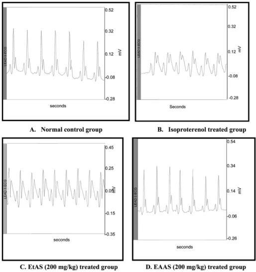
Effect of Argyreia speciosa on ECG parameters Fig. 1. represents the electrocardiographic pattern of control & experimental animals. Normal control and different doses of, EAAS, AQAS and EtAS (100 and 200 mg/kg) alone treated rats showed a normal ECG pattern, where as animals treated with ISO alone showed significant elevation in ST segment, reduction in P wave, QRS complex and R-R interval, in addition there was an increase in heart rate, prolongation of QT interval and cardiac cycles compared to normal control animals. Pretreatment of, EAAS, AQAS and EtAS (200 mg/kg) for 14 days and two doses of ISO (200 mg/kg) administered rats exhibited normal ECG pattern with a slight elevation in ST segment. Furthermore, treatments also resulted in significant (P < 0.001) alterations in P wave, QRS complex and R-R interval, whereas heart rate, QT interval and cardiac cycle were maintained near to normal values especially with EAAS. The data of the experimental animals such as P wave, QRS complex, QT interval, R-R
interval, heart rate & cardiac cycle are represented in Table 1.
Effect of Argyreia speciosa on serum marker enzymes ISO treated rats exhibited significantly (p < 0.001) higher levels of serum myocardial injury marker enzymes such as AST, ALT, LDH, CK, glucose, triglycerides and total cholesterol compared to normal control rats (Table 2). Pretreatment with EtAS and EAAS (200 mg/kg) for 14 days and ISO (200 mg/kg, for two days) administration showed significant (P < 0.05; P < 0.001) reduction in all the tested diagnostic markers compare to ISO alone treated group, where as AQAS is not showing significant attenuation in most of the parameters. However there were no changes in any of these marker enzyme levels in EtAS (200 mg), EAAS (200 mg) and AQAS (200 mg) alone treated groups as compared to normal control group.
Histopathological findings
Histopathological examination of myocardial tissue obtained from normal control animals and animals treated with A. speciosa fractions depicted clear integrity of myocardial membrane and an infiltration Table 1. Effect of Argyreia speciosa root fractions on ECG parameters in ISO-induced Myocardial infarction in rats.
Treatment P Wave QRS Complex Q-T Interval R-R Interval Heart Rate Cardiac Cycle
Normal control 0.03750 ±0.0014 0.04292 ±0.0009 0.08500 ±0.0033 0.1879 ±0.0033 321.2 ±5.87 0.1213 ±0.0037 EtAS (200 mg/kg,p.o) 0.03756 ±0.0012 0.04132 ±0.0007 0.08340 ±0.0032 0.1800 ±0.0079 325.3 ±19.25 0.1203 ±0.0019 EAAS (200 mg/kg,p.o) 0.03889 ±0.0011 0.04278 ±0.0008 0.08278 ±0.0031 0.1837 ±0.0086 328.0 ±17.61 0.1200 ±0.0029 AQAS (200 mg/kg,p.o) 0.03812 ±0.0011 0.04213 ±0.0006 0.08213 ±0.0030 0.1865 ±0.0084 326.1 ±9.2 0.1221 ±0.0023 ISO (200 mg/kg, s.c) 0.03278 ±0.0009a 0.03833 ±0.0014a 0.09944 ±0.0019b 0.1367 ±0.0030b 417.9 ±6.90b 0.1361 ±0.0033b ISO + EtAS (200 mg/kg,p.o) 0.03467 ±0.0089 0.03963 ±0.0011 0.08713 ±0.0012a 0.1215 ±0.0021 378.0 ±8.41 0.1284 ±0.0038 ISO + EAAS (200 mg/kg,p.o) 0.03833 ±0.0014a 0.04250 ±0.0012a 0.08233 ±0.0016a 0.1551 ±0.0052a 365.0 ±9.35b 0.1221 ±0.0040a The data were expressed as Mean ± S.E.M for six rats in each group. Statistical comparisons were performed by one-way ANOVA followed by Tukey’s post-test, The ECG parameters are expressed in seconds (sec) and the Heart rate as Beats per Minute (BPM). aP < 0.05, bP < 0.01 when compared with ISO treated group.
Table 2. Effect of Argyreia speciosa root fractions on serum biochemical parameters in Isoproterenol induced myocardial infracted rats
Groups Dose mg/ kg(p.o., s.c) ALT IU/L AST IU/L LDH IU/L CK IU/L Glucose mg/dl TG mg/dl TC mg/dl Normal control 2 ml/kg 66.40±10.04 97.31±9.437 1021±27.34 389.8±11.74 88.75±2.955 42.25±4.76 37.25±4.51 EAAS 200 63.75±10.52 106.8±5.67 1039±55.66 386.5±5.107 92.00±6.48 96.25±3.25 31.75±3.32 AQAS 200 52.20±6.55 105.3±5.36 995.3±51.69 390.8±5.313 87.3±25.78 322.5±12.61 56.50±6.19 EtAS 200 52.20±6.55 121.5±10.05 836.3±28.10 363.5±17.04 91.50±4.73 176.8±39.94 41.25±2.78 ISO 200 120.6±8.24c 195.2±7.04c 1924±191.2c713.8±21.10c 137.0±2.34c 119.0±3.24c 77.00±3.80c ISO + EAAS 200100 93.20±2.95 128.3±7.07 c 1786±34.82a674.0±43.21b 127.5±4.62a 22.25±4.55c 40.75±5.77b ISO + AQAS 200 100 93.50±5.26 93.75±7.29 1883±137.4 545.5±28.03 120.3±3.09 18.20±4.62 48.00±4.50 ISO +EtAS 200 100 79.75±5.51 a 104.8±8.35c 1535±84.30a591.3±54.80b 99.00±3.67a 20.75±3.902c42.50±5.23a ISO +EAAS 200 200 70.20±9.10 b 130.3±10.1c 1139±204.8b658.3±15.93c105.5±7.75b 31.14±3.96c 57.60±3.26c ISO + AQAS 200200 77.60±4.70 145.8±7.66 2441±87.26 b 517.0±42.59 108.0±8.38 52.96±15.21 67.60±3.20 ISO + EtAS 200 200 76.80±9.383 b 130.3±10.19c1229±204.7b545.3±26.05c100.0±4.56b 89.80±3.90b 65.40±2.42b
The data were expressed as Mean ± S.E.M for six rats in each group. Statistical comparisons were performed by one-way ANOVA followed by Tukey’s post-test, aP < 0.05, bP < 0.01, cP < 0.001 compared to Isoproterenol treated group
Fig. 2. Effect of different fractions of Argyreia speciosa on histopathological changes in rat myocardial tissue (H & E stain, ×100). A. Normal control: clear integrity of myocardial membrane and an infiltration of inflammatory cells were not seen in these experimental groups. B. Isoproterenol induced heart: Various degrees of focal lesions in many sections consisting of molten staining, fragmentation of muscle fibers with confluent retrogressive lesions were observed. In addition marked sequestering mucoid oedema and vacuolar changes along with hyaline necrosis were clearly visible in ISO treated rats. C. EtAS (200 mg/kg) treated group: Pretreatment with EtAS, (200 mg/kg) group demonstrated marked improvement in ISO-induced alterations such as vacuolar changes, edema, capillary dilatation and leukocyte infiltration compared to ISO administered group. D. EAAS (200 mg/kg) treated group: Pretreatment with EAAS (200 mg/kg) group also demonstrated marked improvement in ISO-induced alterations such as vacuolar changes, edema, capillary dilatation and leukocyte infiltration compared to ISO administered group.
of inflammatory cells were not seen in these experimental groups (Fig. 2A). The histological sections obtained from the hearts of animals receiving ISO alone (Fig. 2B) shows various degrees of focal lesions in many sections consisting of molten staining, fragmentation of muscle fibers with confluent retrogressive lesions. In addition marked sequestering mucoid edema and vacuolar changes along with hyaline necrosis were clearly visible in ISO treated rats. Pretreatment with EtAS, and EAAS (200 mg/kg) groups demonstrated marked improvement in ISO-induced alterations such as vacuolar changes, edema, capillary dilatation and leukocyte infiltration compared to ISO administered group (Fig. 2C&D) respectively.
DISCUSSION
Oxidative stress is one of the major concerns in the treatment of ischemic heart diseases. There is ample evidence for a detrimental role of ROS in cardiovascular disease (Hevener et al., 2002; Dhalla et a., 2000). Isoproterenol is well known cardiotoxic agent, destruct the myocardial cells, as a result of this, cytosolic enzymes such as lactate dehydrogenase (LDH), transaminases (ALT and AST) and creatine kinase (CK) were released into blood stream and serve as the diagnostic markers of myocardial tissue damage (Sawyer et al., 2004; Sabeena et al,. 2004). The amount of these cellular enzymes present in blood reflects the alterations in plasma membrane integrity and/or permeability. Drug treatments such as naringin, silibinin, and squalene evidenced by a decline in lactate dehydrogenase, glutamic oxalacetic transaminase and creatine kinase levels indicated their membrane stabilizing action (Sawyer et al., 2004; Sabeena et al., 2004; Gurgan et al., 2008; Rajadurai et al., 2006).
In the present study, ISO treated rats showed significant elevation in the levels of these diagnostic marker enzymes (AST, ALT, LDH and CK). Moreover, elevated levels of these enzymes are an indicator of the severity of ISO-induced
myocardial membrane necrosis, which is in line with an earlier report (Sawyer et al., 2004; Sabeena et al., 2004). The prior administration of EAAS (200 mg/kg p.o) and EtAS (200 mg/kg p.o) showed significant reduction in the ISO-induced elevated serum marker enzymes except CK. This reduction in enzyme levels could be due to its action on maintaining membrane integrity thereby restricting the leakage of these enzymes. It is well known that isoproterenol-induced myocardial injury is mediated primarily via the β1-adrenergic receptor. Acute β-adrenergic receptor stimulation not only rapidly generates reactive oxygen species, but also depresses total cellular antioxidant capacity, down regulates copper–zinc superoxide dismutase enzyme activity, protein and mRNA, and reduces glutathione level, leading to the loss of membrane integrity and inducing heart contractile dysfunction and myocyte toxicity finally producing myocardial necrosis (Zhon et al., 2006; Rathore et al., 1998). In the present study, we found that A. speciosa fractions protected myocardium from isoproterenol-induced myocardial functional and structural injury via normalization levels of diagnostic marker enzymes.
Current epidemiological evidence suggest that inadequate intake of certain nutrients predispose humans to chronic degenerative diseases (Srivastava et al., 2007). In particular it was demonstrated that intake of an adequate diet rich in vegetable and fruit reduces the likelihood of cardiovascular diseases, but the exact mechanisms for this protective effect are inadequately understood. However, increased circulating antioxidants are believed to be important. This is supported by recent trials reporting that the intake of antioxidant flavonols predicts a reduced rate of coronary-heart disease mortality in elderly male, in particular, those epidemiological studies show that dietary intake of flavanoids (quercetin, catechin and epicatechin), notably present in red wine but also in fruits and vegetables is inversely associated with subsequent coronary heart disease (Padmanaban et al., 2007;
Paritha et al., 1999). This effect seems to be in part related to their antioxidant activity. Therefore, the observed myocardial protective effect of EAAS and EtAS could be due to the flavanoids such as quercetin, kaempferol and coumarin like scopoletin (Petra et al., 1999; Srivastava et al., 1998)which are known free radical scavengers.
Electrocardiograph-abnormalities are the main criteria generally used for the definite diagnosis of myocardial infarction. ST-segment elevation was observed either in patient with acute myocardial ischemia (Seveviratine et al., 1999) or in isoproterenol-induced myocardial infarction in rat (Peacock et al., 2007). The study shows significant alterations of ECG patterns in ISO administered rats as compared to normal control rats. The characteristic findings were reductions in the P wave intensity, QRS complex, R-R intervals, QT interval and prolongation of cardiac cycle. We also observed a significant elevation in the ST segment and increase in heart rate. These alterations could be due to the consecutive loss of cell membrane in injured myocardium (Rajdurai et al., 2007). In the present study, we observed an elevation of ST-segments in isoproterenol-induced rat, and pretreatment with A. speciosa fractions markedly inhibited isoproterenol-induced ST-segment elevation suggestive of its cell membrane protecting effects. The appearance of Q wave & ST segment elevation are some of the indicative signs of ischemia. In the present study we did not observe pathological Q wave due to conditions of ischemia. The prominent Q wave were seen only on severe ischemia, infarction and in patients with severe heart diseases (Francis Morris et al., 2003). The consecutive loss of cellular membrane damage due to oxidative stress might be characterized by ST elevation (Holland et al., 1977; Kela et al., 1980). EAAS and EtAS (200 mg/kg p.o) administration showed a protective effect against ISO-induced altered ECG pattern and eliminated the acute fatal comp-lications by protecting the cell membrane damage. Electrocardiograph and biochemical findings were further supported by histopathological studies.
Histopathological examination of myocardial tissue in negative control depicted clear integrity of the myocardial cell membrane. No inflammatory cells infiltration was seen in the rat heart of normal control. In ISO administered group, focal lesions in many sections consisting of molted staining and fragmentation of muscle fibres with confluent retrogressive lesions, hyaline necrosis, sequestering mucoid edema were observed. Pretreatment with A. speciosa EtAS and EAAS fractions at 200 mg/kg demonstrated inhibited focal lesions, fragmentation of muscle fibres and retrogressive lesions with hyaline necrosis seen with ISO treated group. Inflammatory cells were seen with reduced density in EAAS and EtAS treated groups confirming the further cardioprotective activity exerted by A. speciosa. However EAAS and EtAS (200 mg/kg p.o) treated normal rats had no toxic effects on cardiac architecture. Higher dose of isoproterenol induce subendocardial ischemia, hypoxia, necrosis, and finally fibroblastic hyperplasia with decreased myocardial compliance and inhibition of diastolic and systolic function, which closely resembles local myocardial infarction-like pathological changes seen in human myocardial infarction (Karthick and Stanely, 2006). In the present study, we found that A. speciosa root fractions protected myocardium from isoproterenol-induced myocardial functional and structural injury. The phytochemical investigations of EAAS and EtAS revealed the presence of flavanoids, tannins and phenolics. In addition, we reported invitro and invivo antioxidant, antimicrobial, adaptogenic potentiality of flavanoid sulphates and their aglycones (quercetin and kaempferol) from the title plant (Habbu et al., 2008, 2009, 2010). The observed cardioprotective activity of EAAS and EtAS may be attributed to the presence of these bioactive principles and their synergetic properties. Further, the mode of cardioprotective action may be due to the prevention of stimulation of β-adrenergic receptor, there by decreasing the generation of reactive oxygen species which in turn maintains the membrane integrity in myocardial
tissue.
The data of the present study clearly shows that these fractions modulated the most of the electrophysiological, biochemical and histopathological parameters to normal status in experimental rats treated with isoproterenol, suggesting that the beneficial efffect of A. speciosa as a cardioprotective agent.
ACKNOWLEDGEMENTS
Authors are thankful to the president & principal, S.E.T’s college of pharmacy, Dharwad for providing necessary facilities to carry out this experiment. Dr. G.R. Hegde, professor Department of Botany, Karnatak University Dharwad is acknowledged for his help in identification and authentification of plant material.
REFERENCES
Dhalla NS, Temsah RM, Netticadan T. (2000) Role of oxidative stress in cardiovascular diseases. J. Hypertension. 18, 655–673.
Francis Morris, June Edhouse, William J Brady, John Camm. (2003) ABC OF CLINICAL ELECTROCAR-DIOGRAPHY, Chapter 8 Acute myocardial infarction—Part I 29-30. BMJ Publishing Group. Gokhale AB, Damre AS & Saraf MN. (2003)
Investigations in to the Immunomodulatory activity of Argyreia speciosa. J. Ethnopharmacol. 84, 109-114. Gokhale AB, Damre AS, Kulkarni KR & Saraf MN.
(2002) Preliminary evaluation of anti-inflammatory and anti-arthritic activity of S. lappa, A. speciosa and A. aspera. Phytomed. 9, 433-437.
Gürgün C, Ildizli M, Yavuzgil O, Sin A, Apaydin A, Cinar C, Kültürsay H. (2008) The effects of short term statin treatment on left ventricular function and inflammatory markers in patients with chronic heart failure. Int. J. Cardiol. 123, 102–107.
Habbu PV, Mahadevan KM, Kulkarni VH, Marietta P, Pratap V, Thippeswamy BS & Veerapur VP. (2010) Antidiabetic activity of Argyreia speciosa (sweet) (Burm.f.)Boj. in normoglycemic and Streptozotocin-induced diabetic rats. Orient. Pharm. Experment.
Med. 10, 90-102.
Habbu PV, Shastry RA, Mahadevan KM, Joshi HK & Das SK. (2008) Hepatoprotective and antioxidant effects of Argyreia speciosa in rats, Afr.J .Trad. Compl. Altern. Med. 5, 158-164.
Habbu PV, Shastry RA, Mahadevan KM & Sneha C (2009) Antiamnesic activity of Argyreia speciosa in mice. Int. J. Green Pharm. 4, 83-89.
Habbu PV, Shastry RA, Mahadevan KM, Manjunath H. (2009) Antimicrobial activity of flavanoids sulphates and other fractions of Argyreia speciosa (Burm.f.) Boj. Ind. J. Exp. Bio. 47, 121-127.
Hevener A, Reichart D, Janez A, Olefsky J. (2002) Female rats do not exhibit free fatty acid–induced insulin resistance. Diabetes. 51, 1907–1912.
Holland RP, Brooks H. (1977) TQ-ST segment mapping: critical review and analysis of current concepts. Am. J. Cardiol. 40, 110-129.
Karthick M, Stanely, Mainzen Prince P. (2006) Preventive effect of rutin, a bioflavonoid, on lipid peroxides and antioxidants in ISO-induced myocardial infarction in rats. J. Pharm. Pharmacol. 58, 701-707.
Kela AK, Reddy LP, Thrombe DP. (1980) ECG findings in normal rats and after administration of isoproterenol. Ind. J. Physio.l Pharmacol. 24, 84-90. Linz W, Wiemer G, Schaper J, Zimmermann R,
Nagasawa K, Gohlke P, Unger T. (1995) Angiotensin converting enzyme inhibitors, left ventricular hypertrophy and fibrosis. Mol. Cell Biochem. 147, 89–90. Lopez AD, Murray CC. (1998) The global burden
disease, 1990-2020. Nat Med. 4, 1241-1243.
Mochizuki S, Takeda N, Nagano M, Dhalla, NS. (1998) The ischemic heart. In: Mochizuki S. (Ed) Kluwer Academic Publishers.
Padmanabhan M, Stanely, Mainzen Prince. (2007) S-allylcysteine ameliorates isoproterenol-induced cardiac toxicity in rats by stabilizing cardiac mitochondrial and lysosomal enzymes. Life sciences. 80, 972-978. Paritha, Ithayarshi, Padmavathy V., Shyamala, Devi
CS. (1996) Effect of α-tocopherol on isoproterenol-induced Myocardial Infarction in rats-Electrocar-diographic Biochemical and histological evidences. Ind. J. Physiol. Pharmacol. 40, 297-302.
Peacock WF, Hollander JE, Smalling RW, Bresler MJ. (2007) Reperfusion strategies in the emergency treatment of ST-segment elevation myocardial infarction. Am. J. Emerg. Med. 25, 353-366.
Petra M, Britta T, Macki K, Eckart E. (1999) Flavanoid sulphates from Convolvulaceae. Phytochem. 50, 267-271. Rajadurai M, Stanely, Mainzen Prince P. (2006)
Preventive effect of naringin on lipid peroxides and antioxidants in isoproterenol-induced cardiotoxicity in Wistar rats: biochemical and histopathological evidences. Toxicol. 228, 259–268.
Rajadurai M, Stanely, Mainzen Prince P. (2007) Preventive effect of naringin on cardiac markers, electrocardiographic patterns and lysosomal hydrolases in normal and isoproterenol-induced myocardial infarction in Wistar rats. Toxicol. 230, 178-188. Rathore N, John S, Kale M, Bhatnagar D. (1998) Lipid
peroxidation and antioxidant enzymes in isoproterenol induced oxidative stress in rat tissues. Pharmacol. Res. 38, 297–303.
Sabeena, Farvin KH, Anandan R, Kumar SH, Shiny KS, Sankar TV, Thankappan TK. (2004) Effect of squalene on tissue defense system in isoproterenol-induced myocardial infarction in rats. Pharmacol. Res. 50, 231-236.
Sawyer DB, Siwik DA, Xiao L, Pimentel DR, Singh K, Colucci WS. (2002) Role of oxidative stress in myocardial hypertrophy and failure. J. Mol. Cell Cardiol. 34, 379–388.
Seneviratine CK, Timao Li, Khaper N, Singal PK. (1999) Effects of methionine on endogenous antioxidants in the heart. Am. J. Physiol. 277 (Heart Circ Physiol 46), H2122-H2128.
Sharma PC, Yelne, MB & Dennis TJ. (2004) Database on medicinal plants used in Ayurveda. pp 550, Central Council for Research in Ayurveda and Siddha New Delhi.
Srivastava A, Shukla Y. (1998) Aryl esters and a Coumarin from Argyreia speciosa, Ind. J. Chem. 37, 192-194.
Srivastava S, Chandrasekar B, Gu Y, Luo J, Hamid T, Hill BG, Prabhu SD. (2007) Down regulation of Cu, Zn-superoxide dismutase contributes to beta-adrenergic receptor-mediated oxidative stress in the heart. Cardiovasc. Res. 74, 445-455.
Stauss HM, Zhu YC, Redlich T, Adamiak D, Mott A, Kregel KC, Unger T. (1994) Angiotensin-converting enzyme inhibition in infarct-induced heart failure in rats: bradykinin versus angiotensin II. J. Cardiovasc. Risk. 1, 255–262.
Todd GL, Cullan GM. (1980) Isoproterenol-induced
myocardial necrosis and membrane permeability alterations in the isolated perfused rabbit heart. Experim. Mol. Path. 33, 43-45.
Varier PK. (1995) Indian medicinal plants. A compendium of 500 species. Vol. 2, pp235-237 Orient Longman Madras.
Warrier PK, Nambiar VP, Ramankutty C. (1994) Indian medicinal plants-a compendium of 500 species. pp191. Orient Longman New Delhi.
Wexler BC. (1973) Protective effects of propanolol in isoproterenol induced myocardial infarction. Atheroscler. 18, 11-14.
Wexler BC. (1978) Myocardial infarction in young vs old male rats: Pathophysiological changes. Am. Hear.t J. 96, 70-80.
White HD. (1996) Myocardial infarction. In: Julian DG, Camm AJ, Fox KM, Hall RJC, Poole Wilson PA (Ed) Diseases of the heart. pp1042-1075. W.B Saunders Company Ltd London.
Young IS, Purvis JA, Lightbody JH, Adgey AA, Trimble ER. (1993) Lipid peroxidation & antioxidant status following thrombolytic therapy for acute myocardial infarction. Eur. Heart. J. 14, 1027-1033. Zaijun Zhang, Jie Jiang, Pei Yu1, Xiangping Zeng,
James W Larrick. Yuqiang Wang. (2009) Hypoglycemic and beta cell protective effects of andrographolide analogue for diabetes treatment. J. Translat. Med. 7:62, doi:10.1186/1479-5876-7-62.
Zhou B, Wu LJ, Li LH, Tashiro S, Onodera S, Uchiumi F, Ikejima T, (2006) Silibinin protects against isoproterenol-induced rat cardiac myocyte injury through mitochondrial pathway after up-regulation of SIRT1. J. Pharmacol. Sci. 102, 387–395.
Zhu YZ, Zhu YC, Gohlke P, Unger T. (1998) Overview on pharmacological properties of angiotensin converting enzyme inhibitors. In: Hall AS, Unger T (Eds). ACE Inhibition and Target Organ Protection. Euromed Communications Ltd, 1–20.
Zhu YC, Zhu YZ, Gohlke P, Unger T. (1997) Effects of converting enzyme inhibition and angiotensin II AT1 receptor antagonism on cardiac parameters in left ventricular hypertrophy. Am. J. Cardiol. 80, 110A–117A.
Zhu YC, Zhu YZ, Spitznagel H, Gohlke P, Unger T. (1996) Substrate metabolism, hormone interaction and angiotensin converting enzyme inhibition in left ventricular hypertrophy. Diabetes. 45, S59–S65.