PGHN
Case Report
Non-Surgical Management of Gastroduodenal Fistula Caused by Ingested Neodymium Magnets
Claudia Phen*
,†, Alexander Wilsey*
,†, Emily Swan
†, Victoria Falconer
‡, Lisa Summers
§, and Michael Wilsey
∥*Office of Medical Education and ∥Pediatric Gastroenterology, Johns Hopkins All Children’s Hospital, St. Petersburg,
†College of Arts and Sciences, Florida State University, Tallahassee, ‡College of Arts and Sciences, University of Florida, Gainesville, FL, §Pediatric Gastroenterology, Carolines Healthcare System, Charlotte, NC, United States
Foreign body ingestions pose a significant health risk in children. Neodymium magnets are high-powered, rare-earth magnets that is a serious issue in the pediatric population due to their strong magnetic force and high rate of complications. When multiple magnets are ingested, there is potential for morbidity and mortality, including gastro- intestinal fistula formation, obstruction, bleeding, perforation, and death. Many cases require surgical intervention for removal of the magnets and management of subsequent complications. However, we report a case of multiple magnet ingestion in a 19-month-old child complicated by gastroduodenal fistula that was successfully treated by endoscopic removal and supportive care avoiding the need for surgical intervention. At two-week follow-up, the child was asymptomatic and upper gastrointestinal series obtained six months later demonstrated resolution of the fistula.
Key Words: Gastric fistula, Intestinal fistula, Magnets, Neodymium, Endoscopy
Received:August 4, 2017, Revised:December 3, 2017, Accepted:January 8, 2018
Corresponding author: Michael Wilsey, Pediatric Gastroenterology, Johns Hopkins All Children’s Hospital, 601 Fifth Street South, Suite 605, St.
Petersburg, FL 33701, United States. Tel: +1-727-822-4300, Fax: +1-727-456-1399, E-mail: [email protected] Copyright ⓒ 2018 by The Korean Society of Pediatric Gastroenterology, Hepatology and Nutrition
This is an openaccess article distributed under the terms of the Creative Commons Attribution NonCommercial License (http://creativecommons.org/licenses/by-nc/4.0/) which permits unrestricted noncommercial use, distribution, and reproduction in any medium, provided the original work is properly cited.
INTRODUCTION
Ingestion of foreign bodies, particularly magnets, and their subsequent complications have become in- creasingly common with the majority occurring in children less than five years of age [1]. Data from the National Electronic Injury Surveillance System from 2002 to 2011 reveals an estimated 16,386 children presented to emergency centers in the United States with possible magnet ingestions [2]. During this
10-year period, the magnitude of magnetic in- gestions by children has increased eight-and-a-half fold [2]. Increased frequency and morbidity have been noted specifically with the use of neodymium magnets. Neodymium magnets are high-powered, rare-earth magnets that exhibit over five times the force of traditional magnets, and have “demonstrated the tendency to cause gastrointestinal (GI) injury much more readily than their conventional counter- parts” [3]. This poses a significant threat of possible
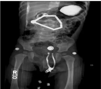
Fig. 1. Upper gastrointestinal series shows a series of foreign bodies attached end-to-end and looping from the stomach through the duodenum.
bleeding, perforation, and enteroenteric fistulae when ingested, which often requires surgical inter- vention [4]. We report the case of a 19-month-old who ingested 13 neodymium magnets, which cre- ated a gastroduodenal fistula. These high-powered, rare-earth magnets created a significant gastro- duodenal fistula, which was successfully treated with endoscopic removal and supportive care, avoid- ing the need for surgery.
CASE REPORT
A 19-month-old male in his usual state of good heath presented with a three-month history of pro- gressive non-bloody, non-bilious emesis. The moth- er denied history of fever, diarrhea, or abdominal pain. His growth parameters plotted above the 75th percentile for height and weight, and there was no previous history of gastroesophageal reflux symptoms.
He was referred to our institution where an upper GI series (UGI) showed 13 ingested foreign bodies con- nected end-to-end and looped from the stomach through the pylorus into the duodenum (Fig. 1). His mother reported that he had received a box of plastic colored magnets as a gift several months earlier.
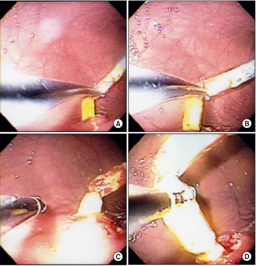
He was brought to the operating room where emergent endoscopy revealed that the most prox- imal, red-colored neodymium magnet in the stom- ach was magnetized to the anterior wall of the stom- ach (Fig. 2). Attempts to grasp the magnet with for- ceps were unsuccessful. The gastric foreign body had tightly adhered to the most distal, yellow-colored ne- odymium magnet located in the small intestine ad- jacent to the stomach. The forceps could not separate the magnets due to the strong magnetic attraction. A loop snare was inserted between the magnets and the proximal, red-colored magnet was tightly grasped. Significant force was applied to separate the proximal magnet from the wall of the stomach; how- ever, this only led to the emergence of the distal, yel- low-colored magnet from the small intestine through the gastric lumen (Fig. 2). This raised the possibility of intestinal perforation. Pediatric surgery was consulted intraoperatively. After much dis- cussion, the decision was made to continue attempts at endoscopic removal prior to considering surgical exploration, as the patient remained clinically stable with a benign abdominal examination.
The proximal magnet was once again tightly grasped with the loop snare and drawn closely to the tip of the scope for better control (Fig. 2). In one smooth motion, the endoscope was torqued in a clockwise fashion and quickly withdrawn with some force. This separated the proximal gastric magnet from the distal small bowel magnet. The magnets were withdrawn through the mouth, magnetized to one another in linear fashion. Repeat endoscopic in- spection of the area revealed a gastroduodenal fistu- la (Fig. 3), but no evidence of perforation.
The patient was admitted to the hospital for observation. He was given nothing by mouth and started on prophylactic intravenous antibiotics while pediatric surgery remained consulted. Follow-up 3-view abdominal films showed no evidence of perforation. He did not develop signs or symptoms of peritonitis. The patient did well during his hospital- ization. His diet was gradually advanced as tolerated, and he was discharged home after three days of observation. The patient did well clinically and re-
Fig. 2. (A) Forceps could not separate the magnets due to the strong magnetic attraction and therefore a loop snare was inserted between the magnets. Attempts to pull the imbedded magnet away from the gastric wall using the loop snare (B, C) only led to the emergence of a distal yellow magnet from the small intestine (D) in to the gastric lumen. This raised the concern for intestinal perforation.
mained asymptomatic at his outpatient clinic visit two weeks later. Follow-up UGI six months later showed no evidence of a persistent gastroduodenal fistula.
Informed patient consent was obtained for pub- lication of the case details.
DISCUSSION
Foreign body ingestions pose a significant health risk to the pediatric population. High-powered mag- net ingestions, in particular, are challenging clinical cases for pediatric gastroenterologists. These types of ingestions are particularly difficult due to their in- sidious presentation and high risk of complications.
The ingestion is often unwitnessed and associated
symptoms can be nonspecific. Initially, ingestion of multiple magnets may not produce physical exami- nation findings [5]. Furthermore, there is a paucity of clinical data in the form of prospective studies to guide management of these patients. A single mag- net may pass without complications; however, mul- tiple magnets usually require either endoscopy or surgical intervention [4]. In the presence of multiple magnets, there is the potential for significant mor- bidity and mortality. These include fistula for- mation, intestinal obstruction, perforation, peri- tonitis, bowel ischemia, pressure necrosis and death [6].
According to the most recent guidelines released in 2015 by the North American Society of Pediatric Gastroenterology, Hepatology and Nutrition
Fig. 3. (A, B) Several magnets are depicted as they emerge from the small intestine into the stomach. (C) A large round magnet is identified in the second portion of the duo- denum. (D) Gastroduodenal fistula is identified in the body of the stomach following magnet removal.
(NASPGHAN), urgent endoscopic removal is recom- mended in the presence of more than one magnet, even in asymptomatic patients [3]. If the magnets are lodged within the esophagus or stomach for less than 12 hours, then pediatric gastroenterology should be consulted. If the magnets have been pres- ent for greater than 12 hours, then consulting pedia- tric surgery is recommended prior to endoscopic removal. Once the magnets proceed beyond the pylo- rus, surgical intervention either by laparoscopy or laparotomy, is strongly recommended [4].
In the literature, there are few reported cases of fistula managed successfully with endoscopic re- moval alone. Hwang et al. [7] report a case of gas- tro-gastro-duodenal fistula formation in a 12-year- old male with autism following ingestion of twen-
ty-two magnets. The magnets were removed by poly- pectomy snare and the patient was treated with two-week course of proton-pump inhibitors. Follow-up endoscopy revealed resolution of the fistulas. Ohno et al. [8] also describe a case of gastroduodenal fistu- la in a 7-year-old autistic child that was successfully removed endoscopically one week after ingestion.
In our patient’s case, there were several in- dications in favor of endoscopic management in lieu of surgery. First and foremost, the chronicity of our patient’s presenting symptoms in the absence of fe- ver, weight loss, or alteration in bowel pattern sug- gests that the patient was a candidate for a less ur- gent approach. According to Sugawa et al. [9],
“surgical management is recommended if severe ab- dominal pain develops.” Those without signs of ob-
struction or acute distress can be handled less urgently. Our patient had a benign abdominal ex- amination and stable vital signs at the time of presentation. The absence of abdominal pain or dis- tension in our patient made it less likely that compli- cations such as bowel ischemia, obstruction, perfo- ration, or peritonitis were present. Review of the lit- erature suggests in the setting of multiple magnet in- gestion, laparotomy should be considered “if signs of intestinal distress develop” and “if the magnetic beads have passed the pylorus and cannot be re- trieved via endoscopy” [10]. In our patient’s case, the magnets were endoscopically retrievable using a loop snare, which successfully removed all but one magnet. Lastly, the abdominal imaging that was ob- tained did not show subcutaneous air, pneumo- mediastinum, free air under the diaphragm, or other radiologic signs that would support that the patient had a bowel perforation that would warrant a more invasive approach such as surgery.
Our case highlights a well-known phenomenon that high-powered, rare earth magnet ingestion can lead to life-threatening complications. There is a need for a high index of suspicion for enteral fistula formation, especially following neodymium magnet ingestion [4]. Our patient’s case also provides an ex- ample of a gastroduodenal fistula successfully treat- ed with endoscopic removal and supportive care, avoiding the need for surgery.
ACKNOWLEDGEMENTS
The authors would like to acknowledge the con- tributions of Alexander Kim, MD, Department of Medical Genetics, Johns Hopkins University School of Medicine, Baltimore, MD, USA for his critical re- view of the manuscript.
REFERENCES
1. Oestreich AE. Worldwide survey of damage from swal- lowing multiple magnets. Pediatr Radiol 2009;39:142-7.
2. Abbas MI, Oliva-Hemker M, Choi J, Lustik M, Gilger MA, Noel RA, et al. Magnet ingestions in children pre- senting to US emergency departments, 2002-2011. J Pediatr Gastroenterol Nutr 2013;57:18-22.
3. Kramer RE, Lerner DG, Lin T, Manfredi M, Shah M, Stephen TC, et al.; North American Society for Pediatric Gastroenterology, Hepatology, and Nutrition Endoscopy Committee. Management of ingested for- eign bodies in children: a clinical report of the NASPGHAN Endoscopy Committee. J Pediatr Gastro- enterol Nutr 2015;60:562-74.
4. Waters AM, Teitelbaum DH, Thorne V, Bousvaros A, Noel RA, Beierie EA. Surgical management and mor- bidity of pediatric magnet ingestions. J Surg Res 2015;199:137-40.
5. Dutta S, Barzin A. Multiple magnet ingestion as a source of severe gastrointestinal complications requir- ing surgical intervention. Arch Pediatr Adolesc Med 2008;162:123-5.
6. Bauman B, McEachron K, Goldman D, Louiselle A, Zheng E, Mills D, et al. Emergency management of the ingested magnet: an algorithmic approach. Pediatr Emerg Care 2017. doi: 10.1097/PEC.0000000000001168.
[Epub ahead of print]
7. Hwang J, Park M, Choi S, Park W, Kim AS. How strong construction toy magnets are! A gastro-gastro-duodenal fistula formation. J Pediatr Gastroenterol Nutr 2007;
44:291-2.
8. Ohno Y, Yoneda A, Enjoji A, Furui J, Kanematsu T.
Gastroduodenal fistula caused by ingested magnets.
Gastrointest Endosc 2005;61:109-10.
9. Sugawa C, Ono H, Taleb M, Lucas C. Endoscopic man- agement of foreign bodies in the upper gastrointestinal tract: a review. World J Gastrointest Endosc 2014;6:
475-81.
10. Tsai J, Shaul DB, Sydorak RM, Lau ST, Akmal Y, Rodriguez K. Ingestion of magnetic toys: report of seri- ous complications requiring surgical intervention and a proposed management algorithm. Perm J 2013;17:
11-4.