Abstract :
Key Words :
Two Surgical Cases of Squamous Cell Carcinoma of Temporal Bone
Joong Gahng Kim, M.D. , Eun Deok Kim, M.D. , In Hyuk Song, M.D. , Young Jin Nam, M.D. , Su Gil Sohn, M.D.
Department of Otolaryngology, Keimyung University School of Medicine , and Deafness Research Center, Dongsan Hospital , Daegu, Korea
117
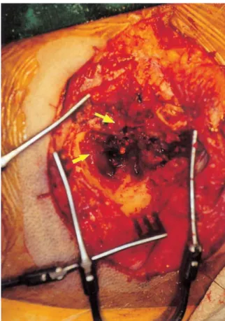
Fig. 1. Intraoperative finding in case 1 shows easily bleeding, exophytic tumor mass, occupying the left external auditory canal, middle ear cavity and mastoid air cells (arrows).
118
Fig. 2. Excised tumor mass in case 1.
Fig. 3. Intraoperative finding in case 2 shows a friable, well-encapsulated tumor mass (2 x 3 cm), occupying the right middle ear cavity and mastoid air cells with its adhesion to the dura mater (arrows).
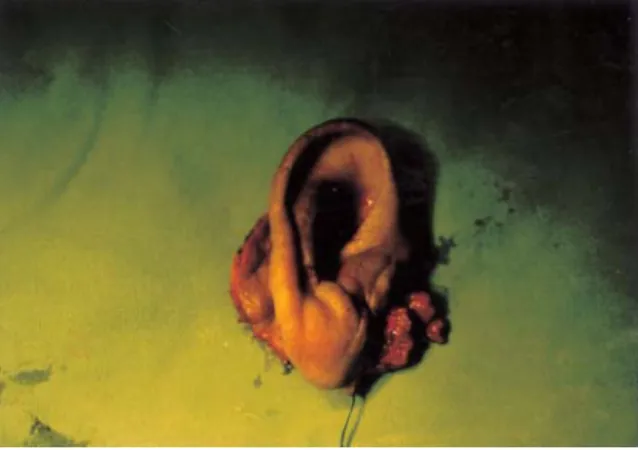
Fig. 4. Excised tumor mass including the right auricle in case 2.
119
120
1. Lewis JS. Squamous carcinoma of the ear. Arch Otolaryngol 1973;97:41-2.
2. Whitaker SR. Surgery for carcinoma of the ear. In:
Wiet RJ, Causse JB, editors. Complications in Otolaryngology Head and Neck Surgery. Vol 1.
Philadelphia: Decker Inc;1986, p.161-78.
3. Crabtree JA, Britton BH, Pierce MK. Carcinoma of the external auditory canal. Laryngoscope 1976;86:405-15.
4. Kim TH, Choi IS, Park JY, Jun BH. A case of adenoid cystic carcinoma of external auditory canal.
Korean J Otolaryngol 1996;39:1714-9.
5. Ahn CM, Chun YH, Chung DH, Choi JO. Five cases of squamous cell carcinoma in external auditory canal. Korean J Otolaryngol 1992;35:500-4.
6. Suh HK, Choi G, Lee KS, Hwang SJ. Adenoid cystic carcinoma of external auditory canal. Korean J Otolaryngol 1990;33:617-22.
7. Lee WS, Park YE, Lee K, Lee KB. A case of adenoid cystic carcinoma. Korean J Otolaryngol 1985;28;
473-7.
8. CS Kim, SO Chang, SH Oh, JW Koo, JH KIm, WS Yoo. Subtotal temporal bone resection for malignancies of the temporal bone. Korean J Otolaryngol 1998;41:1406-12.
9. Kinney SE. Tumors of the external auditory canal, middle ear, mastoid and temporal bone. In: Thawley SE, Panje WR, Batsakis JG, Lindberg RD, editors.
Comprehensive Management of Head and Neck Tumors. Vol 2. Philadelphia: Saunders;1987, p.182.
10. Wagenfeld DJH, Keane T, Nostrand AWP, Bryce DP. Primary carcinoma involving the temporal bone: analysis of twenty-five cases. Laryngoscope 1980;90:912-9.
11. Michaels L, Wels M. Squamous cell carcinoma of the middle ear. Clin Otolaryngol 1980;5:235-48.
12. Spector JG. Management of temporal bone carcinomas: a therapeutic analysis of two groups of patients and long-term followup. Otolaryngol Head Neck Surg 1991;104:58-66.
13. Gloria-Cruz TI, Schachern PA, Paparella MM.
Metastases to temporal bones from primary nonsystemic malignant neoplasms. Arch Otolaryngol Head Neck Surg 2000;126:209-14.
14. Arriaga M, Curtin H, Takahashi H, Hirsch BE, Kamerer DB. Staging proposal for external auditory meatus carcinoma based on preoperative clinical examination and computer tomography findings.
Ann Otol Rhinol Larynogol 1990;99:714-21.
121
15. Adams GL. Cancers involving the middle ear and temporal bone. In: Head and Neck Cancer. Chicago:
Yearbook medical publishers;1986, p.387-99.
16. Kinney SE. Malignancies of the temporal bone- limited temporal bone resection. In: Brackmann DE, Shelton C, Arriaga MA, editors. Otologic Surgery. Philadelphia: Saunders;1994, p.37-47.
17. Prasad S, Janecka IP. Efficacy of surgical treatments for squamous cell carcinoma of the temporal bone : a literature review. Otolaryngol Head Neck Surg 1994;110:270-80.
18. Kenyon GS, Marks PV, Scholtz CL, Dhillon R.
Squamous cell carcinoma of the middle ear: a 25
year retrospective study. Ann Otol Rhinol Laryngol 1985;94:273-7.
19. Kinney SE, Wood BG. Malignancies of the external ear canal and temporal bone: surgical techniques and results. Laryngoscope 1987;97:158-64.
20. Myers EN, Jamess YS. Cancer of the Head and Neck. 2nd ed. Churchill Livingstone; 1989, p.691- 709.
21. Goodwin WJ, Jesse RH. Malignant neoplasms of the external auditory canal and temporal bone. Arch Otolaryngol 1980;106:657-79.
22. Coleman CC. Removal of the temporal bone for cancer. Am J Surg 1966;79:583-90.