서 론
진주종성 중이염의 치료 원칙은 진주종의 완전 제거, 재발의 방지 그리고 청력 보전에 있으며, 이를 위해 주로 개방형 유양동 삭개술(canal wall down mastoidectomy)
등을 시행하고 있다. 개방형 유양동 삭개술은 진주종을 제거할 수 있는 충분한 시야를 제공하여, 진주종 제거 및 재발을 방지하는데 유리하지만,1) 수술 후 회복기간이 길 며, 필연적으로 공동을 형성하게 되어, 정기적인 공동 청 소가 필요한 공동 문제(Cavity problem)가 생긴다. 또한
논문접수일: 2020년 4월 3일 / 논문수정일: 2020년 4월 21일 / 심사완료일: 2020년 5월 12일 교신저자 : 강명구, 49201 부산광역시 서구 대신공원로 26 동아대학교 의과대학 이비인후과학교실 전화: (051) 240-5428・전송: (051) 253-0712・E-mail: [email protected]
J Clinical Otolaryngol 2020;31:63-69 증 례
외이도 재건을 위해 잔존 외이도 후벽을 보존한 채 시행한 개방형 유양동 삭개술 4예
동아대학교 의과대학 이비인후과학교실
김대연
·
김상준·
이동근·
강명구Canal Wall Down Mastoidectomy with Preservation of Remnant Posterior Canal Wall for Mastoid Obliteration and Canal Wall Reconstruction: 4 Cases
Daeyeon Kim, MD, Sangjun Kim, MD, Dong Kun Lee, MD, PhD, and Myung Koo Kang, MD, PhD Department of Otorhinolaryngology, Head and Neck Surgery, Dong-A University,
College of Medicine, Busan, Korea
- ABSTRACT -
The aim of cholesteatoma surgery is to completely eradicate cholesteatoma and to reconstruct the normal anatomy of the middle ear. Canal wall down mastoidectomy (CWD) is often applied for the complete eradication of choles- teatoma and prevention of recurrence in patients with cholesteatoma. However, CWD involves a significant de- struction of the middle ear and the mastoid anatomy, that leads to potential complications including chronically draining cavity, retention of debris that requires frequent cleaning, and difficulty in fitting a hearing aid. To resolve these problems, surgeons have attempted to reconstruct posterior external auditory canal wall with a variety of techniques and materials. We recently experienced four cases of patients who had tympanomastoid surgery be- cause of cholesteatoma. In the operation, superior segment of posterior canal wall was removed but remnant infe- rior segment of posterior canal wall was preserved. CWD with preservation of the remnant canal wall was per- formed unlike the conventional CWD operation. Surgical view was sufficient to eradicate the entire cholesteatoma.
Canal wall reconstruction was done with remnant canal wall and conchal cartilage, and then mastoid obliteration was done with Tissue glue mixed demineralized bone matrix. This procedure was enough to eradicate cholesteato- ma without lowering facial ridge. Saucerization, mastoid tip amputation and meatoplasty were not even needed. We report these four cases with a review of literature. (J Clinical Otolaryngol 2020;31:63-69)
KEY WORDS: MastoidectomyㆍEar canalㆍCholesteatomaㆍReconstructive surgical procedure.
개방형 유양동 삭개술을 시행한 뒤 유양동 폐쇄술과 외 이도 후벽 재건술을 성공적으로 시행한 4예(Table 1)를 치험 하였기에 문헌고찰과 함께 보고하고자 한다.
증 례
증례 1
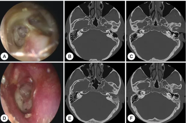
70세 남자가 1년 전부터 좌측 귀에 이물이 들어갈 시 어지러움이 발생하였으며, 내원 3일 전부터는 좌측 안 면마비가 발생하여 내원하였다. 고혈압 외 다른 과거력 은 없었다. 신체 진찰에서 House-brackmann grade 3 의 좌측 안면마비가 있었으며, 고막 내시경 검사에서 좌 측 외이도 후벽을 침범한 진주종이 관찰되었다(Fig.
1A). 측두골 전산화단층촬영상 상고실(epitympanum) 및 유양동(mastoid cavity)에 진주종으로 의심되는 연부 조직 음영이 관찰되었으며, 안면신경 침범이 동반되어 있었고, 수평반규관누공 소견도 관찰되었다(Fig. 1B, C). 진주종의 제거를 위해 개방형 유양동 삭개술을 계획 하였다. 수술은 전신 마취 하에 후이개 절개술을 시행한 후 고막 재건 및 유양동 폐쇄를 위해 측두 근막을 채취 하였고, 유양동 폐쇄를 위해 전방 기저 근골막 피판(an- terior based musculoperiosteal flap)과 이개 연골을 준 비하였다. 이 후 개방형 유양동 삭개술 및 상고실절제술 (epitympanectomy)을 시행하였다. 개방형 유양동 삭개 술은 고전적인 개방형 유양동 삭개술과는 다르게 진주 종을 충분히 제거할 수 있을 정도로만 외이도 후벽을 제 거하고 가능한 외이도 후벽의 하부를 남겨두었다(Fig.
3A). 유양동을 Tissue glue(TisseelⓇ, Baxter Healthcare) 을 섞은 동종 골기질(demineralized bone matrix: Re- genafilⓇ; Regeneration Technologies Inc., FL, DBM) 로 폐쇄하며 외이도 후벽의 모양을 만든다. 진주종 제거 Table 1.
Patients demographic data PatientSex/ ageDiagnosisPast historyLesion of cholesteatomaSurgery17) Reconstruction 1M/70Attic cholesteatoma [L], Labyrinthine fistula [L]NoneEpitympanum, aditus, mastoid cavity, lateral semicircular canal
CWD c POR [L] fistula repair [L]Concha cartilage, DBM, Ant.based flap 2M/58Recur.Cholesteatoma [L]CWD state [L]Epitympanum, aditus, mastoid cavityCWD [L]Concha cartilage, DBM, bone chip, fascia, Ant.based flap 3F/74
Recur.Cholesteatoma [R] Labyrinthine fistula [R]
CWU state [R]Mesotympanum, epitympanum, aditus, mastoid cavity lateral semicircular canal
CWD c T3 [R] fistula repair [R]Concha cartilage, DBM, megaderm, Ant.based flap 4M/35Attic Cholesteatoma [R]NoneCWD c T1 [R]Concha cartilage, DBM, fascia, Ant.based flap DBM
: Demineralized bone matrix, CWD : Canal wall down mastoidectomy, Ant.based flap
: Anterior based musculoperiosteal 1, T3
: Tympanoplasty type 3, PORP : Partial ossicular replacement prosthesis, fascia : temporalis muscle fascia
를 위해 제거된 외이도 후벽의 결손 부위는 연골을 덮어 외이도의 모양을 완성하였다(Fig. 3B, C). 연골 위는 측 두 근막으로 덮고 그 위에 근골막 피판과 외이도 피부로 덮었다(Fig. 3D). 외이도 안에는 Silastic sheet 삽입 및 거즈패킹을 통해 외이도의 모양을 유지하였다. 술 후 4 개월 뒤 고막 내시경 검사에서 고막 천공이 있으나, 좌 측 외이도 후벽이 유지되있음을 확인하였다(Fig. 1D).
측두골 전산화단층촬영상 유양동 폐쇄술 및 외이도 후 벽이 유지되있음을 확인하였다(Fig. 1E, F).
증례 2
59세 남자로 10년 전에 좌측의 진주종성 중이염으로 타 병원에서 개방형 유양동 삭개술을 받았던 과거력이 있으며, 최근 이루가 다시 발생하여 내원하였다. 고막
내시경 검사에서 좌측 외이도 후벽을 침범한 진주종이 관찰되었다(Fig. 2A). 측두골 전산화단층촬영에서 상고 실 및 유양동에 진주종으로 의심되는 연부조직 음영이 관찰되었으며, 외이도 후벽은 보전되어 있었다(Fig. 2B, C). 재발성 진주종 제거를 위해 개방형 유양동 삭개술 을 시행하였다. 유양동 삭개술의 방법은 증례 1의 방법 과 동일하게 이루어졌다. 술 후 5개월 뒤 확인 시 고막 내시경 검사에서 좌측 외이도 후벽이 유지되있음을 확 인하였다(Fig. 2D). 측두골 전산화단층촬영상 유양동 폐쇄술 및 외이도 후벽이 유지되있음을 확인하였다 (Fig. 2E, F).
증례 3
74세 여자로 3년 전 우측의 진주종성 중이염으로 타 병 Fig. 1. Representative case. Pre- and Postoperative findings in patients with cholesteatoma in the left ear. A: Preop- erative endoscopic finding showing cholesteatoma protruding external auditory canal. B, C: Preoperative comput- ed tomography scan showing attic cholesteatoma involving epitympanum, mesotympanum and mastoid cavity with lateral semicircular canal fistula and facial nerve dehiscence in the left ear (white arrow) with remnant canal wall (white circle). D: Four-month postoperative endoscopic finding showing external auditory canal reconstruction in the left ear. E, F: Four-month postoperative computed tomography scan showing posterior canal wall reconstruc- tion and mastoid obliteration with remnant canal wall (white circle).
A
D
B
E
C
F
Fig. 2. Representative case. Pre- and Postoperative findings in patients with cholesteatoma in the left ear. A: Preop- erative endoscopic finding showing canal wall down state with tympanic membrane adhesion. B, C: Preoperative computed tomography scan showing canal wall down mastoidectomy state with remnant mastoid mucosa in the left ear with remnant canal wall (white circle). D: Five-month postoperative endoscopic finding showing external au- ditory canal reconstruction in the left ear. E, F: Five-month postoperative computed tomography scan showing pos- terior canal wall reconstruction and mastoid obliteration with remnant canal wall (white circle).
A
D
B
E
C
F
원에서 개방형 유양동 삭개술을 받았던 과거력이 있으며, 최근 우측의 이루 및 어지러움으로 내원하였다. 고막 내 시경 검사에서 우측 상고실의 연부조직 부종이 및 고막 천공이 관찰되었다. 측두골 전산화단층촬영에서 상고실, 중고실 및 유양동에 진주종으로 의심되는 연부조직 음영 이 관찰되었으며, 안면신경 침범과 수평반규관누공이 확 인되었다. 진주종 제거를 위해 개방형 유양동 삭개술을 시행하였다. 유양동 삭개술의 방법은 증례 1의 방법과 동 일하게 이루어졌다. 술 후 2개월 뒤 확인 시 고막 내시경 검사에서 우측 외이도 후벽이 유지되있음을 확인하였다.
증례 4
35세 남자가 증상없이 우연히 발견된 우측의 상고실 진주종성 중이염으로 내원하였다. 고막 내시경 검사에 서 우측의 상고실을 침범한 진주종이 관찰되었다. 측두
골 전산화단층촬영에서 상고실 및 유양동에 진주종으 로 의심되는 연부조직 음영이 관찰되었다. 진주종 제거 를 위해 개방형 유양동 삭개술을 시행하였다. 유양동 삭개술의 방법은 증례 1의 방법과 동일하게 이루어졌 다. 술 후 2개월 뒤 확인 시 고막 내시경 검사에서 우측 외이도 후벽이 유지되있음을 확인하였다
고 찰
진주종 치료의 이상적인 목표는 진주종을 완전히 제 거하면서 재발을 방지하며 소리가 잘 전달되는 중이강 을 형성하고, 외이도를 재건하는 것이다. 이를 위하여 다양한 수술 방법이 개발되어 왔으며 진주종의 위치나 파급 정도에 맞춰 다양하게 적용되었다. 개방형 유양동 삭개술은 귀의 정상적인 구조를 파괴하는 수술이지만,
진주종을 제거하기 위한 충분한 시야를 제공할 수 있어 진주종 제거 및 재발 방지에 유리한 수술이다.1) 하지만 개방형 유양동 삭개술은 진주종 제거 및 시야 확보를 위해 외이도 후벽을 제거해야 하며, 이로 인해 공동을 만들게 된다. 외이도 후벽의 제거로 인해 공동에서 이 구의 자연 배출 장애가 발생하여 지속적인 이비인후과 치료가 필요한 부작용이 있으며, 수술 후 회복시간이 길며, 반규관 노출로 인한 어지러움, 보청기 착용 제한 등의 문제 등으로 환자의 삶의 질을 저하시킨다.2,3) 수 술 후 발생하는 공동 문제를 줄이기 위해 배상형성술
(Saucerization), 충분한 안면신경릉(facial ridge) 낮추 기, 외이도성형술(Meatoplasty), 유돌첨(Mastoid tip)의 제거, 유양동 폐쇄술(Mastoid obliteration) 등이 시행되 고 있으나, 앞서 언급한 부작용들에서 완전히 자유로울 수는 없었다.7) 그래서 진주종 제거를 위한 충분한 시야 를 얻으면서도 개방형 유양동 삭개술 후 외이도 후벽 재건을 위해 여러가지 재료를 사용하였고,4,5) 외이도 후 벽을 microsagittal saw로 절제하여 보존한 뒤 진주종 제거 후 외이도 후벽을 재건하는 등의 시도가 있다.8) 외 이도 재건을 위한 유양동 폐쇄 재료로 근골막 피판 과 Fig. 3. Intraoperative findings. A: Canal Wall Down Mastoidectomy preserving remnant posterior canal wall was per- formed (arrow). B: Posterior canal wall reconstruction was done by using piece of concha cartilage (white arrow- head). C: Mastoid cavity was obliterated and covered with DBM with tissue glue complex (arrowhead). D: Anterior based musculoperiosteal flap (white arrow) was repositioned into epitympanium.
A
C
B
D
되고, 높은 안면신경릉에 의해 유돌첨에 주머니 공간을 형성한다. 직립보행에 따라 중력에 의해 각질(Keratin), 이구등이 유돌첨의 공간에 쌓이게 되고 배출의 지장이 생기게 된다.10) 그리하여 유양동 폐쇄 시 부피의 제한이 없고 흡수가 적게 일어나는 재료들을 사용하려고 하였 으며, 대표적으로 골분, Hydroxyapatite cement, DBM, 실리콘 블록(Silicone block) 등이 있다.11,12)
골 이식 시 재료가 가져야 할 이상적인 세가지 속성은 골전도(Osteoconduction), 골유도(Osteoinduction), 골 융합(Osteointergration)이다. 세가지 속성 다 높을수록 좋은 재료이나 그 중에서도 순서상 가장 늦게 일어나는 골융합이 가장 중요하며 필수적이다.13) 재료별로 살펴보 면 자가골 이식의 경우 골전도, 골유도, 골융합의 효과를 모두 가지고 있어 가장 좋으나, 충분한 양을 얻기 어렵 다는 단점이 있다. 골 시멘트(bone cement)는 뼈와 인공 관절을 고정하기 위해 사용하는 물질로 Polymethyl methacrylate로 이루어져 있으며 사용 시 골전도가 약하 게 있고 골유도, 골융합의 기능이 없다. 실리콘 블록은 유양동 폐쇄를 위한 충분한 부피를 제공할 뿐, 골전도, 골유도, 골융합의 효과는 모두 다 없고 향후 실리콘 제 거가 필요할 수도 있다. Hydroxyapatite cement는 골전 도, 골유도, 골융합의 효과가 다 있으나 DBM 이식보다 효과가 적다.14) DBM의 경우 사체에서 탈무기질 화 시킨 동종이식 골 조직으로 골전도 효과는 약하지만 골유도 및 골융합 기능이 강하여 가장 좋은 골 이식 재료라고 할 수 있다.15) 동종 DBM은 골 조직 및 Collagen으로 구 성되어 다른 물질보다 생체 이식 시 염증 및 이물 반응 이 적은 것으로 알려져 있어 술 후 감염에 유리하다고 볼 수 있다. 하지만 이식 후 염증 반응을 줄이기 위해 술 후 배액관 삽입 및 항생제를 사용할 수 있다.14) DBM을 사용 할 때 외이도 성형을 위해 Tisseel을 혼합하여 사용
이도 재건이 어려워지기 때문이다. 또한 DBM의 적용으 로 인해 외이도 후벽의 재건에 충분한 부피를 확보할 수 있게 되어, 유양동 폐쇄 시 근골막 피판의 사용을 줄일 수 있게 된다. 본 술식에서는 근골막 피판을 연골 앞에 두어 연골이 확산에 의한 영양공급이 잘될 수 있도록 하 였으며 이는 외이도 형태 유지에 도움이 될 것으로 예상 하고 있다. 폐쇄 물질로 DBM이 상당히 유용 하다고 생 각되며 추후 장기적 추적 관찰과 연구가 필요하다. 외이 도 후벽을 모두 제거하지 않고 일부만 제거한 상태에서 진주종 제거를 위한 충분한 시야를 확보한다면, 잔존 외 이도 후벽이 남아 있어 외이도 재건 시 흡수될 가능성도 적고 모양의 유지에도 유리하다고 생각된다.
본 사례에서는 4명의 환자 모두 잔존 외이도 후벽을 남긴 상태에서 개방형 유양동 삭개술을 시행하였고 이 후 동종 골기질, 연골 등을 이용하여 외이도 재건술 및 유양동 폐쇄술을 시행하였다. 수술 후 진주종의 재발은 보이지 않았고 외이도 후벽이 재건된 것을 확인할 수 있었다. 진주종 수술 후 합병증을 막기 위해서는 진주 종의 제거가 가장 중요하며 이를 위해 진주종 제거를 위해 개방형 유양동 삭개술 및 외이도 재건이 많이 사 용된다. 다만 본 사례와 같이 외이도 후 아래벽을 남기 고 진주종을 제거할 수 있다면, 술 후 외이도 재건이 유 리하다. 성공적인 외이도 재건은 술 후 합병증을 줄일 수 있고, 환자의 삶의 질을 높일 수 있다. 본 사례에서는 새로운 술식의 발표로 인하여 2~5개월의 추적기간을 가지고 중간발표로 논문을 발표하였다. 5년 이상의 장 기간의 진주종의 재발의 확인이 필요하며 외이도 모양 의 유지에 대해서도 추적관찰이 필요하다.
중심 단어: 진주종・유양동・삭개술・외이도.
REFERENCE
1) Karmarkar S, Bhatia S, Saleh E, DeDonato G, Taibah A, Russo A, et al. Cholesteatoma surgery: the individualized technique. Ann Otol Rhinol Laryngol 1995;104(8):591-5.
2) Palmgren O. Long-term results of open cavity and tympa- nomastoid surgery of the chronic ear. Acta Otolaryngol 1979;88(5-6):343-9.
3) Sade J, Weinberg J, Berco E, Brown M, Halevy A. The marsupialized (radical) mastoid. J Laryngol Otol 1982;96 (10):869-75.
4) Kim BG, Kim HJ, Lee SJ, Lee E, Lee SA, Lee JD. Out- comes of modified canal wall mastoidectomy and mastoid obliteration using autologous materials. Clin Exp Otorhi- nolaryngol 2019;12(4):360-6.
5) Mobashir MK, Basha WM, Mohamed AES, Elmaghawry ME. Posterior canal wall reposition for management of cho- lesteatoma: technique and results. Auris Nasus Larynx 2018;
45(2):254-60.
6) Yoon SW, Hong JC, Jeong SW, Kang MK. The two cases of the canal wall down mastoidectomy & mastoid oblit- eration using Tachosil in recurrent huge cholesteatoma of middle ear. J Clinical Otolaryngol 2016;27:322-6.
7) Min GM, Park G, Moon TO, Hong SB. Partial mastoid obliteration using inferior based musculoperiosteal flap and autogenous conchal cartilage chips. Korean J Otolaryngol 1999;42(7);843-8.
8) Mobashir MK, Basha WM, Mohamed AES, Elmaghawry ME. Posterior canal wall reposition for management of cholesteatoma: technique and results. Auris Nasus Larynx 2018;45(2):254-60.
9) Skoulakis C, Koltsidopoulos P, Iyer A, Kontorinis G. Mas- toid obliteration with synthetic materials: a review of the lit- erature. J Int Adv Otol 2019;15(3):400-4.
10) Wormald PJ, Nilssen EL. The facial ridge and the dis- charging mastoid cavity. Laryngoscope 1998;108(1 Pt 1):92-6.
11) Han CS, Kim HB, Park JR, Jeong EH, Oh JG, Lee WY, et al. Reconstruction of the posterior canal wall with mastoid obliteration after canal wall down mastoidectomy. Korean J Otolaryngol 2008;51(1):33-40.
12) Cho SW, Cho YB, Cho HH. Mastoid obliteration with sili- cone blocks after canal wall down mastoidectomy. Clin Exp Otorhinolaryngol 2012;5(1):23-7.
13) Albrektsson T, Johansson C. Osteoinduction, osteocon- duction and osseointegration. Eur Spine J 2001;10 Suppl 2:S96-101.
14) Gu TW, Jang YS, Kim SH, Kim SJ, Hong SH, Kang MK.
Histopathologic study on the obliteration of the temporal dorsal bullae in rat using hydroxyapatite cement and de- mineralized bone matrix. Korean J Otolaryngol 2008;
51(9):777-82.
15) Kirk JF, Ritter G, Waters C, Narisawa S, Millán JL, Talton JD. Osteoconductivity and osteoinductivity of Nano- FUSE(®) DBM. Cell Tissue Bank 2013;14(1):33-44.
16) Lee HJ, Chao JR, Yeon YK, Kumar V, Park CH, Kim HJ, et al. Canal reconstruction and mastoid obliteration using floating cartilages and musculoperiosteal flaps. Laryngo- scope 2017;127(5):1153-60.
17) Wullstein H. Theory and practice of tympanoplasty. La- ryngoscope 1956;66(8):1076-93.