Open Access
Tumor Stage-Related Role of Radiotherapy in Patients with an External
Auditory Canal and Middle Ear Carcinoma
Original Article
Purpose
The purpose of this study was to evaluate the clinical outcomes of patients treated with radiotherapy (RT) for a carcinoma of the external auditory canal (EAC) and middle ear. Materials and Methods
The records of 32 patients who received RT from 1990 to 2013 were reviewed retrospec-tively. The Pittsburgh classification was used to stage all the cancers (early stage, T1/T2 [n=12]; advanced stage, T3/T4 or N positive [n=20]). Twenty-one patients (65.6%) were treated with postoperative RT and 11 patients (34.4%) were treated with definitive RT. The median radiation doses for postoperative and definitive RT were 60 Gy and 64.8 Gy, respectively. Chemotherapy was administered to seven patients (21.9%).
Results
The 5-year overall survival and disease-free survival rates for all patients were 57% and 52%, respectively. The disease control rates for the patients with early stage versus advanced stage carcinoma were 55.6% (5/9) and 50% (6/12) in the postoperative RT group and 66.7% (2/3) and 37.5% (3/8) in the definitive RT group, respectively. Overall, 15 cases (14 patients, 46.7%) experienced treatment failure; these failures were classified as local in four cases, regional in one case, and distant in 10 cases. The median follow-up period after RT was 51 months (range, 7 to 286 months).
Conclusion
Patients with early stage carcinoma achieved better outcomes when definitive RT was used. Advanced stage carcinoma patients experienced better outcomes with postoperative RT. The high rate of distant failure after RT, with or without surgery, reflected the lack of a con-sensus regarding the best therapeutic approach for treating carcinoma of the EAC and mid-dle ear.
Key words
Radiotherapy, Ear canal, Middle ear
Jinhyun Choi,
MD1Se-Heon Kim,
MD, PhD2Yoon Woo Koh,
MD, PhD2Eun Chang Choi,
MD, PhD2Chang Geol Lee,
MD, PhD3Ki Chang Keum,
MD, PhD31Department of Radiation Oncology,
Gangnam Severance Hospital,
Yonsei University College of Medicine, Seoul,
2Department of Otorhinolaryngology,
Yonsei University College of Medicine, Seoul,
3Department of Radiation Oncology,
Yonsei Cancer Center, Yonsei University College of Medicine, Seoul, Korea
+ + + + + + + + + + + + + + + + + + + + + + + + + + + + + + + + + + + + + + + + + + + + + + + + + + + + + + + + + + + + + + + + + + + + + + + + + + + + + + + + + + + + + + + + + + + + + + + + + + + + + + + + + + + + + + + + + + + + + + + + + + + + + + + + + + + + + + + + + + + + + + + + + + + + + + + + + + + + + + + + + + + + + + + + + + + + + + + + + + + + + + + + + + + + + + + + + + + + + + + + + + + + + + + + + + + + + + + + + + + + + + + + + + + + + + + + + + + + + + + + + + + + + + + + + + + + + + + + + + + + + + + + + + + + + + + + + + + + + + + +
Correspondence: Ki Chang Keum, MD, PhD Department of Radiation Oncology, Yonsei Cancer Center, Yonsei University College of Medicine, 50-1 Yonsei-ro, Seodaemun-gu, Seoul 03722, Korea Tel: 82-2-2228-8112
Fax: 82-2-312-9033 E-mail: [email protected] Received April 20, 2016 Accepted June 3, 2016 Published Online July 4, 2016
Introduction
Cancer of the external auditory canal (EAC) and middle ear is rare, with a prevalence of one to six patients per 1 mil-lion people [1-3]. The disease presents variable patterns of clinical practice in terms of management. Although several treatment modalities have been investigated, there are no standard guidelines for treating cancers of the EAC and mid-dle ear due to a lack of prospective randomized studies. A
surgical resection with a clear tumor margin is usually rec-ommended for resectable disease [4]. On the other hand, the role of adjuvant and definitive radiotherapy (RT) in treating unresectable disease is less well-defined.
The prognosis of an EAC carcinoma depends on the dis-ease stage and the primary treatment approach used [5]. Investigators have reported the 5-year survival rates ranging from 10% for advanced stage to as high as 85% for early stage disease [4,6-9]. In the literature, the 5-year locoregional con-trol rates range from 20% for advanced stage to 70% for early
stage disease [10]. As there is no widely accepted staging sys-tem, using the results from different reports to compare the treatment outcomes can be difficult [3,11]. Moreover, although several articles describe the efficacy of RT for EAC tumors, there is little information on the relationships between the extent of disease and the clinical outcomes for different types of RT [4,12,13]. To help fill this void, this study evaluated the treatment outcomes for 32 patients with a carcinoma of the EAC and middle ear who were treated with postoperative or definitive RT, with or without chemo-therapy, at the authors’ institution over a 20-year period.
Materials and Methods
1. PatientsThe records of 32 patients treated with postoperative or definitive RT at the Yonsei Cancer Center for primary carci-noma of the EAC and middle ear between 1990 and 2013 were reviewed retrospectively. The Institutional Review Board of Severance Hospital, Korea (IRB No. 4-2015-0884) approved this retrospective study in accordance with the eth-ical guidelines and the Declaration of Helsinki. Initially, all patients had previously untreated primary cancer. All patients had received computed tomography (CT) or mag-netic resonance imaging (MRI) scans, or both, prior to treat-ment. The tumor stage was established according to the University of Pittsburgh system [14]. Four, eight, five, and 15 patients were classified as having stage I (T1N0), stage II (T2N0), stage III (T3N0), and stage IV (T3N1, T4N0, T4N1) carcinoma, respectively. Early stage carcinoma and advanced stage carcinoma was defined as T1/T2 (n=12) and T3/T4 or N positive (n=20), respectively. Four patients had regional node involvement (12.5%). Surgical procedures were chosen at the discretion of the surgeon in accordance with the tumor extent and nodal status. Twenty-one patients (65.6%) were treated surgically. Six of these patients received a total or subtotal temporal bone resection (TBR), eight received a par-tial or lateral TBR, and seven received a wide excision. A neck dissection was performed only in patients with clini-cally positive neck nodes. Postoperative RT was used to treat 21 (65.6%) patients and definitive RT was used to treat 11 (34.4%). Chemotherapy was administered to seven patients (21.9%).
All patients were treated using external beam RT with cur-ative intent. With the exception of two patients treated before the year 2000, all patients underwent CT-based simulation. The structures were contoured manually on the CT scan slices. An immobilization device was used during RT. In the
postoperative setting, radiation was given once per day at 1.8 to 2.25 Gy. The total doses applied to the primary tumor bed were 59.4 or 60 Gy for patients with negative tumor mar-gins and between 54 and 71.4 Gy for those patients with pos-itive tumor margins. The prescribed doses in definpos-itive RT were 51 to 75.6 Gy at 1.8 to 2.5 Gy per fraction for the primary tumor bed. Based on the pretreatment CT or MRI scan results, the treatment volumes were drawn to include the pri-mary tumor bed or ipsilateral lymph nodes, or both, with 1-2 cm margins. No patient received prophylactic clinically negative neck irradiation. Most patients were treated using three or more fields. Two patients received treatment using a wedge-pair field arrangement with megavoltage irradia-tion. Eleven patients (34.4%) were treated with intensity-modulated RT.
2. Statistical analysis
The differences in patient characteristics between the two radiation groups were assessed using a chi-square test. The overall survival (OS) rates, disease-free survival (DFS) rates, disease control rates, and failure patterns were evaluated. The survival time was defined as the interval from the date of the completion of RT to the date of the last follow-up or death. The survival data were analyzed using the Kaplan-Meier method. The sites of failure were recorded as local, regional, or distant. Statistical analysis was carried out using SPSS ver. 18.0 (SPSS Inc., Chicago, IL). A p 0.05 was con-sidered significant.
Results
1. Patient characteristics
Table 1 lists the patient and tumor characteristics. The median age was 51 years (range, 26 to 87 years) at the time of diagnosis. Twenty patients were male, and 12 patients were female. The primary tumor site was the EAC and mid-dle ear in 31 and one patient, respectively. A histological examination revealed squamous cell carcinoma (SCC) in 21 patients (65.6%). The other histological types present in the other patients were adenoid cystic carcinoma (ACC, 28.1%), adenocarcinoma (3.1%), and malignant melanoma (3.1%). Most of the patients had Eastern Cooperative Oncology Group performance scores of 0 or 1.
The most frequent symptoms and signs were otorrhea (n=12, including two patients with hemorrhagic otorrhea), preauricular mass (n=8), otalgia (n=7), and facial palsy (n=5). Two patients complained of hearing loss. An analysis of the
treatment patterns showed that 59% (n=19) of the patients had been treated with combined surgery and RT, 22% (n=7) with chemoradiotherapy, and 19% (n=6) with RT alone. Nine of the 12 patients with stage I-II disease received surgery with RT; three patients received RT alone. None the patients with stage I-II carcinoma received chemotherapy. Ten of the 20 patients with stage III-IV disease underwent surgery with RT; seven underwent combined chemoradiotherapy and three were treated using RT alone (Fig. 1). Concurrent chemoradiotherapy was delivered only to those patients with stage III-IV disease.
2. Treatment outcomes
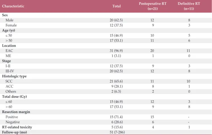
The median post-RT follow-up period was 51 months (range, 7 to 286 months). Twenty-four of the 32 patients (75%) experienced a complete response after RT; five patients (15.6%) had a partial response. The 5-year OS and DFS rates for all patients were 57% and 52%, respectively. Fig. 2 pres-ents the survival outcomes according to the stage and RT Table 1. Comparison of the patient and tumor characteristics according to the type of radiotherapy
Characteristic Total Postoperative RT Definitive RT
(n=21) (n=11) Sex Male 20 (62.5) 12 8 Female 12 (37.5) 9 3 Age (yr) 50 15 (46.9) 10 5 > 50 17 (53.1) 11 6 Location EAC 31 (96.9) 20 11 ME 1 (3.1) 1 0 Stage I-II 12 (37.5) 9 3 III-IV 20 (62.5) 12 8 Histologic type SCC 21 (65.6) 11 10 ACC 9 (28.1) 8 1 Others 2 (6.3) 2 0
Total dose (Gy)
60 15 (46.9) 12 3 > 60 17 (53.1) 9 8 Resection margin Positive 15 (71.4) 15 -. Negative 6 (28.6) 6 -. RT-related toxicity 5 (15.6) 4 1 Follow-up (mo) 51 (7-286)
Values are presented as number (%) or median (range). RT, radiotherapy; EAC, external auditory canal; ME, middle ear; SCC, squamous cell carcinoma; ACC, adenoid cystic carcinoma.
Ap op to si s (% ) 10 12 6 4 8 2 0 Surgery+RT RT alone CRT Stage I-II Stage III-IV 10 9 3 3 0 7
Fig. 1. Patterns of care for 32 patients with external audi-tory canal and middle ear carcinoma according to the tumor stage. RT, radiotherapy; CRT, chemoradiation ther-apy.
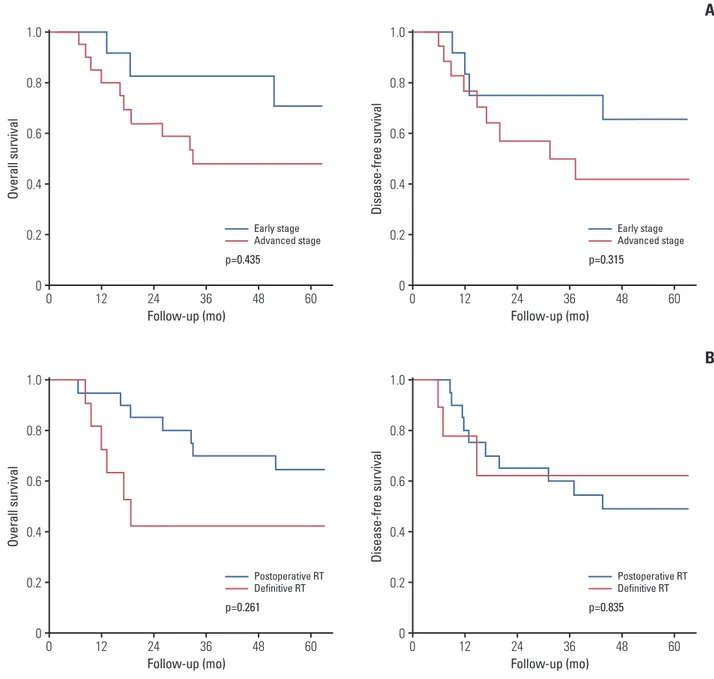
type. The 5-year OS and DFS rates for early stage versus advanced stage disease were 70.7% versus 48% (p=0.315) and 65.6% versus 41.4% (p=0.435), respectively. The 5-year OS rate was 64.8% and 42.4% for the postoperative and defini-tive RT group, respecdefini-tively (p=0.261). The 5-year DFS rate was 49.1% for the postoperative RT group and 62.2% for the definitive RT group (p=0.835). No patient experienced grade 3 or higher acute or chronic radiation-induced toxicity.
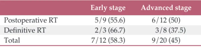
Table 2 lists the stage-related disease control rates. Sixteen patients (50%) showed no evidence of a disease status
with-out recurrence at the last follow-up visit. The disease control rates for patients with early stage versus advanced stage car-cinoma were 55.6% (5/9) versus 50% (6/12) in the postoper-ative RT group and 66.7% (2/3) versus 37.5% (3/8) in the definitive RT group, respectively. Eleven patients (52.4%) and five patients (45.5%) in the postoperative RT and defin-itive RT group, respectively, were considered cured.
Ov er al l s ur vi va l 1.0 0.8 0.6 0.4 0.2 0 0 Follow-up (mo) 12 24 36 48 60 Early stage Advanced stage p=0.435 Di se as e-fre e su rv iv al 1.0 0.8 0.6 0.4 0.2 0 0 Follow-up (mo) 12 24 36 48 60
A
Early stage Advanced stage p=0.315 Ov er al l s ur vi va l 1.0 0.8 0.6 0.4 0.2 0 0 Follow-up (mo) 12 24 36 48 60 Postoperative RT Definitive RT p=0.261 Di se as e-fre e su rv iv al 1.0 0.8 0.6 0.4 0.2 0 0 Follow-up (mo) 12 24 36 48 60B
Postoperative RT Definitive RT p=0.835Fig. 2. Overall survival and disease-free survival rates for patients with early stage versus advanced stage external auditory canal and middle ear carcinoma (A) and for treatment with postoperative radiotherapy (RT) vs. definitive RT (B).
3. Patterns of failure
Two patients experienced disease progression after the ini-tial treatment, and were excluded from the analysis of the failure patterns. Overall, 14 patients (46.7%) experienced treatment failure; four of these were classified as local failure and one as regional failure. Ten distant failures were observed as the first site of recurrence. The most common metastatic site was the lung (seven cases), followed by the spine (two cases) and bone (one case). One patient had simultaneous lung and spine metastases at the time of fail-ure. Table 3 lists the results of an analysis of the failure pat-terns according to the RT type. The “any recurrence” rate was 52.4% (11/21) in the postoperative RT group and 44.4% (4/9) in the definitive RT group.
Discussion
Primary cancer of the EAC and middle ear is extremely rare and tends to be locally advanced at presentation and has a poor prognosis [1]. Surgery (e.g., lateral TBR for T1/T2 dis-ease and subtotal or total TBR for T3/T4 disdis-ease) is generally
the recommended treatment. In most clinical settings, how-ever, less invasive surgeries are undertaken to treat this dis-ease, even when T3/T4 disease is present, because the EAC is located adjacent to the critical organs, such as the brain and major vessels. Nevertheless, the use of less invasive surgical techniques is the main reason for local failure after surgical treatment [15].
A comparison of the treatment strategies for EAC and mid-dle ear cancer is hindered by treatment and staging classifi-cation heterogeneity due to the small patient populations in published series. According to the Pittsburgh staging system [14], standard therapies include lateral TBR or definitive RT for T1 EAC tumors, lateral TBR combined with postoperative RT for T2 tumors, and subtotal or total TBR with postopera-tive RT for T3 and T4 tumors. On the other hand, despite these standardized treatment recommendations, the prog-noses remain poor [15]. At a single institutional experience, seven patients were treated using definitive RT and 22 patients were treated with postoperative RT. Better local and local-regional control was achieved for the early stage (T1/2) tumors than for the late-stage (T3) tumors, even though less than half of the patients were cured [16]. This suggests that a suboptimal treatment approach is being used for the local management of EAC cancers.
The use of meta-analysis is important for an optimal eval-uation of the treatment strategies because it has been difficult for a single institution to obtain a sufficient number of cases to achieve satisfactory statistical power. A review of 26 pub-lications containing information on 144 patients showed that patients with carcinoma confined to the EAC had similar sur-vival times, regardless of the type of surgery performed (e.g., mastoidectomy, lateral TBR, and subtotal TBR) [17]. Patients with an extension of the disease into the middle ear had longer survival times if they received a subtotal TBR. On the other hand, the addition of radiation therapy to lateral TBR did not improve the survival rates [17]. Another multi-insti-tutional review of 87 records that focused primarily on the Table 2. Stage-related disease control rates for external
auditory canal and middle ear cancer patients treated with RT
Early stage Advanced stage Postoperative RT 5/9 (55.6) 6/12 (50)
Definitive RT 2/3 (66.7) 3/8 (37.5)
Total 7/12 (58.3) 9/20 (45)
Values are presented as number (%). RT, radiotherapy.
Table 3. Patterns of failure for patients with external auditory canal and middle ear cancer according to the RT type Recurrence site All patients Postoperative RT Definitive RT
(n=30) (n=20) (n=9) Any recurrence 15 11 4 Local failure 4 3 1 Regional failure 1 0 1 Distant 10 8 2 Lung 7 6 1 Spine 2 1 1 Bone 1 1 0 RT, radiotherapy.
roles of surgery and RT in these patients found that the 5-year OS and DFS rates for all patients were 55% and 54%, respectively [13]. Moreover, the T stage and treatment modality were significant prognostic factors. The 5-year DFS rates for the T1, T2, and T3 patients were 83%, 45%, and 0% in the RT group, and 75%, 75%, and 46% in the surgery with RT group, respectively. They concluded that a radical RT is the treatment of choice for stage T1, and surgery combined with RT is recommended for T2 or T3 stage cancer. The role of chemotherapy, however, remains unclear [13].
Many authors have suggested that postoperative RT is effective for the control of residual tumors at incomplete resection margins [3,4,18]. The present study compared treat-ment outcomes between early stage and advanced stage tumors according to the treatment modality (e.g., postoper-ative and definitive RT). Although the results were not sta-tistically significant, they suggested that the prognosis of patients with early stage tumors is more favorable than for patients with advanced stage tumors. Tumor extension is an important prognostic factor for patients with these cancers. These results also indicated that patients with early stage cancer achieve better outcomes with definitive RT rather than postoperative RT. The stage-related disease control rates for definitive and postoperative RT were 66.7% and 55.6%, respectively. Therefore, this study suggests that patients with early stage cancer can undergo less invasive treatment. These results and conclusions are consistent with those of a previ-ous report [19]. Some authors have suggested that small lesions without bony erosion can be treated with RT alone [17,20]. On the other hand, the disease control rates for advanced stage tumors treated with postoperative and definitive RT were 50% and 37.5%, respectively. Therefore, patients with advanced stage tumors show better outcomes when postoperative RT is used. Although the optimal radi-ation dose is unclear, the median doses for tumor control in this study were 59.4 Gy for postoperative RT and 68.4 Gy for definitive RT. A previous study indicated that total doses of 65 to 75 Gy are needed to control the disease [21].
One limitation of this study was that it was a retrospective, single institution study that included a 20-year time period. Heterogeneity among the patient characteristics (e.g., tumor histologic type) might have confounded the results. The most common histologic type of carcinoma of the EAC and middle ear is SCC. This type was also the most common type (65.6%) in the present study population. ACC (28.1%) was the second most common histologic type. ACC is a special entity among tumors of the EAC; it has a slow proliferation rate and a low tendency towards lymphatic spread. On the other hand, lesions recur frequently when they are not excised com-pletely, which contributes to the poor prognosis of ACC patients [4]. Therefore, more aggressive surgery is recom-mended for patients with incompletely resected ACC and
patients with late-stage diseases [19]. Paradoxically, this study revealed 5-year OS rates of 76.2% and 50% for patients with ACC and SCC, respectively. Secondly, although the MRI findings can be used to evaluate the parotid, soft tissue, infratemporal fossa, and temporal dura mater involvement [22], the patients in the present study were staged according to the clinical and CT-based radiologic findings so that the results would be comparable to those in other studies that used the Pittsburgh staging system. This staging system was applied to all histologic types of tumors. Thirdly, 14 patients (46.7%) experienced treatment failure; the incidence of dis-tant failure was particularly high. Further investigation will be needed to identify the role of chemotherapy for these patients. Physicians from several institutions have applied chemoradiation therapy (CRT) to improve the patients’ prog-nosis. They found preoperative CRT to be effective [3,22]. The results of meta-analysis to assess the role of CRT for EAC SCC [15] suggested that preoperative CRT may improve the survival of surgically treated patients with EAC SCC. The meta-analysis results also suggested that definitive CRT may be an effective alternative to surgical resection.
Surgical approaches to treat advanced stage EAC and mid-dle ear cancer often cause severe complications [17,23]. In addition, because the recurrence rates are high, a minimum of 66 Gy is recommended for patients with positive margins [4]. Newer treatment techniques, such as 3-dimensional con-formal and intensity-modulated RT, can be used to deliver higher doses to the tumor and lower doses to the surround-ing critical organs.
Conclusion
Patients with early stage EAC and middle ear carcinoma had better outcomes when definitive RT was used. Patients with advanced stage cancer experienced better outcomes with postoperative RT. The high rate of distant failure after RT, with or without surgery, reflected the lack of a consensus on the best therapeutic approach for treating carcinomas of the EAC and middle ear.
Conflicts of Interest
1. Koto M, Hasegawa A, Takagi R, Sasahara G, Ikawa H, Mizoe JE, et al. Carbon ion radiotherapy for locally advanced squa-mous cell carcinoma of the external auditory canal and middle ear. Head Neck. 2016;38:512-6.
2. Kuhel WI, Hume CR, Selesnick SH. Cancer of the external auditory canal and temporal bone. Otolaryngol Clin North Am. 1996;29:827-52.
3. Nakagawa T, Kumamoto Y, Natori Y, Shiratsuchi H, Toh S, Kakazu Y, et al. Squamous cell carcinoma of the external auditory canal and middle ear: an operation combined with preoperative chemoradiotherapy and a free surgical margin. Otol Neurotol. 2006;27:242-8.
4. Pfreundner L, Schwager K, Willner J, Baier K, Bratengeier K, Brunner FX, et al. Carcinoma of the external auditory canal and middle ear. Int J Radiat Oncol Biol Phys. 1999;44:777-88. 5. Boamah H, Knight G, Taylor J, Palka K, Ballard B. Squamous
cell carcinoma of the external auditory canal: a case report. Case Rep Otolaryngol. 2011;2011:615210.
6. Korzeniowski S, Pszon J. The results of radiotherapy of cancer of the middle ear. Int J Radiat Oncol Biol Phys. 1990;18:631-3. 7. Gabriele P, Magnano M, Albera R, Canale G, Redda MG, Krengli M, et al. Carcinoma of the external auditory meatus and middle ear: results of the treatment of 28 cases. Tumori. 1994;80:40-3.
8. Waldemar E, Sorensen T, Bretlau P, Hansen HS. Cancer in the middle ear and the auditory canal. Ugeskr Laeger. 1995;157: 2139-42.
9. Spector JG. Management of temporal bone carcinomas: a ther-apeutic analysis of two groups of patients and long-term fol-lowup. Otolaryngol Head Neck Surg. 1991;104:58-66. 10. Chen WY, Kuo SH, Chen YH, Lu SH, Tsai CL, Cheng JC, et al.
Postoperative intensity-modulated radiotherapy for squa-mous cell carcinoma of the external auditory canal and middle ear: treatment outcomes, marginal misses, and perspective on target delineation. Int J Radiat Oncol Biol Phys. 2012;82:1485-93.
11. Arriaga M, Curtin H, Takahashi H, Hirsch BE, Kamerer DB. Staging proposal for external auditory meatus carcinoma based on preoperative clinical examination and computed tomography findings. Ann Otol Rhinol Laryngol. 1990;99 (9 Pt 1):714-21.
12. Pemberton LS, Swindell R, Sykes AJ. Primary radical
radio-therapy for squamous cell carcinoma of the middle ear and external auditory cana: an historical series. Clin Oncol (R Coll Radiol). 2006;18:390-4.
13. Ogawa K, Nakamura K, Hatano K, Uno T, Fuwa N, Itami J, et al. Treatment and prognosis of squamous cell carcinoma of the external auditory canal and middle ear: a multi-institutional retrospective review of 87 patients. Int J Radiat Oncol Biol Phys. 2007;68:1326-34.
14. Moody SA, Hirsch BE, Myers EN. Squamous cell carcinoma of the external auditory canal: an evaluation of a staging sys-tem. Am J Otol. 2000;21:582-8.
15. Takenaka Y, Cho H, Nakahara S, Yamamoto Y, Yasui T, Ino-hara H. Chemoradiation therapy for squamous cell carcinoma of the external auditory canal: a meta-analysis. Head Neck. 2015;37:1073-80.
16. Prabhu R, Hinerman RW, Indelicato DJ, Morris CG, Werning JW, Vaysberg M, et al. Squamous cell carcinoma of the exter-nal auditory caexter-nal: long-term clinical outcomes using surgery and external-beam radiotherapy. Am J Clin Oncol. 2009;32: 401-4.
17. Prasad S, Janecka IP. Efficacy of surgical treatments for squa-mous cell carcinoma of the temporal bone: a literature review. Otolaryngol Head Neck Surg. 1994;110:270-80.
18. Kinney SE. Squamous cell carcinoma of the external auditory canal. Am J Otol. 1989;10:111-6.
19. Chang CH, Shu MT, Lee JC, Leu YS, Chen YC, Lee KS. Treat-ments and outcomes of malignant tumors of external auditory canal. Am J Otolaryngol. 2009;30:44-8.
20. Arthur K. Radiotherapy in carcinoma of the middle ear and auditory canal. J Laryngol Otol. 1976;90:753-62.
21. Hashi N, Shirato H, Omatsu T, Kagei K, Nishioka T, Hashimoto S, et al. The role of radiotherapy in treating squa-mous cell carcinoma of the external auditory canal, especially in early stages of disease. Radiother Oncol. 2000;56:221-5. 22. Gillespie MB, Francis HW, Chee N, Eisele DW. Squamous cell
carcinoma of the temporal bone: a radiographic-pathologic correlation. Arch Otolaryngol Head Neck Surg. 2001;127: 803-7.
23. Leong SC, Youssef A, Lesser TH. Squamous cell carcinoma of the temporal bone: outcomes of radical surgery and postop-erative radiotherapy. Laryngoscope. 2013;123:2442-8.