Case Report : Temporomandibular Joint Involvement in Rheumatoid Arthritis
Hyun-Dae Lim,D.D.S.,M.S.D., You-Mee Lee, D.D.S.,M.S.D.,Ph.D.
Department of Oral medicine, College of Dentistry, Wonkwang University,
Rheumatoid arthritis(RA) is an of autoimmune inflammatory systemic disease. It is characterized by uncontrolled proliferation of synovial tissue and a wide array multisystem comorbidities. The disease may involve any joint of the body, but often statrs in the peripheral joints.
It was reported that more than 50% of RA patients exhibit clinical involvement of TMJ.
This report is a case report of dental management and progression for 16 months in patients who had severe bony change in TMJ involved rheumatoid arthritis
Dental management was included palliative treatment such as interocclusal splints, physical therapy, mouth opening exercise.
Although it was progressed rapidly osteolytic bone change during follow-up, no more advanced occulsal change and improved symptom and jaw motion.
Further investigations about rule of dentistry in TMJ involvement in RA maybe needed.
Key words: TMJ, Rheumatoid Arthritis, Osteolytic change
1)
I. INTRODUCTION
Rheumatoid arthritis(RA) is an autoimmune inflammatory systemic disease in which the inflamed and hypertrophic synovial membrane grow onto the articulating surfaces. The disease may involve any joint of the body, but often starts in the peripheral joints.
Prevalence of RA has varied from 0.1% to 1.8%
depend on geographic distributions, and more high prevalence in women than in men.
1,2)The disease
Corresponding Author : Prof. You-Mee Lee
Department of Oral medicine, College of Dentistry, Wonkwang University, 344-2 Shinyoung-dong, Iksan, Jeonbuk 570-749, Korea
E-mail: [email protected] received: 2006-05-08
accepted: 2006-07-23
can occur at any age, but it is most common among those aged 40-70 years, its incidence increasing with age. In Korea, Prevalence of this disease was reported as 1.4%.
3)The wrist are nearly always involved, as are the proximal interphalangeal joints and metacarpophalangeal joints. The distal interphalangeal joints and sacroiliac joints tend not to be affected. Although the earliest symptoms of rheumatoid arthritis occasionally may occur in the TMJ, generally other joints are involved first. Also, TMJ complaints may be overshadowed by RA symptoms elsewhere in the body.
However, in this case, there are more severe osteolytic change on radiological findings of TMJ than in other joints.
This report is a case report of dental
management and progression in patients who had
severe bony change in TMJ involved rheumatoid
arthritis
Ⅱ. CASE REPORT 1. Chief complain & History
34 years old man was referred from department of rheumatology in medical center of Wonkwang university, Ik-san, Korea. Before 4 months, He had been diagnosed as rheumatoid arthritis in there. He mainly complained pain of Rt elbow & posterior neck pain and it's duration was 6months.
His dental chief complain was pain of both TMJ.
When opening and chewing he complained pain of both TMJ, especially Rt TMJ was more prominent.
He had been history of locking closed before a few years. There are no familiar history.
2. Clinical Examination
He was observed mild limitaion of jaw movement with pain of TMJ. Maximum mouth opening length was 42 mm and comfortable mouth opening length was 36 mm. He also complained TMJ pain during protrusion and lateral excursion of mandible. In Head and Neck muscle palpation, he was seen tenderness of ant of Rt. masseter.
It was discluded in canine to the other side canine. There was much difference of 4 mm in RCP(retruded contact position)-ICP(inter cuspal position).
3. Radiographic findings
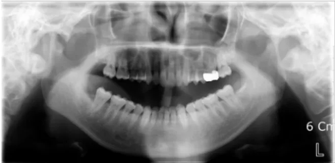
Severly flattening and irregularity finding was seen both TMJ on panoramic view and transcranial view.
4. Laboratory test
Laboratory finding was increased erythrocyte sedimentation rate(17 mm/hr), the presence of C-reactive protein(10.12 mg/L), positive rheumatoid factor(10.12 mg/L).
5. Progress of treatment
He had been medication in department of rheumatology. Therefore addictive medication didn't need. To improve symptom association with TMJ of patient and decrease load in TMJ, interocclusal stabilization splint was set, and did supplemental physical and self-exercise therapy. He recorded visual analog scale(VAS) for his symptom every visit. At first visit of VAS was 5,4 in each Rt and Lt TMJ pain. He was improved gradually.
After 3months, mouth opening length was regain in normal(45 mm). After 5months, he complained mild discomfort as 1 in VAS. However, on 8 months after first visit of radiographic finding, it was seen more advanced aggressive osteolytic change. In 14 months after first visit, radiographic was additionally taken, slightly progression of bony change than second radiograph. But, it was seen to more degree stably. Although there was severe bony change in TMJ rapidly, no advanced occlusal change more than.
Ⅲ. DISCUSSION
Rheumatoid arthritis is characterized by persistent joint synovial tissue inflammation. The predominant symptom of RA are pain, stiffness, and swelling of peripheral joints.
4,5)Over time, bone erosion, destruction of cartilage, and complete loss of joint integrity can occur. Eventually, multiple organ systems may be affected.
8)Joints destruction from synovitis can occur rapidly and early in the course of the disorder:
radiographic evidence is present in the more than 70% of patients with in first 2 years.
4)The TMJ may be involved in patients with RA
and occasionally is the initial symptomatic joint in
the body. The reported frequency of clinical TMJ
involvement has varied from 4.7%
6)to 84%.
7)This
variation may be due to different type of
examination, difference in the study population or
diagnostic criteria. When the TMJ is involved, there
is usually pain, tenderness and swelling, crepitation,
stiffness on opening the mouth, and limitation of jaw motion, or it may be asymptomic.
9,10)The change seen radiologically, especially cortical erosion or flattening, reduced joint space and subcodylar cystic destruction. The incidence of TMJ lesions found increased with the duration of RA, and there is a positive correlation between the severity of RA and the severity of involvement in TMJ.
11)It has known that radiologic evaluation is difficult because of the location of the joint and its relation to other cranial structures.
12)But, CT(computerized tomography) or MRI(magentic resonance imaging) was reported as being extremely effective in diagnosis of the early changes in patients with RA.
13,14)In this case, it is CT or MRI expensive, so we had to make to do radiograph. But, it was enough to find bone change due to so severe bone change in TMJ.
It was observed ant. open bite when we had examed this patient first. Ant open bite has been reported as a clinical sign in RA.
15)This clinical sign developed as a result of progressive loss of
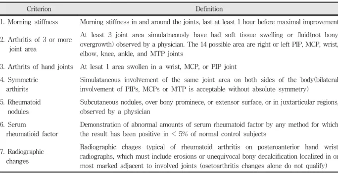
Criterion Definition
1. Morning stiffness Morning stiffness in and around the joints, last at least 1 hour before maximal improvement
2. Arthritis of 3 or more joint area
At least 3 joint area simulatneously have had soft tissue swelling or fluid(not bony overgrowth) observed by a physician. The 14 possible area are right or left PIP, MCP, wrist, elbow, knee, ankle, and MTP joints
3. Arthrits of hand joints At lesat 1 area swollen in a wrist, MCP, or PIP joint 4. Symmetric
arthirits
Simulataneous involvement of the same joint area on both sides of the body(bilateral involvement of PIPs, MCPs or MTP is acceptable without absolute symmetry)
5. Rheumatoid nodules
Subcutaneous nodules, over bony prominece, or extensor surface, or in juxtarticular regions, observed by a physician
6. Serum
rheumatioid factor
Demonstration of abnormal amounts of serum rheumatoid factor by any method for which the result has been positive in < 5% of normal control subjects
7. Radiographic changes
Radiographic chages typical of rheumatoid arthritis on posteroanterior hand wrist radiographs, which must include erosions or unequivocal bony decalcification localized in or most marked adjacent to involved joints (osetoarthritis changes alone do not qualify) At least 4 of these 7 criteria. Criteria 1 through 4 must have been present for at least 6 weeks.
Table 1. The 1987 revised criteria for the classification of rheumatoid arthritis
mandibular rami height secondary to destruction of the condylar surface and hence the inability to bring incisors into occlusion.
Many studies
9,16)are reported Masticatory muscle tenderness or myalgia in TMJ involvement in RA, but in this case there is only Rt MM tenderness. It may be due to medication before TMJ examination.
Rheumatoid arthritis primarily is a clinical diagnosis. No laboratory tests exist that are pathognomonic or diagnostic of RA. Laboratory findings most commonly seen in RA include an increased erythrocyte sedimentation rate, the presence of C-reactive protein, positive rheumatoid factor in 85% of affected patients, and hypochromic microcystic anemia.
17)Table 1 is showed the 1987 revised criteria for the classification of rheumatoid arthritis.
18)Pharmacotherapy for RA involves NSAIDs for control of pain, with selective use of low-dose oral or intra-articular glucocorticoids, and DMARDs.
DMARD(Disease modifying anti rheumatic drugs)
included methotrexate, gold salts, hydroxychlo-
roquine, sulfasalazine, ciclosporin, and azathioprine.
Fig. 1. Panoramic view of first examination Severly flattening and irregularity of both condyle of mandible.
Because joint destruction in rheumatoid arthritis begins within a few weeks of symptom onset; early treatment decreases the rate of disease progression, in todays " a reversed pyramidal" approach is favored, in which DMARDs are initiated quickly to slow disease progression as early as possible.
5,19,20,21)In recent studies reported the biological agents etanercept and infliximab, both TNF-α inhibitors, were shown to be very effective for treating early rheumatoid arthritis when compared with methotrexate.
22,23)Although It was progressed rapidly osteolytic bone change during follow-up, no more advanced occlusal change and improvement of symptom and jaw motion. In TMJ involvement in RA, palliative treatment such as interocclusal splints, physical therapy, and medication may prove to be helpful,
8)and mouth opening exercise were helpful in maintaining jaw function.
24)Unfortunately, we still don't know why is more prominent in TMJ than other joints and rapidly progress bone destruction during management in spite of clinical improvement. Further investigation maybe needed.
It is well known as clinical and radiographic finding of the TMJ in rheumatoid arthritics. But, It is still lack of study about rule of dentistry in TMJ involvement in RA. The more studies about this may be need. It is helpful to improve quality of life of these patient.
Fig. 2. Panoramic view of 8 months after first examination The more advanced aggressive bony change than first examination.
Fig. 3. Panoramic view of 14 months after first examination. Slightly progression of bony change than second radiograph. But, it was seen to more degree stably.
REFERENCES
1. Chaiamnuay P, Daramawan J, Muriden KD, Assawa- tanabodee P. Epidermiology of rheumatic disease in rural Thailand: a WHO-ILAR COPCORD study.
Community Oriented Programme for the Control of Rheumatic Disease. J Rheumatol 1998;25:1382-1387.
2. Boyer GS, Benevolenskaya LI, Templin DW et al.
Prevalance of Rheumatoid Arthritis in circumpolar native populations. J Rheumatol 1998;25:23-29.
3. Park NG, K WK, Shin DH et al. Prevalence of Osteoarthritis and Rheumatoid Arthritis in Two Communities in Korea. Journal of the Korean Rheumatism Association 2003;10(2):151-157.
4. Lee DM, Weinblatt ME. Rheumatoid Arthritis. The Lancet 2001;358:903-911.
5. Rindfleisch JA, Muller D. Diagnosis and Management of Rheumatoid Arthritis. American Family Physician 2005;72:1037-1047,1049-50.
6. Ragan C. General management of Rheumatoid arthitis.
JAMA 1949;141:124.
7. Friez L, Le Goc Y. Arthicualtions Tempro- Maxillarires et polyarthrite rheumatoide. Rheuma- tologie 1982;34:193-196.
8. Little JW, Falace DA, Miller CS, Rhodus NL. Dental Management of the Medically Compromised Patient.
6th ed., Philadelpia, 2002, Mosby pp.478-486.
9. Koh ET, Yap AU, Koh CK, Chee TS, Chan SP, Boudville IC. Temporomandibular Disorders in Rheumatoid Arthritis. J Rheumatol 1999;26:1918-22.
10. Ettala-Ylitalo UM, Syrjänen S, Halonen P. Functional disturbances of the masticatory system related to temporomandibular joint involvement by rheumatoid arthritis. Journal of Oral Rehabilitation 1987;14:415- 427.
11. Çeliker R, Gökçe-Kutsal Y, Eryilmaz M. Temporo- mandibular joint involvement in Rheumatoid Arthritis Relationship with Disease Activity. Scand J Rheumatol 1995;24:22-5.
12. Goupille P, Fouquet B, Goga D, Cotty P, Valat JP.
The temporomandbular joint in rheumatoid arthritis:
correlations between clinical and tomographic features. J Dent 1993;21:141-146.
13. Bayar N, Kara SA, Keles I, Koç MC, Altinok D, Orkun S. Temporomandibular joint involvement in Rheumatoid Arthritis: a radiological and Clinical Study. Journal Of Craniomandibular Practice 2002;20 (2):105-110.
14. Suenaga S. Ogura T, Matusuda T, Noikura T.
Severity of synovium and bonemarrow abnormalities of the temporomandibular joint in early rheumatoid arthritis: role of gadolinium-enhanced fat-suppressed T1-wedge spin echo MRI. J Comput Assist Tomogr 2000;24(3):461-465.
15. Bush FM, Dolwick MF: The temporomandibular joint and realted orofacial disorders. philadelphia: JB Lippincott; 1995: 35-86.
16. Helenius LMJ, Hallikainen D, Helenius I et al. Clinical and radiographic findings of the temporomaddibular joint in patients with various rheumatic disease. A case-control study. Oral Surg Oral Med Oral Pathol Oral Radiol Endod 2005;99:455-63.
17. Lane SK, Gravel JW. Clinical utility of common serum rheumatologic tests. American Family Physician 2002;65(6):1073-1080.
18. Arnett FC, Edworthy SM, Bloch DA et al. The American rheumatism association 1987 revised criteria for the classification of rheumatoid arthritis.
Arthritis and Rheumatism 1988;31(3):315-324.
19. Brooks P. Clinical review Recent advances Rheuma- tology. British Medical Journal 1998;316:1810-1812.
20. Simon HKH. Rheumatology: 7. Basic of Therapy.
Canadian Medical Association Journal 2000;163(4):
417-423.
21. Diane Lacaille. Rheumatology: 8. Advanced Therapy.
Canadian Medical Association Journal 2000;163 (4):721-728.
22. Baton J et al. A comparison of etanercept and methotrexate in patients with early rheumatoid arthritis. N engl J Med 2000;343:1586-1593.
23. Lipsky P et al. Infliximab and mehotrexate in the treatment of patients with early rheumatoid arthritis.
N engl J Med 2000;343:1594-1602.
24. Telberg A, Kopp S. A three-year follow up of temporomandibular disorders in rheumatoid arthritis and ankylosing spondylitis. Acta Odontol Scand 1996;54:14-18.
국문요약
증례보고: 류마티스 관절염 환자에서 측두하악관절의 이환
원광대학교 치과대학 구강내과학 교실