Activation of the M1 Muscarinic Acetylcholine Receptor Induces GluA2 Internalization in the Hippocampus
Keun Oh Ryu1 and Heon Seok1,2*
1Department of Biomedical Science, Collage of Science and engineering, Jungwon University, Goesan 367-805, Korea
2Department of Biological Sciences, Ulsan National Institute of Science and Technology (UNIST), Ulsan 689-798, Korea Received September 23, 2015 /Revised October 6, 2015 /Accepted October 8, 2015
Cholinergic innervation of the hippocampus is known to be correlated with learning and memory.
The cholinergic agonist carbachol (CCh) modulate synaptic plasticity and produced long-term synaptic depression (LTD) in the hippocampus. However, the exact mechanisms by which the cholinergic sys- tem modifies synaptic functions in the hippocampus have yet to be determined. This study introduces an acetylcholine receptor-mediated LTD that requires internalization of alpha-amino-3-hydroxy-5- methylisoxazole-4-propionate (AMPA) receptors on the postsynaptic surface and their intracellular mechanism in the hippocampus. In the present study, we showed that the application of the chol- inergic agonist CCh reduced the surface expression of GluA2 on synapses and that this reduction was prevented by the M1 muscarinic acetylcholine receptor antagonist pirenzepine in primary hippo- campal neurons. The interaction between GluA2 and the glutamate receptor-interacting protein 1 (GRIP1) was disrupted in a hippocampal slice from a rat upon CCh simulation. Under the same con- ditions, the binding of GluA2 to adaptin-α, a protein involved in clathrin-mediated endocytosis, was enhanced. The current data suggest that the activation of LTD, mediated by the acetylcholine receptor, requires the internalization of the GluA2 subunits of AMPA receptors and that this may be controlled by the disruption of GRIP1 in the PDZ ligand domain of GluA2. Therefore, we can hypothesize that one mechanism underlying the LTD mediated by the M1 mAChR is the internalization of the GluA2 AMPAR subunits from the plasma membrane in the hippocampal cholinergic system.
Key words : Alpha-amino-3-hydroxy-5-methylisoxazole-4-propionate receptor, endocytosis, hippocampus, long-term depression, muscarinic acetylcholine receptor
*Corresponding author
*Tel : +82-43-830-8608, Fax : +82-43-830-8579
*E-mail : [email protected]
This is an Open-Access article distributed under the terms of the Creative Commons Attribution Non-Commercial License (http://creativecommons.org/licenses/by-nc/3.0) which permits unrestricted non-commercial use, distribution, and reproduction in any medium, provided the original work is properly cited.
Journal of Life Science 2015 Vol. 25. No. 10. 1103~1109 DOI : http://dx.doi.org/10.5352/JLS.2015.25.10.1103
Introduction
Long-term synaptic depression (LTD) can be defined as an activity-dependent decrease in the efficacy of synaptic de- pression [3, 4]. The mechanism of LTD illustrates that the activation of the N-methyl-D-aspartate (NMDA) receptor- mediated loss of the amino-3-hydroxy-5-methylisoxazole-4- propionate (AMPA) receptors in the synapse is the main mo- lecular event of synaptic depression [1, 4]. However, it has also been found that LTD can occur through alternative mechanisms that do not require NMDAR activation [11].
Recent studies have shown that the activation of muscarinic acetylcholine receptors (mAChRs) with the agonist CCh can induce LTD in a rat hippocampus [10]. Nonetheless, the ex-
act mechanisms of AMPA receptor regulation in CCh-LTD have remained largely unknown.
The predominant mechanism of NMDAR-dependent LTD is the removal of the AMPA receptors from the synaptic sur- face [18]. The synaptic loss of AMPA receptors during NMDAR-dependent LTD has been intensively studied. It seems that the interaction of GluA2 with the PDZ domain in GRIP1 requires the synaptic targeting of the AMPA receptors. The GluA2 serves as a scaffolding protein to link AMPA receptors to other neuronal proteins, such as PSD-95 [7], synapse-associated protein-97(SAP) [22], ABP [27], star- gazin [28] and the clathrin adaptor, AP-2 [12]. In particular, phosphorylation at ser880 in the PDZ ligand domain of GluA2 is an important site for the dissociation of AMPARs from the GRIP/ABP complex that allows AMPA receptor internalization during LTD induction [16, 29]. These mecha- nisms are likely to be critically important to understanding the AMPA receptor regulation in CCh-LTD. In the present study, we used cultured hippocampal neurons and brain sli- ces to show that CCh stimuli that induce LTD cause the in- ternalization of post-synaptic GluA2 and the modulation of
GluA2 clustering proteins for the internalization.
Materials and Methods
Primary neuronal culture
Four post-natal day P2 Wistar rats were used for each cell culture. Each hippocampus was carefully excised and washed twice with HBSS and incubated in HBSS plus tryp- sin (750 μg/ml; Gibco, UK) for 7 min at 37°C in a heated water bath. The cells were carefully dissociated with a fire-polished long glass Pasteur pipette. The dissociated cells were centrifuged (800× g, 10min, 4°C) and the supernatant aspirated off. The pellet was re-suspended in a 3-ml plating medium. Fifty thousand cells were plated onto coverslips coated with poly-D-lysine (10 mg/ml) and laminin (1 mg/ml) in 0.5 M borate buffer. The cells were placed into a 5% CO2 incubator set at 37°C and maintained with a Neurobasal-B27-based feeding medium. Every three days, 50% of the feeding medium was changed.
Fluorescence internalization assay
Live hippocampal neurons at 25-28 days in vitro were la- beled for 10 min at 37°C with antibodies directed against the extracellular region of GluA2 (Chemicon, USA). Neurons were incubated at 37°C in a conditioned growth medium containing CCh (50 μM for 10 min), pirenzepine (500 nM for 10 min) with CCh, then fixed for 5 min in 4% paraf- ormaldehyde/ 4% sucrose without permeabilization, and stained with FITC-conjugated secondary antibodies (Jackson Immuno Research Laboratories, USA), for 1 hr in order to visualize the pre-labeled surface receptors. Neurons were then permeabilized for 1 min in 100% methanol at -20°C and stained with Texas red-conjugated secondary antibodies (Jackson Immuno Research Laboratories, USA), for 1 hr in order to visualize the pre-labeled internalized receptors.
Fluorescent images were acquired from two to five platings of neurons using a monochrome CCD camera. For quantifi- cation, fluorescent puncta in a representative field (50×25 μm) around each neuron were traced and the puncta were identi- fied based on their having a fluorescence intensity that ex- ceeded a threshold set to maximize the discrimination of the puncta from the background. The threshold values of the puncta were obtained using an Image J program (National Institute of Mental Health, Bethesda, Maryland, USA). The mean ± s.e.m. was calculated from pooled data, and t-tests were carried out to determine the significant differences be-
tween different groups.
Brain sample preparation
Hippocampi were excised from 6-8-week old male Wistar rats and transferred into ice-cold artificial cerebrospinal fluid (aCSF) containing the following: (mM) NaCl, 124; KCl, 3;
NaHCO3, 26; NaH2PO4, 1.25; CaCl2, 2; MgSO4, 1; D glu- cose, 10. Parasagittal hippocampal slices of 400 μm thickness were prepared using a McIllwain tissue chopper (Mickle Laboratory Engineering Co. Ltd., Gomshall, UK). The slices were then submerged in aCSF saturated with 95% O2/5%
CO2 at room temperature for 1 hr before CCh stimulation.
This study was approved by the Animal Care and Use Committee of UNIST (UNISTIACUC-14-005).
Western Blots
CCh-stimulated hippocampal slices were placed into a 1.5-ml tube and 50 μl of ice cold protein extraction buffers (50 mM Tris-HCl, 150 mM NaCl, 1% NP-40, 10 μl protease inhibitor cocktail per 1 ml) were added, then homogenized using a motorized homogenizer (Sigma, USA) for 15 seconds. A protein extraction buffer (100 mg per 200 μl) was immediately added to the brain homogenates. The samples were then incubated at 4°C for 30 min while rotated at 50 r.p.m. for further lysis. A crude fraction of the hippocampal homogenates were obtained by centrifugation (1,000× g, 10 min). For western blotting, a hippocampal lysate from each treatment condition was separated with SDS-PAGE, trans- ferred onto a PVDF membrane, and then probed with the relevant antibodies. For sequential re-probing of the same blots, the membranes were stripped off using a reblotting solution (Chemicon, USA) and subjected to immunoblotting with another type of target antibodies. Blots were developed using enhanced chemiluminescent detection (Amersham, USA). Band intensities were quantified using NIH ImageJ software, and normalized to the quantity of relevant anti- bodies in each sample lane.
Co-immunoprecipitation
The hippocampi were treated in three different ways: con- trol (no stimulation), CCh only and pirenzepine with CCh.
Crude fractions of the extracts were prepared. The samples were centrifuged (1,000×g, 10 min, 4°C) to remove non-solu- ble components. The protein concentration was then ana- lyzed by a Bradford assay and 500 μg from each sample was transferred to a fresh tube and 1 ug of GluA2 or AP2
A
B
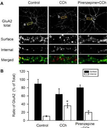
Fig. 1. The GluA2 AMPAR subunit is internalized following M1 mAChR stimulation in the hippocampal neurons.
(A) DIV 27 hippocampal cultured neurons were live-la- belled with an antibody recognizing the extracellular N-terminal domain of GluA2 before treatment with CCh (50 μM for 10 min) and with Pirenzepine plus CCh (500 nM Pirenzepine + 50 μM CCh for 10 min). The cells not stimulated with any drug served as a control. Bound GluA2 antibodies were visualized with secondary anti- bodies conjugated to FITC (extracellular) or Texas red (intracellular). (B) Quantitative analysis of pooled data from 5 independent experiments. The internalized GluA2 ratio was obtained by expressing internalized GluA2 as a percentage of the total GluA2 signals (green and red).
antibodies were added to immobilize the proteins. The tubes were then placed on a rotator (40 r.p.m) and incubated for 4 hr at 4°C. Then, 30 μl of the 50% slurry of protein G-sephar- ose beads in PBS was added to each tube, and rotated at the same speed for an additional 4 hrs. After incubation, the co-immunoprecipitated complexes were centrifuged (800×g, 2 min, 4°C) and washed four times with a protein extraction buffer. Then, the western blots were processed with the appropriate antibodies.
Results
CCh-LTD-induced GluA2 internalization
A previous study demonstrates AMPAR internalization using a “live”-labeling assay with antibodies recognizing the extracellular epitopes of GluA2 in the NMDA-dependent LTD [12, 14]. This study examined whether CCh stimulation could occur for GluA2 internalization in the hippocampal neurons. For this, a fluorescence-based internalization assay was performed as described [14]. Live cells were labelled with monoclonal antibodies that bind to the epitopes in the extracellular N-terminal domain of GluA2. Cells were then stimulated with CCh (50 μM for 10 min) or pirenzepine (500 nM for 10 min), then with CCh (50 μM for 10 min).
Extracellular GluA2 labelling was visualized using the FITC-conjugated secondary antibodies, whereas intracellular GluA2 labelling was visualized (following fixation and per- meabilization of the cells) with Texas Red-conjugated secon- dary antibodies (Fig. 1A). The control cells showed abundant surface expression of GluA2 (89.5±10.9%, left column, sur- face in Fig. 1Aa) as well as a small amount of intracellular GluA2 expression (10.5±1.5%, p<0.05, left column, internal in Fig. 1Aa). The application of 50 μM of CCh resulted in increases in internalized GluA2 immunoreactivity (36.0±5.1%, p<0.05, internal in Fig. 1Ab). However, this in- ternalization of GluA2 was inhibited by the blockade of the muscarinic acetylcholine receptors (mAChR) M1 by pir- enzepine (19.9±3.9%, p<0.05, internal in Fig. 1Ab). These ex- periments showed that CCh stimulation on the hippocampal neurons results in GluA2 internalization, and this internal- ization is specifically required for the activation of mAChR subunits of M1.
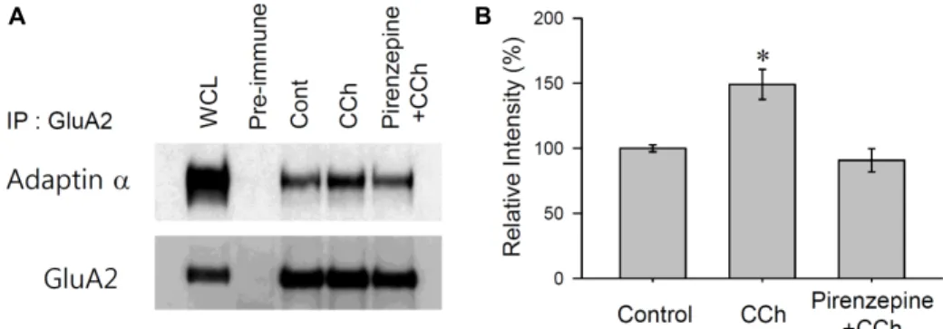
GluA2 interactions with adaptin-α increased in CCh-LTD for its internalization
We hypothesised that an interaction between adaptin-α
and the C-terminal region of GluA2 could be required for the formation of clathrin-coated vesicles (CCVs) containing GluA2 receptors for its internalisation in CCh-LTD. Co-im- munoprecipitation (Co-IP) was performed to investigate if CCh stimulation regulated an interaction between adaptin-α and GluA2 during CCh-LTD in M1 mAChR in a specific manner. Rat (P25-P35) hippocampi were treated in three dif- ferent ways: control (no stimulation), CCh only and pir- enzepine with CCh. In order to block the activation of M1 mAChR, the M1 antagonist pirenzepine (500 nM) was ap- plied for 10 min prior to CCh application. The interaction property of adaptin-α to GluA2 in the hippocampus was confirmed by co-immunoprecipitation (Fig. 2A). After stim-
A B
Fig. 2. GluA2-adaptin-α interaction enhanced by M1 mAChR stimulation in hippocampus. (A) The association of adaptin-α and GluA2 under the CCh stimuli was determined by Co-IP in a rat hippocampus. Six-week old rat hippocampal slices were submerged in aCSF with CCh (50 μM for 10 min), Pirenzepine plus CCh (500 nM 10 min + 50 μM CCh for 10 min), and no stimulation as a control, then solubilized with a protein extraction buffer. GluA2 was immobilized with an anti-GluA2 antibody. GluA2 associated adaptin-α was determined by a western blot using anti-adaptin-α antibodies. (B) Quantified data obtained from three independent experiments. The interaction ratios of the intensity of the adaptin-α was calculated against the intensity of GluA2, then normalized to the control slices.
ulation with CCh, the interaction of adaptin-α to GluA2 was significantly increased (149.1±11.5% of control, p<0.05, n=3, Fig. 2B). However, when M1 mAChRs were blocked with pirenzepine, CCh stimulation failed to promote the inter- action between adaptin-α and GluA2 (90.7±8.9% of control, p>0.05, n=3, Fig. 2B). These data confirm that the increased association of GluA2 with adaptin-α could be required for the initial formation of CCVs in the process of M1 mAChR-mediated AMPAR internalization.
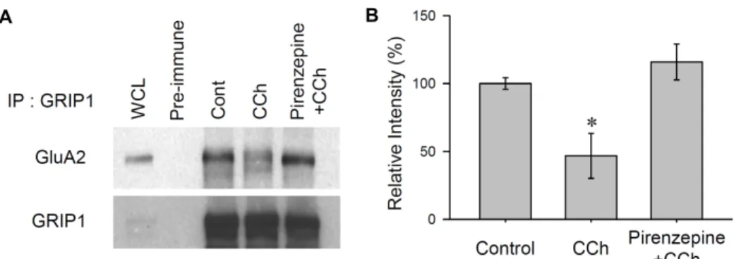
Activation of mAChRs disrupts the interaction between GluA2 and GRIP1
A previous study showed that the C-terminal region of GluA2 directly binds to the glutamate receptor interaction protein 1 (GRIP1) via its PDZ domain [7], and is thus stabi- lized at the synapse [21]. GRIP is closely related to multi- PDZ domain-containing proteins that are thought to scaffold receptors [6, 30]. Recently, it has been suggested that the interaction of GluA2 with GRIP1 is critical in AMPAR regu- lation [15]. Therefore, we investigated if the interaction be- tween GRIP1 and GluA2 had been affected during CCh-in- duced LTD.
To examine the changes in the association of GluA2 with GRIP1 in CCh-LTD, we prepared hippocampal lysates and incubated them under the three different conditions (control, CCh only, pirenzepine + CCh). The GRIP1 protein complex was immobilized using rabbit polyclonal GRIP1 antibodies (2 μg per reaction). The presence of co-immunoprecipitated GluA2 was determined by using western blots with rabbit polyclonal GluA2 C-terminal antibodies (1:3,000 dilution,
Fig. 3A). Hippocampal slices treated with CCh showed a substantially reduced interaction between GluA2 and GRIP1 (46.7±16.5% of control, p<0.05, Fig. 3B). However, when the M1 mAChR was inhibited by the application of pirenzepine, CCh did not affect the interaction between GRIP1 and GluA2 (115.8±13.2% of control, p>0.05, Fig. 3B). These results sug- gest that under basal conditions, GRIP1 interacts with the C-terminal region of GluA2 and supports its synaptic surface expression. However, the disruption of the GluA2-GRIP1 in- teraction following CCh stimulation could be a potential mechanism that promotes the internalization of GluA2 from the surface of the post-synaptic region.
Discussion
Several functional roles have been proposed for the inter- action between AMPARs and scaffolding/anchoring pro- teins [9, 16, 25]. In particular, these scaffolding/anchoring proteins are thought to be important for regulating AMPAR trafficking [5, 21, 23, 31]. Following the finding that the acti- vation of the M1 mAChR leads to the induction of LTD in the hippocampus [10], we have found that this form of LTD may involve the internalization of the GluA2 AMPAR subunits. The basal rate of AMPA receptor cycling is surpris- ingly fast in cultured hippocampal neurons [13]. Even in im- mature neurons that have formed numerous synapses, around 10-20% of surface AMPARs are internalized from the surface within 10 min [8, 14]. In chemically-induced LTD in primary cultured neurons, the application of NMDA [2]
and DHPG [24] induce the internalization of 79% and 40%
A B
Fig. 3. GluA2-GRIP1 interactions disrupted by M1 mAChR stimulation in hippocampus. (A) Changes in the endogenous association of GRIP1 and GluA2 under CCh stimuli were determined by Co-IP. Six-week old rat hippocampal slices were submerged in aCSF with CCh (50 μM for 10 min), Pirenzepine plus CCh (500 nM 10 min + 50 μM CCh for 10 min) and no stimulation as a control, then solubilized with protein extraction buffer. GRIP1 was immobilized with polyclonal anti-GRIP1 antibodies.
GRIP1-associated GluA2 was determined by western blots using anti-GluA2 antibodies (B) Quantified data obtained from the three independent experiments. The interaction ratios of the intensity of the GluA2 were calculated against the intensity of GRIP, then normalized to the control slices.
of GluA2 subunits, respectively. Here, we found that the CCh application resulted in 36% of GluA2 subunits of internalization. This was blocked by the M1 mAChR antago- nist pirenzepine. Considering that 10% of AMPA receptors are constitutively recycled [8, 14], the CCh application had probably induced the internalization of approximately an ex- tra 26%.
Since NMDAR activation is a crucial step in hippocampal LFS-LTD, adaptin-α recruitment to AMPAR is likely to be a key event linking NMDAR activation to AMPAR endocy- tosis and synaptic depression. In earlier studies, the pep2m peptide was used to block the NSF-GluA2 interaction [17], causing a run-down of basal AMPA EPSCs [19, 20, 26] and leading to reduced surface expression of the AMPARs [20].
A more recent study showed that adaptin-α associates with a region of GluA2 that closely overlaps with the NSF binding site, thus suggesting that pep2m application also disrupts adaptin-α binding to GluA2 [12]. We found that the activa- tion of the M1 mAChR increases the association of the adap- tin-α subunits of adaptin-α with GluA2 by 49%. This sug- gests that during the endocytosis of AMPARs, adaptin-α could be recruited to AMPAR via intermediary proteins that bind elsewhere on GluA2. However, this study did not ad- dress the intracellular signalling after the activation of M1 mAChR-mediated LTD. A recent study showed that tyrosine de-phosphorylation of the AMPAR was necessary in group I mGluR-mediated LTD, and that PKC-dependent regulation of AMPAR phosphorylation regulates AMPAR internalization.
Therefore, it would be interesting to investigate the involve- ment of PKC or protein tyrosine phosphatase as a direct sig-
nalling mechanism affecting AMPARs during LTD.
Acknowledgment
This research was supported by Basic Science Research Program through the National Research Foundation of Korea (NRF) funded by the Ministry of Education (NRF- 2013R1A1A4A01010683).
References
1. Bear, M. F. and Abraham, W. C. 1996. Long-term depression in hippocampus. Annu. Rev. Neurosci. 19, 437-462.
2. Beattie, E. C., Carroll, R. C., Yu, X., Morishita, W., Yasuda, H., Zastrow, M. and Malenka, R. C. 2000. Regulation of AMPA receptor endocytosis by a signaling mechanism shared with LTD. Nat. Neurosci. 3, 1291-1300.
3. Bliss, T. V. and Collingridge, G. L. 1993. A synaptic model of memory: long-term potentiation in the hippocampus.
Nature 361, 31-39.
4. Collingridge, G. L., Isaac, J. T. and Wang, Y. T. 2004. Receptor trafficking and synaptic plasticity. Nat. Rev. 5, 952-962.
5. Craven, S. E., El-Husseini, A. E. and Bredt, D. S. 1999.
Synaptic targeting of the postsynaptic density protein PSD-95 mediated by lipid and protein motifs. Neuron 22, 497-509.
6. DeSouza, S., Fu, J., States, B. A. and Ziff, E. B. 2002.
Differential palmitoylation directs the AMPA receptor-bind- ing protein ABP to spines or to intracellular clusters. J.
Neurosci. 22, 3493-3503.
7. Dong, H., O'Brien, R. J., Fung, E. T., Lanahan, A. A., Worley, P. F. and Huganir, R. L. 1997. GRIP: a synaptic PDZ do- main-containing protein that interacts with AMPA receptors.
Nature 386, 279-284.
8. Ehlers, M. D. 2000. Reinsertion or degradation of AMPA receptors determined by activity-dependent endocytic sorting.
Neuron 28, 511-525.
9. Hirbec, H., Perestenko, O., Nishimune, A., Meyer, G., Nakanishi, S., Henley, J. M. and Dev, K. K. 2002. The PDZ proteins PICK1, GRIP, and syntenin bind multiple gluta- mate receptor subtypes. Analysis of PDZ binding motifs.
J. Biol. Chem. 277, 15221-15224.
10. Jo, J., Son, G. H., Winters, B. L., Kim, M. J., Whitcomb, D.
J., Dickinson, B. A., Lee, Y. B., Futai, K., Amici, M., Sheng, M., Collingridge, G. L. and Cho, K. 2010. Muscarinic re- ceptors induce LTD of NMDAR EPSCs via a mechanism involving hippocalcin, AP2 and PSD-95. Nat. Neurosci. 13, 1216-1224.
11. Lee, H. K., Min, S. S., Gallagher, M. and Kirkwood, A. 2005.
NMDA receptor-independent long-term depression corre- lates with successful aging in rats. Nat. Neurosci. 8, 1657- 1659.
12. Lee, S. H., Liu, L., Wang, Y. T. and Sheng, M. 2002. Clathrin adaptor AP2 and NSF interact with overlapping sites of GluA2 and play distinct roles in AMPA receptor trafficking and hippocampal LTD. Neuron 36, 661-674.
13. Lee, S. H., Simonetta, A. and Sheng, M. 2004. Subunit rules governing the sorting of internalized AMPA receptors in hippocampal neurons. Neuron 43, 221-236.
14. Lin, J. W., Ju, W., Foster, K., Lee, S. H., Ahmadian, G., Wyszynski, M., Wang, Y. T. and Sheng, M. 2000. Distinct molecular mechanisms and divergent endocytotic pathways of AMPA receptor internalization. Nat. Neurosci. 3, 1282- 1290.
15. Liu, S. J. and Cull-Candy, S. G. 2005. Subunit interaction with PICK and GRIP controls Ca2+ permeability of AMPARs at cerebellar synapses. Nat. Neurosci. 8, 768-775.
16. Lu, W. and Ziff, E. B. 2005. PICK1 interacts with ABP/GRIP to regulate AMPA receptor trafficking. Neuron 47, 407-421.
17. Luthi, A., Chittajallu, R., Duprat, F., Palmer, M. J., Benke, T. A., Kidd, F. L., Henley, J. M., Isaac, J. T. and Collingridge, G. L. 1999. Hippocampal LTD expression involves a pool of AMPARs regulated by the NSF-GluA2 interaction.
Neuron 24, 389-399.
18. Malinow, R. and Malenka, R. C. 2002. AMPA receptor traf- ficking and synaptic plasticity. Annu. Rev. Neurosci. 25, 103-126.
19. Nishimune, A., Isaac, J. T., Molnar, E., Noel, J., Nash, S.
R., Tagaya, M., Collingridge, G. L., Nakanishi, S. and
Henley, J. M. 1998. NSF binding to GluA2 regulates synaptic transmission. Neuron 21, 87-97.
20. Noel, J., Ralph, G. S., Pickard, L., Williams, J., Molnar, E., Uney, J. B., Collingridge, G. L. and Henley, J. M. 1999.
Surface expression of AMPA receptors in hippocampal neu- rons is regulated by an NSF-dependent mechanism. Neuron 23, 365-376.
21. Osten, P., Khatri, L., Perez, J. L., Kohr, G., Giese, G., Daly, C., Schulz, T. W., Wensky, A., Lee, L. M. and Ziff, E. B.
2000. Mutagenesis reveals a role for ABP/GRIP binding to GluA2 in synaptic surface accumulation of the AMPA receptor. Neuron 27, 313-325.
22. Schluter, O. M., Xu, W. and Malenka, R. C. 2006. Alternative N-terminal domains of PSD-95 and SAP97 govern activ- ity-dependent regulation of synaptic AMPA receptor function.
Neuron 51, 99-111.
23. Sheng, M. and Lee, S. H. 2001. AMPA receptor trafficking and the control of synaptic transmission. Cell 105, 825-828.
24. Snyder, E. M., Philpot, B. D., Huber, K. M., Dong, X., Fallon, J. R. and Bear, M. F. 2001. Internalization of ionotropic gluta- mate receptors in response to mGluR activation. Nat.
Neurosci. 4, 1079-1085.
25. Song, I. and Huganir, R. L. 2002. Regulation of AMPA re- ceptors during synaptic plasticity. Trends. Neurosci. 25, 578- 588.
26. Song, I., Kamboj, S., Xia, J., Dong, H., Liao, D. and Huganir, R. L. 1998. Interaction of the N-ethylmaleimide-sensitive fac- tor with AMPA receptors. Neuron 21, 393-400.
27. Srivastava, S. and Ziff, E. B. 1999. ABP: a novel AMPA re- ceptor binding protein. Ann. N. Y. Acad. Sci. 868, 561-564.
28. Vandenberghe, W., Nicoll, R. A. and Bredt, D. S. 2005.
Stargazin is an AMPA receptor auxiliary subunit. Proc. Natl.
Acad. Sci. USA 102, 485-490.
29. Xia, J., Chung, H. J., Wihler, C., Huganir, R. L. and Linden, D. J. 2000. Cerebellar long-term depression requires PKC- regulated interactions between GluA2 and PDZ do- main-containing proteins. Neuron 28, 499-510.
30. Yamazaki, M., Fukaya, M., Abe, M., Ikeno, K., Kakizaki, T., Watanabe, M. and Sakimura, K. 2001. Differential palmitoy- lation of two mouse glutamate receptor interacting protein 1 forms with different N-terminal sequences. Neurosci. Lett.
304, 81-84.
31. Ziff, E. B. 2007. TARPs and the AMPA receptor trafficking paradox. Neuron 53, 627-633.
초록:쥐 해마에서 M1 무스카린 아세틸콜린 수용체의 활성에 의한 GluA2 세포내이입 연구
류근오 · 석 헌*
(중원대학교 의생명과학과)
뇌 해마의 콜린성 신경분포는 학습과 기역에 연관성이 있는 것으로 알려져 있으며 이의 작용제인 carbachol 투여 시 장기기억 저하가 유도됨이 알려져 왔다. 그러나 이러한 콜린성 자극에 의한 해마 신경세포의 시냅스 내 변화기작은 완전히 알려지지 않고 있다. 본 연구에서는 아세틸콜린 수용체의 활성에 의하여 유도되는 장기기억 저하 현상에 있어 alpha-amino-3-hydroxy-5-methylisoxazole-4-propionate (AMPA) 수용체가 후시냅스 표면으로 부터 사라지는 현상과 이의 조절기작에 대하여 알아보고자 한다. 이를 위하여 쥐 해마의 일차세포를 추출하고 체외에서 배양한 성숙 신경세포에 carbachol 을 투여하여 장기기억 저하를 유도 하였으며, 후시냅스의 표면으로부 터 AMPA 수용체의 아단위체인 GluA2가 M1 무스카린 수용체의 길항제에 의하여 저해 되었다. 또한 콜린성 자극 에 의한 GluA2의 내재화 현상의 작용기작 연구를 위하여 쥐 해마 절편에 carbachol 투여 후 GluA2와 직접적인 상호작용을 하는 Glutam내재화 되었음을 확인하였다. 이러한 현상은 ate receptor-interacting protein 1 (GRIP1) 과 clathrine 단백질이 매개하는 세포내이입 작용을 하는 adaptin-α 단백질의 결합 변화를 관찰하였다. GluA2는
carbachol 자극에 의해 세포내이입 과정에서 adaptin-α 와의 결합이 증가하였으며 반대로 GRIP1과는 해리되었다.
이는 아세틸콜린의 수용체의 자극에 의하여 GluA2의 내제화 작용이 수반되며, 이의 작용기작으로 GluA2의 후시 냅스 표면 발현시에 결합하고 있는 GRIP1과 해리 되면서 장기기억 저하 현상이 유도됨을 의미한다.