1연세대학교 의과대학 내과학교실, 2인체상피장벽연구소
이창률1, 정재희1, 장윤수1,2, 김세규1, 김형중1,2, 장준1, 김성규1, 안철민1,2
Clinical Features of Patients with Lung Cancer and Upper Aerodigestive Tract Cancer
Chang Youl Lee, M.D.1, Jae Hee Chung, M.D.1, Yoon Soo Chang, M.D.1, Se Kyu Kim, M.D.1,2,3, Hyung Jung Kim, M.D.1,2, Joon Chang, M.D.1,2, Sung Kyu Kim, M.D.1,2, Chul Min Ahn, M.D.1,2
1Department of Internal Medicine, 2THuman Barrier Research Center, Yonsei University College of Medicine, Seoul, Korea
Background: To define the clinical features of patients with lung and upper aerodigestive tract cancer through a review of the histopathology, clinical features and follow-up results.
Methods: Patients with lung and upper aerodigestive tract cancer who were diagnosed in Young dong Severance Hospital from 1992 to 2005, were retrospectively reviewed. The clinical data, radiologic findings, pathologic findings, treatment modalities were evaluated.
Result: There was a total of 20 patients with aerodigestive tract cancer who were diagnosed with lung cancer over a 13 years period. The mean age was 58.45 ±15.09 years and 19 cases were male. There were 14 smokers with an average pack year of 46 years. Twelve patients had aerodigestive tract cancer and later developed lung cancer, and 5 lung cancer patients were later diagnosed with aerodigestive tract cancer.
Conclusion: These results suggest that cancers of the aerodigestive tract and lung can arise as either dependent or independent events and most aerodigestive tract cancer patients who developed lung cancer are not treated properly.
Therefore, regular low dose chest CT with close suspicion is needed to properly manage upper aerodigestive tract cancer patients. (Tuberc Respir Dis 2007; 62: 284-289)
Key words: Aerodigestive tract cancer, Lung cancer, Clinical feature.
Adress for correspondence: Chul Min Ahn, M.D.
Yongdong Severance Hospital, Department of Internal Medicine, Yonsei University College of Medicine, Kangnam P.O. Box 1217, Seoul, Korea.
Phone: 82-2-2019-3317, Fax: 82-2-3463-3882 E-mail: [email protected]
Received: Feb. 8. 2007 Accepted: Mar. 26. 2007
서 론
유병률에 따른 우리나라 7대 암은 위암, 간암, 폐암, 대장암, 자궁암, 유방암, 두경부암이다. 이 중 서로 장 기간에 연속성을 가지고 있으며 흡연과 관계가 있는 상부소화호흡기계(upper aerodigestive tract, UADT) 와 폐에서 발생하는 다발성 종양(multiple primary cancer)의 발생기원에 대한 논의가 있어왔다.
상부소화호흡기계 환자에 있어서 이차암(second primary tumors, SPT)의 위험성이 증가한다는 것이 잘 알려져 있다1-4. 소위 ‟Field Cancerization” 이론으 로 상부소화호흡기계에서 다발성 종양의 빈도가 높
은 현상을 설명하는 적절한 이론으로 받아들여져 왔 다5,6. 일반적으로 상부소화호흡기계 환자에서 평균 매 해당 이차암이 발생할 위험률은 4-6% 정도라 알려져 있고 흡연량과 음주량, 돌연변이유발성(mutageni- city)과 밀접한 관계가 있다7,8. 따라서 이러한 환자에 게 핵형분석(karyotype analysis), p53 유전자 검사 등 분자유전학적 검사를 통해 이차암 발생을 예측할 수 있을 거라 하였다9. 우리나라에서 폐암은 암 발생률에 있어서는 1위가 아니지만 다른 암에 비해서 치료가 잘 되지 않으므로 암 중에서 폐암으로 사망한 환자의 수가 가장 많으며 상부 소화호흡기계암의 경우, 환자 가 매년 얼마나 발생하고 있는지 정확한 통계는 알 수 없으나, 한국중앙암등록본부에서 발표한 자료에 의하 면, 매년 후두암 환자가 1,096명, 구강암 환자가 800여 명이 새로 발생한다고 한다10. 상부 소화호흡기계암이 란 후두, 구강, 구인두, 비강, 부비동, 비인두, 하인두, 타액선 등의 다양한 신체부위에서 발생하는 종양을 총괄하는 것이 타당하나, 국가적 통계에서는 개별발 생부위별로 집계되어 중요성이 낮게 평가되어 왔다.
Sex Age Smoking(py) UADT cancer lung cancer feature of lung cancer M 41 15 tongue base, squamous cell carcinoma* T4N3M0 Squamous Multiple lung lesion
M 72 20 larynx, squamous cell carcinoma T4N3M0 Squamous single lung lesion T4N2M0 M 46 20 larynx, squamous cell carcinoma* T4N2M0 Squamous single lung lesion T4N2M0
M 61 5 larynx, squamous cell carcinoma‡ Multiple lung lesion
M 72 40 larynx, squamous cell carcinoma* T2N1M0 Multiple lung lesion
M 68 40 larynx, squamous cell carcinoma‡ Multiple lung lesion
M 70 50 larynx, squamous cell carcinoma* T1N3M0 Multiple lung lesion
M 73 30 larynx, squamous cell carcinoma T4N2M0 Squamous single lung lesion T1N0M0 M 62 60 larynx, squamous cell carcinoma* T3N2M0 Multiple lung lesion
M 46 20 oropharyx, squamous cell carcinoma* T2N0M0 Squamous single lung lesion T4N1M0 M 60 20 larynx, squamous cell carcinoma T3N2M0 Adenocarcinoma† single lung lesion T3N0M0 M 83 50 larynx, squamous cell carcinoma T4N3M0 Squamous single lung lesion T4N2M0 M 60 missing oropharyx, squamous cell carcinoma* T2N2M0 Multiple lung lesion
M 57 missing nasopharynx, squamous cell carcinoma* T2N1M0 Squamous single lung lesion T3N1M0 M 65 missing nasopharynx, squamous cell carcinoma‡ Multiple lung lesion
M 45 missing oropharyx, squamous cell carcinoma* T3N2M0 Squamous Multiple lung lesion M 57 90 oropharyx, squamous cell carcinoma* T1N1M0 Multiple lung lesion
M 72 120 nasopharynx, squamous cell carcinoma T4N3M0 Squamous single lung lesion T2N0M0 M 63 40 oropharyx, squamous cell carcinoma* T2N1M0 Small cell
carcinoma single lung lesion T4N3Mx M 34 missing oropharyx, squamous cell carcinoma* T1N2M0 Squamous single lung lesion T3N0M0
*First UADT cancer and second lung cancer †First lung cancer and second UADT cancer ‡First UADT cancer and lung cancer Table 1. Clinical fearures of UADT and lung cancer, 20 cases
하지만 갑상선암을 제외한 두경부에 발생하는 종양을 통합하면, 발생빈도에서 중요한 암이며 국가적인 관 심이 요망된다. 후두암과 인두암은 조기발견 및 치료 가 최선이나 눈으로 종양을 들여다보기에 용이치 않 기 때문에 목이 아프거나, 음식물을 삼키는데 곤란한 경우, 목 속에서 덩어리가 만져지거나, 목소리가 변하 고 귀에 통증을 느끼는 등의 자각증상이 매우 중요하 며 이런 증상이 있을 때는 자세한 진찰과 내시경을 이 용하여 의심되는 부위를 관찰하고 종양이 발견되면 조직검사를 시행하여 암 여부를 확인하여야 한다11,12. 상부소화호흡기암과 폐암의 연관관계가 있다는 것 은 알려져 있지만 국내 임상발현 양상에 대한 보고는 부족한 상황이다. 상부소화호흡기암은 조직형이 대부 분 편평상피세포암(Squamous cell cancer)으로 최근 치료법의 발달에 힘입어서 과거에는 상부소화호흡기 암의 주 사망원인이 원발암의 치료실패였으나 현재에 는 환자들이 상당기간 생존하게 되므로 원격전이나 중복암(multiple primary cancer)를 경험하게 되는 환
자의 수가 증가 되고 있고 이들이 사망원인의 중요 부 분을 차지하고 있다. 따라서 조기에 발견하는 것이 중 요하나13 실제 임상적으로 상부소화호흡계 암 발생 후 폐에 다발성 전이 양상으로 보이는 경우를 볼 수 있고 이들 중 상당수의 환자가 이차암으로 여겨진다고 하 였다14.
이에 저자들은 폐암과 상부소화호흡기암이 동반된 경우 환자들이 어떠한 조직학적 유형과 임상경과를 보이는지 알고자 하였다.
대상 및 방법
1992년 1월부터 2005년 12월까지 13년동안 연세대 학교 의과대학 영동세브란스병원에서 진단된 상부소 화호흡기암 환자는 122명이며 이 중 상부소화호흡기 암과 폐암이 진단된 20예(16.39%)가 있었다(Table 1).
조직학적 진단을 위해 병리의사에 의해 조직절편을 10% 중성 포르말린에 고정하고, 파라핀 포매한 후 5
Diagnosis sequence Case (%) Interval time(aver.) First UADT cancer and
second lung cancer 12 (60%) 36.8 months First lung cancer and
second UADT cancer 5 (25%) 16.2 months First UADT cancer and
lung cancer 3 (15%)
p<0.05
Table 2. Diagnosis sequence and interval time of UADT and lung cancer
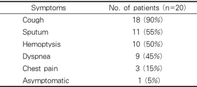
Symptoms No. of patients (n=20)
Cough 18 (90%)
Sputum 11 (55%)
Hemoptysis 10 (50%)
Dyspnea 9 (45%)
Chest pain 3 (15%)
Asymptomatic 1 (5%)
Table 3. Clinical symptoms
㎛의 두께로 박절하여 일률적으로 Hematoxylin- Eosin 염색을 시행하였으며, 필요에 따라서는 특수 면 역 염색을 시행하였다.
상부소화호흡기암과 폐암이 동시에 진단된 20예에 대한 의무기록 열람을 통하여 연령, 성병, 과거력, 흡 연력, 증상, 흉부 전산화 단층촬영 소견, 병리학적 소 견, 수술 후 치료방법, 추적 관찰 여부 및 생존 여부를 조사하였다.
단변량 통계분석을 위해 위험인자에 대한 비연속 변수의 비교는 chi-square test로, 연속 변수의 비교 는 independent T test를 이용하여 검증하였다. 유의 수준 0.05 미만일 때 통계적으로 유의한 것으로 판정 하였으며, 통계 프로그램은 SPSS 12.0을 사용하였다.
결 과 1. 임상 소견
1992년 1월부터 2005년 12월까지 13년동안 연세대 학교 의과대학 영동세브란스병원에서 상부소화호흡 기암이 122예 진단되었으며 이 중 상부소화호흡기암 과 폐암이 동시에 진단된 예가 20예(16.39%) 있었다.
이 중 남자 19예, 여자 1예로 평균연령은 58.45±15.09 세, 흡연자는 14예(70.0%), 평균 흡연량은 46갑년이었 다(Table 1. p<0.05). 폐암과 상부소화호흡기암의 발 생 순서에서 상부소화호흡기암 발생 후 폐암의 발생 은 12예(9.84%), 폐암 발생 후 상부소화호흡기암의 발 생은 5예(4.10%)이었고 동시에 발견된 경우도 3예 (2.46%)에서 관찰되었다(Table 2. p<0.05). 상부소화 호흡기암 발생 후 폐암의 발생 양상은 흉부 전산화 단
층 촬영 소견으로 보았을 때 이차암 양상 5예, 다발성 전이 양상이 7예이었다(Table 1). 폐암 진단 당시의 증상은 기침이 18예(90%)로 가장 많았고 이어서 객 담, 객혈증상이 그 뒤를 이었다(Table 3). 상부소화호 흡기암 진단 후 폐암이 진단되는 기간은 평균 36.8개 월이고 폐암 진단 후 상부소화호흡기암 진단이 되는 기간은 16.2개월 이었다(Table 2. p<0.05).
2. 병리학적 소견
폐암과 상부소화호흡기암 모두 조직학적으로 확인 된 경우, 대부분인 18예에서 편평상피암이였으나 폐 에서 선암과 소세포암도 각각 1예씩 보고되었다 (Table 1). 폐암과 상부소화호흡기암의 조직학적 차 이를 확인해 볼 수 있었던 5예에서 시행한 면역 조직 화학 염색 결과를 보면 두 암에서 모두 CK7/CK20 +/+로 조직학적으로 동일하다고 여겨진 경우가 3예 있었으며, 상부소화호흡기암에서는 CK7/CK20 -/-인 반면 폐암에서는 +/- 보여 발생기원이 다르다고 여겨 진 경우가 2예에서 관찰되었다(data not shown.
p>0.05).
3. 경 과
상부소화호흡기암 진단 후 폐암이 진단된 12환자의 경우 수술할 수 있는 폐암 병기의 환자도 환자의 거부 로 인해 수술한 예는 없으며 항암제치료 2예, 항암제 치료와 방사선 치료 병행은 4예이었고 나마지 경우 6 예에서 환자의 불량한 전신상태와 치료거부 등으로 보전적 치료를 행하였다. 항암치료와 방사선치료를
Treatment modality of second lung
cancer No. of patients(%)
Operation 0
Chemotherapy 2(16.7%)
Chemotherapy & Radiotherapy 4(33.3%) Conservative treatment 6(50.0%) Table 4. Treatment of second lung cancer
하지 않고 보전적 치료만을 받은 환자 6예 모두 1년 이내에 사망하였다(Table 4). 대상 증례의 부족으로 인해 생존율, 예후 등과 관련된 인자에 대한 분석은 이루어지지 못하였다.
고 찰
상부소화호흡기계 암 환자에 있어서 이차암의 발생 은 치료실패와 사망의 중요한 원인이다15-17. 최근 상 부소화호흡기계암과 폐암 등에서 흔히 발생하는 소위 다발성 종양(multiple primary cancer)의 발생 기원에 대한 논의가 활발하다. 1953년 Slaughter 등은 126명 의 두경부암 환자의 조직 소견을 관찰한 결과 다발성 종양의 빈도가 11%로 매우 높았고, 암 주위의 점막 조직이 이미 암화 과정이 진행된 상태임을 보고하였 다5. 따라서 암화 과정이 어느 정도 진행된 암 주위의 전암병변에서 새로운 종양이 발생하였을 것이라는 이 론을 발표하였다. 이러한 결과는 소위 “Field Can- cerization” 이론으로 상부소화호흡기계에서 다발성 종양의 빈도가 높은 현상을 설명하는 적절한 이론으 로 받아들여져 왔다5,6,18. 한편 1993년 Chung 등은 31 명의 상부소화호흡기계암 환자의 일차암과 이차암의 p53 유전자의 변이를 조사하였고, 변이가 있었던 21 명 모두에서 일차암과 이차암의 p53 유전자의 변이가 일치하지 않다는 사실을 보고하였다19. 이러한 결과는 이차암이 일차암과 무관하게 독립적으로 발생하였을 가능성이 큰 것으로 설명되었다. 그러나 p53 유전자 의 변이는 상부소화호흡기계암 발생 과정의 후기에 발생하고, 동일한 하나의 세포에서 기원한 종양이라 도 p53 유전자의 변이 양상이 다를 수 있다는 주장도 제기되었다18. 또한 상부소화호흡기계암이 소위 상피 세포내이동(intraepithelial spread)하거나, 원발 장소
에서 분리되어 다른 장소로 물리적으로 이동(flot and seed)하여 두 번째 암을 발생시킬 수 있다는 의견도 제시되었다18. Bedi 등은 8명의 여성 다발성 상부소화 호흡기계암 환자를 조사하여, 4명에서 다발성 암의 X-chromosome inactivation, 9p와 3p LOH 양상이 정 확히 일치함을 발표하였다20. 이러한 결과는 상부소화 호흡기계의 다발성 암이 독립적으로 생겨난다기보다 는 일차암이 재발하거나, 물리적으로 이동하여 이차 암이 발생함을 시사하였다. 한편 1996년 Califano 등 은 상부소화호흡기계의 암조직과 암주변의 다양한 전 암병변을 조사하여, 암조직과 전암병변에서 동일한 LOH 양상이 나타나고 있음을 관찰하였다21. 이는 Slaughter 등이 조직학적으로 관찰한 것과 같은 현상 이 분자 유전학적 방법으로도 관찰된 것으로 생각된 다. 최근 연구에서는 다양한 시간적 간격을 두고 검사 한 다발성의 전암병변도 동일한 유전적 변화를 보이 는 것으로 보고 되었다22. 이러한 결과를 어떻게 해석 할 것인지에 대한 논란이 아직 계속 중인 것으로 생각 된다. 즉 상부소화호흡기계에서 이차암 혹은 다발성 암의 발생 기원이 동일한 하나의 세포에서 발생한 암 의 재발 혹은 전이인지, 아니면 동일한 발암 과정에 노출된 또 다른 점막세포가 독립적으로 암화 과정을 거쳐 새로운 암이 발생하였는지에 대한 논란은 아직 유효한 것으로 여겨지며 다발성암의 기원에 대한 논 란의 결과는 향후 상부소화호흡기계암의 예방과 치료 방침에 적지 않은 영향을 미칠 것으로 생각된다.
이번 연구를 통해본 결과도 비록 유전자 검사를 시 행하지 않은 제한이 있으나 면역 조직 화학염색법을 이용할 수 있었던 일차암과 이차암의 조직을 비교해 보았을 때 동일한 경우와 동일하지 않은 경우가 통계 학적으로 의의가 없어 일차암과 이차암의 발생에 있 어서 저자들은 폐암과 상부소화호흡계 암의 발생의 기원이 동일한 경우와 독립적으로 발생하는 경우 모 두 가능한 것으로 생각되어 진다.
상부소화호흡기암 진단 후 폐암이 진단되는 경우가 폐암 진단 후 소화호흡기암이 진단되는 경우 보다 더 많은 시간이 걸리며 이는 상부소화호흡기암의 증상이 폐암에 비해 더 일찍 나타나기 때문으로 여겨진다. 또 한 일차암 진단 후 이차암이 진단될 당시 대부분의 환
자에 있어서 전신 상태가 불량한 경우가 대부분을 차 지하며 주치의의 치료 권유에도 적극적인 치료 의지 를 보이지 않는 경우가 많았다. 또한 대부분의 상부소 화호흡기암 환자에 있어서 정기적으로 폐암의 조기 진단을 위해 노력한 경우는 없었으며 호흡기계 증상 이 있어도 상부소화호흡기암에 의한 증상일 경우라 여겨 적극적인 폐암검사가 이루어지지 않았고 이후 시행한 흉부 검사상 이미 수술적 치료가 불가능한 경 우가 많았다. 매년 정기적으로 후두암 환자에게 이차 암을 조기 진단하기 위해 단순 흉부 방사선 촬영을 시 행하는 것과 기관지 내시경검사와 객담검사는 유용하 지 않은 결과가 있다23-25. 따라서 상부소화호흡기암 환자에게 조기에 폐암을 진단 할 수 있도록 진찰 시 적극적으로 주기적인 저선량 흉부 전산화단층촬영을 시행하는 것이 의미가 있을 것으로 사료된다.
요 약
연구배경: 상부소화호흡기암과 폐암의 연관관계가 있다는 것은 알려져 있지만 국내 임상발현 양상에 대 한 보고는 부족한 상황이다. 이에 저자들은 폐암과 상 부소화호흡기암이 동반된 경우 환자들이 어떠한 조직 학적 유형과 임상경과를 보이는지 알고자 하였다.
방 법: 1992년 1월부터 2005년 12월까지 13년동안 연세대학교 의과대학 영동세브란스병원에서 진단된 상부소화호흡기암과 폐암이 진단된 20예를 대상으로 후향적으로 조직학적 유형과 임상 양상을 조사하였 다.
결 과: 1) 13년 동안 연세대학교 의과대학 영동세 브란스병원에서 상부소화호흡기암과 폐암이 진단된 20예 중 남자 19예, 여자 1예로 평균연령은 58.45세, 흡연자는 14예, 평균 흡연량은 46갑년이었다. 폐암과 상부소화호흡기암의 발생 순서에서 상부소화호흡기 암 발생 후 폐암의 발생은 12예, 폐암 발생 후 상부소 화호흡기암의 발생은 5예이었다. 2) 폐암과 상부소화 호흡기암의 조직학적 차이를 확인해 볼 수 있었던 5 예에서 면역 조직 화학 염색을 통해 조직학적으로 동 일한 경우 3예, 다르다고 여겨진 경우가 2예에서 관찰 되었다. 상부소화호흡기암 진단 후 폐암이 진단되는
기간은 평균 36.8개월이고 폐암 진단 후 상부소화호흡 기암 진단이 되는 기간은 16.2개월이었다. 3) 상부소 화호흡기암 진단 후 폐암이 진단된 경우 폐암의 치료 로 수술을 시행한 예는 없으며 항암제치료 2예, 항암 제치료와 방사선 치료 병행은 4예이었고 나마지 경우 는 환자의 불량한 전신상태와 치료거부 등으로 보전 적 치료를 행하였다.
결 론: 폐암과 상부소화호흡계 암의 발생의 기원 이 동일한 경우와 독립적으로 발생하는 경우 모두 가 능한 것으로 여겨지며 상부소화호흡기암 환자에게 조 기에 폐암을 진단 할 수 있도록 진찰 시 적극적으로 주기적인 저선량 흉부 전산화단층촬영을 시행하는 것 이 의미가 있을 것으로 사료된다.
참 고 문 헌
1. X wu YH, Lippman SM, ed. Upper aerodigestive tract cancers. Philadelphia: Lippincott Williams & Wilkins;
1999.
2. Schwager K, Nebel A, Baier G, Hoppe F. [Second primary carcinomas in the upper aerodigestive tract in different locations and age groups]. Laryngor- hinootologie 2000;79:599-603.
3. Gao X, Fisher SG, Mohideen N, Emami B. Second primary cancers in patients with laryngeal cancer: a population-based study. Int J Radiat Oncol Biol Phys 2003;56:427-35.
4. Vokes EE, Weichselbaum RR, Lippman SM, Hong WK. Head and neck cancer. N Engl J Med 1993;
328:184-94.
5. Slaughter DP, Southwick HW, Smejkal W. Field cancerization in oral stratified squamous epithelium:
clinical implications of multicentric origin. Cancer 1953;6:963-8.
6. Strong MS, Incze J, Vaughan CW. Field cancerization in the aerodigestive tract--its etiology, manifestation, and significance. J Otolaryngol 1984;13:1-6.
7. Hemminki K, Boffetta P. Multiple primary cancers as clues to environmental and heritable causes of cancer and mechanisms of carcinogenesis. IARC Sci Publ 2004;(157):289-97.
8. Spitz MR, Hoque A, Trizna Z, Schantz SP, Amos CI, King TM, et al. Mutagen sensitivity as a risk factor for second malignant tumors following malignancies of the upper aerodigestive tract. J Natl Cancer Inst 1994;86:1681-4.
9. Ha PK, Califano JA. The molecular biology of mucosal field cancerization of the head and neck. Crit Rev Oral Biol Med 2003;14:363-9.
10. Ministry of Health Welfare. 2005 Annual report of the Korea Central Cancer Registry. Gwacheon: Ministry of Health Welfare Republic of Korea; 2005.
11. Marioni G, Marchese-Ragona R, Cartei G, Marchese F, Staffieri A. Current opinion in diagnosis and treatment of laryngeal carcinoma. Cancer Treat Rev 2006;32:504-15.
12. Chan AT, Teo PM, Johnson PJ. Nasopharyngeal carcinoma. Ann Oncol 2002;13:1007-15.
13. Dhooge IJ, De Vos M, Van Cauwenberge PB. Multiple primary malignant tumors in patients with head and neck cancer: results of a prospective study and future perspectives. Laryngoscope 1998;108:250-6.
14. Geurts TW, Nederlof PM, van den Brekel MW, van't Veer LJ, de Jong D, Hart AA, et al. Pulmonary squamous cell carcinoma following head and neck squamous cell carcinoma: metastasis or second primary? Clin Cancer Res 2005;11:6608-14.
15. Ogden GR. Second malignant tumours in head and neck cancer. BMJ 1991;302:193-4.
16. Muir C, Weiland L. Upper aerodigestive tract cancers.
Cancer 1995;75:S147-53.
17. Lippman SM, Hong WK. Second malignant tumors in head and neck squamous cell carcinoma: the overshadowing threat for patients with early-stage disease. Int J Radiat Oncol Biol Phys 1989;17:691-4.
18. Carey TE. Field cancerization: are multiple primary cancers monoclonal or polyclonal? Ann Med 1996;
28:183-8.
19. Chung KY, Mukhopadhyay T, Kim J, Casson A, Ro JY, Goepfert H, et al. Discordant p53 gene mutations in primary head and neck cancers and corresponding second primary cancers of the upper aerodigestive tract. Cancer Res 1993;53:1676-83.
20. Bedi GC, Westra WH, Gabrielson E, Koch W, Sidransky D. Multiple head and neck tumors:
evidence for a common clonal origin. Cancer Res 1996;56:2484-7.
21. Califano J, van der Riet P, Westra W, Nawroz H, Clayman G, Piantadosi S, et al. Genetic progression model for head and neck cancer: implications for field cancerization. Cancer Res 1996;56:2488-92.
22. Califano J, Westra WH, Meininger G, Corio R, Koch WM, Sidransky D. Genetic progression and clonal relationship of recurrent premalignant head and neck lesions. Clin Cancer Res 2000;6:347-52.
23. Engelen AM, Stalpers LJ, Manni JJ, Ruijs JH, van Daal WA. Yearly chest radiography in the early detection of lung cancer following laryngeal cancer.
Eur Arch Otorhinolaryngol 1992;249:364-9.
24. Fontana RS, Sanderson DR, Taylor WF, Woolner LB, Miller WE, Muhm JR, et al. Early lung cancer detection: results of the initial (prevalence) radiologic and cytologic screening in the Mayo Clinic study. Am Rev Respir Dis 1984;130:561-5.
25. Rachmat L, Vreeburg GC, de Vries N, Hordijk GJ, Lubsen H, Manni JJ, et al. The value of twice yearly bronchoscopy in the work-up and follow-up of patients with laryngeal cancer. Eur J Cancer 1993;29A:1096-9.