The Role of Bmi1 in Pilocarpine-induced Status Epilepticus in Mice
Hae-In Pyeon1, Jia Bak1 and Yun-Sik Choi1,2*
1Department of Pharmacy, Kyungsung University, Nam-Gu, Busan 48434, Korea
2Convergence Research Center for Smart Healthcare, Kyungsung University, Nam-Gu, Busan 48434, Korea Received May 8, 2020 /Revised May 25, 2020 /Accepted June 3, 2020
B-cell-specific Moloney murine leukemia virus integration site 1 (Bmi1) is a polycomb group protein and a core component of polycomb repressive complex 1. Initial research into Bmi1 has focused on its role in tumorigenesis, and it is generally accepted that it is important for the proliferation and sur- vival of cancer cells. However, more recent studies have revealed that Bmi1 is downregulated in brains with neurodegenerative disease and that it regulates the function of mitochondria and reactive oxygen species levels. In this study, we tested the therapeutic potential of Bmi1 in pilocarpine-induced seizures in Bmi1-knockout mice. Bmi1 expression transiently increased in the hippocampal CA1 and CA3 and the dentate gyrus following pilocarpine-induced status epilepticus (SE). In terms of seizure behavior, SE induction was 43.14% and 53.57% for Bmi1+/+ and Bmi1+/- mice, respectively. However, there was no significant difference in mortality or hippocampal damage between the two groups. Two months after SE induction, the frequency of epileptic seizures in the Bmi1+/- mice was 50% lower than in the control group, although the difference was not statistically significant. In addition, mossy fiber outgrowth in the Bmi1+/- mice was significantly higher than in their wild-type littermates. Taken to- gether, these data indicate that reduced Bmi1 activity increases pilocarpine-induced seizure probability and mossy fiber outgrowth.
Key words : Bmi1, mossy fiber sprouting, pilocarpine, recurrent seizure, status epilepticus
*Corresponding author
*Tel : +82-51-633-4890, Fax : +82-51-663-4809
*E-mail : [email protected]
This is an Open-Access article distributed under the terms of the Creative Commons Attribution Non-Commercial License (http://creativecommons.org/licenses/by-nc/3.0) which permits unrestricted non-commercial use, distribution, and reproduction in any medium, provided the original work is properly cited.
Introduction
Antiepileptic drugs (AEDs) are medications usually taken long-term to prevent the occurrence of seizures in epileptic patients. AEDs can also be used to prevent seizures in pa- tients with meningitis or in the early period following either neurosurgery or traumatic brain injury. In addition, certain AEDs are used to terminate ongoing seizures, such as in status epilepticus (SE) or prolonged febrile seizures [17]. The goals of AEDs are to reduce the number of seizures or to enhance the patient’s quality of life. Bromide was intro- duced as the first AED in 1857, but because of adverse ef- fects, it is no longer in use [25]. Depending on the mecha- nism of action, currently prescribed AEDs are classified as those that enhance gamma aminobutyric acid (GABA)- mediated inhibitory neurotransmission, modulate volt- age-gated ion channels, or reduce synaptic glutamate trans-
mission [8]. However, although more than 40 AEDs have been developed, epilepsy is not controlled in 20-30% of pa- tients with any of the currently approved AEDs [31].
Therefore, the identification of new drug targets involved in epileptogenesis and the control of seizure initiation and/
or propagation are urgently required.
B-cell-specific Moloney murine leukemia virus integration site 1 (Bmi1) is a member of polycomb group (PcG) proteins and is a core component of polycomb repressive complex 1 (PRC1) [33]. Bmi1 is a constituent of the multimeric protein complex and is involved in regulating the cell cycle [36].
Initial studies have focused on the role of Bmi1 in tumori- genesis. For example, Bmi1 plays a crucial role in tumori- genesis by regulating the tumor suppressor proteins of p16Ink4a andp19Arf [23]. Bmi1 also regulates p53 stability by binding with the p53 complex, which leads to p53 inactiva- tion [3]. Furthermore, Bmi1 is overexpressed in cancer cells such as those of prostate, breast, lung, ovarian, and bladder cancers [37, 38].
Recent findings suggest that Bmi1 is involved in neuro- protection through, for example, its role in regulating mi- tochondrial function, reactive oxygen species (ROS) levels, and DNA damage processes [22]. In addition, Bmi1 is down- regulated in aging brains and in brains with neurodegener-
ative diseases, such as Alzheimer’s disease [5]. Furthermore, Bmi1 might play an important role in the repair of DNA double-strand breaks [13]. Since these devastating events are common following epileptic seizures, including SE, and DNA double-strand breaks are the main inducer of neuronal death, Bmi1 could be an important therapeutic target against epilepsy.
In this study, we evaluated the role of Bmi1 in the patho- genesis of pilocarpine-induced SE in mice. We used a mouse model of SE with atropine and pilocarpine. The mice used in this study are heterozygous knockout mice for Bmi1 since the homozygous knockout mice are lethal during develop- ment. This strain has reduced expression levels of Bmi1 in the whole brain. With this animal model, we investigated neuronal damage and epileptogenesis and evaluated Bmi1 as a novel target against epilepsy.
Materials and Methods
Animals
Male Bmi1+/-(Bka.Cg-PtprcbBmi1tm1IlwThy1a/J) mice were obtained from The Jackson Laboratory (BarHarbor, ME, USA) and crossed with wild-type females (C57BL/6). The genotypes of the pups were determined by polymerase chain reaction (PCR) analysis according to Jackson Laboratory guidelines. The specific primers used in this study were 5’-GAGAATCCAGCTGTCCAGTGT-3’ for common forward primer and 5’-TACCCTCCACACAGGACACA-3’ or 5’-GAA CTTCAGGGTCAGCTTGC-3’ for wild type and mutant re- verse primers, respectively. All mice were housed in stand- ard temperature (22±1℃) and humidity (50% ±5%) con- ditions, with the light-controlled from 8:00 a.m. to 8:00 p.m.
(12 hr interval), and they had ad libitum access to food and water. The facility for the Living Modified Organism (LMO) was approved by the Ministry of Science, ICT, and Future Planning (LML 15-537). The animal experimental protocol was approved by the University Institutional Animal Care and Use Committee (IACUC-2014-015).
Real-time quantitative PCR analysis
To determine the expression level of Bmi1 in the hippo- campus, real-time quantitative PCR was performed. The RNA was extracted from the hippocampus using the Mini BEST Universal RNA Extraction kit (TaKaRa Dalian Biotechnology Co., Ltd. Dalian, China) according to the manufacturer's protocol. The RNA concentration was ad-
justed with a NanoDrop spectrophotometer (Maestrogen Inc., Las Vegas, NV). Real-time PCR amplification was per- formed using gene-specific primers and One-step SYBR® Prime Script™ RT-PCR Kit (TaKaRa Dalian Biotechnology Co., Ltd) with Bio-Rad CFX-96 thermocycler (Bio-Rad La- boratories Inc., Hercules, CA). Cycling conditions were as follows: 42℃ for 5 min, 95℃ for 10 sec, and 40 cycles. The primer sets were 5’-TGGCTCCAATGAAGACCGAG-3’ (for- ward) and 5’-TGCTGGGCATCGTAAGTACC-3’ (reverse) for Bmi1, or 5’-TACTGCCCTGGCTCCTAGCA-3’ (forward) and 5’TGGACAGTGAGGCCAGGATAG-3’ (reverse) for β-actin.
Pilocarpine-induced status epilepticus in mice Mice were administered with atropine (2 mg/kg, i.p.) and terbutaline hemisulfate salt (2 mg/kg, i.p.) and after 30 min they were injected with pilocarpine hydrochloride (295 mg/kg, i.p.) dissolved in saline. Convulsive behaviors were closely monitored for approximately 6 hr. The stage of seiz- ure was determined according to Racine’s scale: Stage 1, facial clonus; Stage 2, head nodding; Stage 3, forelimb clo- nus; Stage 4, rearing; and Stage 5, rearing and falling [27].
Animals that showed stage 5 generalized tonic-clonic seiz- ures (rearing and falling) or higher were selected for further study.
Tissue processing
Mice were anesthetized with 15% chloral hydrate and transcardially perfused with saline, followed by 4% paraf- ormaldehyde in 0.1 M phosphate buffer solution (PBS, pH 7.4). Brains were postfixed for 4 hr at 4℃ and then cry- oprotected in a 30% sucrose solution (0.1 M PBS). The se- quential coronal sections (30 µm) through the hippocampus (bregma -1.28 ~ -2.92 mm) were prepared using a freezing cryotome (SM2010R; Leica Biosystems, Germany).
Immunohistochemistry
For immunohistochemical labeling, sections were blocked with 10% normal goat serum in PBS, followed by overnight incubation (4℃) with rabbit polyclonal anti-Bmi1 antibodies (1:500; Santa Cruz, CA). Sections were then processed using the ABC staining method (Vector Laboratories, Burlingame, CA) and diaminobenzidine (DAB; Vector Laboratories) was used to visualize the signal.
Cresyl violet staining
Cell viability was evaluated using cresyl violet. The mice
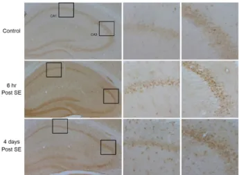
Fig. 1. Immunohistochemistry of Bmi1 in the hippocampus.
Compared to control, Bmi1 expression is transiently in- creased in the CA1, CA3 and dentate gyrus at 6 hr post SE. At 4 days post SE, the expression level of Bmi1 is observed in the glial cells throughout the hippocampus.
were euthanized at 3 days post SE, and the tissues were processed as described above. The sections were mounted on gelatin-coated slides. Sections were serially rehydrated in alcohol (100% to 70%) and tap water, then incubated in 0.1% cresyl violet solution (Sigma, St. Louis, MO, USA) for 20 min. After destaining with 95% ethanol containing 0.1% glacial acetic acid, the sections were dehydrated, dried, and mounted with Permount (Sigma, St. Louis, MO, USA).
The severity of the neuronal damage in the hippocampus was evaluated by an examiner blinded to the study groups.
The severity was semiquantitatively assessed according to the method described previously with minor modification [18]. Neuronal damage was measured on the following scale: no neuronal damage, grade 0; less than 20% neuronal damage, grade 1; 21 ~ 50% neuronal damage, grade 2; 51–
100% neuronal damage, grade 3.
Neuronal toxicity assay
Hippocampi were isolated from postnatal day 1 mice and put into a 1.5 mL polyethylene tube. Hippocampi were dis- sociated with trypsin and isolated cells were seeded on a 24-well plate. Isolated hippocampi from one pup were not mixed with the tissue from another, and each tail was cut and genotyping was carried out to determine their geno- type, as described above. The cells were maintained in Neurobasal media supplemented with 2% B27, 1% pen- icillin/streptomycin, and 0.25 mM glutamine (all culture media were from Gendepot, Katy, TX) for 10 days. N-meth- yl-D-aspartate (NMDA, 25 μM) was added to the culture for 1 hr, and the cell culture media was collected at 12 hr later for the measurement of lactate dehydrogenase (LDH) release as a measure of cell membrane integrity.
Behavioral assessment of spontaneous recurrent seizures
Spontaneous recurrent seizures were monitored at 1 or 2 months after SE induction. For this study, animal behavior was recorded for 10 hr per day for 1 week. Frequency and duration of spontaneous recurrent seizures were analyzed on the screen.
Timm’s staining
After the last monitoring for recurrent seizures, mice were euthanized, as previously described [20]. Mice were transcardially perfused with saline followed by sodium phosphate buffer containing 1.2% Na2S. Brains were post-
fixed for 2 days in 10% neutral-buffered paraformaldehyde (pH 7.4), then transferred to a 3% glutaraldehyde solution for 90 min, and finally cryoprotected in a 30% sucrose sol- ution (0.1 M PBS). Sequential coronal sections (30 µm) through the whole hippocampus were prepared using a cryotome. The sections were mounted on gelatin-coated slides and dried. After rehydration in a graded alcohol ser- ies, the sections were stained with a solution containing 30% gum arabic, 1.7% hydroquinone, and 0.085% silver ni- trate in a citrate buffer. After dehydration, the sections were covered with Permount. The optical density of Timm- stained axonal terminals in the dentate gyrus was measured using Photoshop® software.
Statistical analysis
Data are presented as mean ± SEM, and the significance was assessed using GraphPad Prism (version 3.0). Statistical significance was accepted when p<0.05.
Results
Expression of Bmi1 following pilocarpine-induced status epilepticus
Pilocarpine-induced SE induces neuronal cell death in CA1 and hilus. On the other hand, granule cells in the den- tate gyrus are resistant to SE and only minor cell damage is observed in the CA3 region. We examined the expression pattern of Bmi1 in the hippocampus of normal mice. The expression level of Bmi1 is quite low in the whole hippo-
Table 1. Seizure behaviors following pilocarpine injection
Genotypes No. animals % of SV seizure Onset time of SV
seizure (min)* % of SE seizure Onset time of SE seizure (min)*
Mortality (%) Bmi1+/+
Bmi1+/-
51 28
80.4 81.2
44.17±2.75 40.48±2.56
43.13 53.57
58.91±3.78 55.07±2.80
13.7 14.3 SV: stage V, SE: status epilepticus
*Onset time of SV or SE seizure means time of stage V or SE seizure onset taken after pilocarpine injection.
A
B
Fig. 2. Expression of Bmi1 mRNA in Bmi1 heterozygous mice.
(A) Targeted allele was identified by PCR products of DNA extracted from tails. (B) The expression level of Bmi1 mRNA in the hippocampus from each genotype.
Data were represented as relative ratio to Bmi1+/+ (mean
± SEM, n = 4 for each group). ** p<0.005 compared to control.
campus (Fig. 1). However, robust expression of Bmi1 is ob- served in the pyramidal cell layer of the CA1 and CA3 region and dentate gyrus at 6 hr after SE. At 4 days after SE, Bmi1 expression was remained in the CA3 pyramidal cell layer, but not in the CA1 area. In addition, Bmi1 ex- pression was also observed in the glial cells at 4 days post SE. These data suggest that Bmi1 may be involved in the hippocampal pathogenesis of SE. To test this hypothesis, we used Bmi1 knockout mice.
Phenotypic verification of Bmi1 knockout mice Male Bmi1+/- (Bka.Cg-Ptprcb Bmi1tm1Ilw Thy1a/J) mouse was crossed with female C57BL/6J wild type mice. Genomic DNA of pups was isolated from mouse tail tissue, and the DNA template was amplified using PCR. The mutant allele was confirmed as a heterozygous knockout in mice (Fig.
2A). In this study, heterozygous knockout mice were used
because mice homozygous for the Bmi1 knockout allele die before birth or soon after they are born (provider’s in- formation, The Jackson Laboratory. Bar Harbor, ME). The expression level of Bmi1 mRNA in heterozygous knockout mice was reduced by more than 50% (Fig. 2B).
Neuronal cell death in the Bmi1 knockout mice following status epilepticus
SE was induced in Bmi1+/- mice and its wild type litter- mates, and 3 days later neuronal damage was examined with cresyl violet staining. First, there was no significant difference in seizure behavior (stage V seizure, SE, and mor- tality) between the two groups (Table 1). However, the per- cent of SE induction in Bmi1+/- mice was 10.44% higher than that in wild type littermates. At 3 days after SE, neuro- nal damage in the CA1 and hilus regions was induced in both groups, and there was no significant difference in cell death between the two groups (Fig. 3). Next, we tested neu- ronal vulnerability in cultured hippocampal neurons from Bmi1+/- pups and wild type littermates. In this experiment, there was no significant difference in NMDA-induced neu- ronal damage between the two groups (Fig. 4). These in vivo and in vitro data indicate that reduced expression of Bmi1 does not affect neuronal damage in SE.
Epileptic behavior in Bmi1 knockout mice following status epilepticus
Pilocarpine-induced SE is a well-known animal model of epilepsy. In this study, we monitored the behavior of epi- lepsy and recurrent seizures in mice that showed SE.
Although there was no significant difference, the frequency of recurrent seizure was lower in Bmi1+/- mice compared to the wild type littermates. On the other hand, the duration of recurrent seizure was similar between two groups (Table 2). SE induces mossy fiber outgrowth in the dentate gyrus, and it has been reported that mossy fiber outgrowth is in- volved in epileptogenesis. In our study, mossy fiber out- growth was significantly increased in Bmi1+/- mice com-
A
B
Fig. 3. Neuronal damage in the hippocampus following pilocarpine-induced status epilepticus. (A) Representative images of cresyl violet stained-hippocampal sections. Neuronal damage is observed in the CA1, CA3 and the hilus. DG; dentate gyrus, Hil;
hilus. (B) Semi-quantitative analysis of hippocampal damage indicates that there is no significant difference between Bmi1+/- mice and wild type littermates (n=5 for each group).
Table 2. Frequency and duration of recurrent seizure
Genotypes No. animals 1 month after SE 2 months after SE
Frequency (events/hr) Duration (sec) BFrequency (events/hr) Duration (sec) Bmi1+/+
Bmi1+/-
7 6
1.00±0.18 0.75±0.15
18.97±3.35 17.71±3.62
0.33±0.08 0.22±0.06
20.99±8.57 17.97±7.34
pared to wild type animals (Fig. 5).
Discussion
Bmi1 is the core component of PRC1 and has been identi- fied as a Myc cooperating oncogene in murine B- and T-cell lymphomas. Bmi1 has also been identified as an oncogene promoting carcinogenesis in cancers, including prostate, lung, ovarian, urinary bladder, and breast cancer, and lym- phoma, mesothelioma, medulloblastoma, glioma, acute mye-
loid leukemia [16]. Likewise, Bmi1 inhibitors promoted cell cycle arrest in cervical cancer cell lines, induced a temporal decrease in ATP, and compromised the mitochondrial redox balance resulting in caspase-dependent apoptosis [10, 21].
On the other hand, during development, Bmi1 plays an im- portant role in primitive endoderm formation and the devel- opment of hematopoietic cells and the axial skeleton and cerebellum [7, 11, 19]. Combined, it is believed that Bmi1 is an essential component for embryonic development and cell proliferation and survival.
Fig. 4. NMDA-induced cytotoxicity in culture hippocampal neurons. Although neuronal damage increased by NMDA incubation, there was no significant difference between cultured neurons from Bmi1+/- pups and wild type littermates. Data were represented as mean ± SEM, (n=4 for each group).
A B
Fig. 5. Mossy fiber sprouting. (A) Representative images of mossy fiber sprouting from control or 2 months post SE. (B) Quantitative analysis of mossy fiber sprouting. Relative density from Bmi1+/- mice was significantly higher compared to that of wild type littermates. Data are represented as mean ± SEM. *p<0.05 and ***p<0.001 compared to control animals and ##p<0.01 compared to the epileptic wild type littermates (Bmi1+/+, 2 months post SE).
Recently, the roles of Bmi1 in the central nervous system has been realized. For example, Bmi1 hemi-deficiency (Bmi1+/-) mice showed reduced median and maximal life- spans and showed impaired learning and memory ability at age 15 months [5, 12]. In addition, tau levels increased in the cortex of 22-24 month-old Bmi1+/- mice [12]. In the eyes, cone photoreceptors and bipolar neurons are normally generated, but these undergo rapid degeneration in Bmi1-/- mice caused by Rip3-associated necroptosis [2]. Interestingly, there is increasing evidence showing that Bmi1 may be in- volved in neuroprotective effects in the central nervous system. First, Bmi1 transcription has been shown to be tran-
siently increased in the CA3 region when focal-onset SE was induced by the stereotaxic microinjection of kainic acid into the amygdala. However, there was no significant difference in the CA1, which is the region most vulnerable to pilo- carpine-induced SE [29]. Second, Bmi1 expression increased in the ischemic-preconditioned brain and knocking down Bmi1 with siRNA ablated the tolerance to oxygen-glucose deprivation in cultured cells and increased tissue infarction in animal models of strokes [34]. Third, increased expression of Bmi1 in cultured cortical neurons conferred robust pro- tection against DNA-damage-induced cell death or mi- tochondrial poisoning through activation of antioxidant re- sponse genes [1]. Taken together, these data suggest that Bmi1 could be the therapeutic target against a diverse range of neuronal diseases. Therefore, we aimed to elucidate Bmi1’s therapeutic potential in an animal model of epilepsy using Bmi1+/- mice.
In our study, Bmi1 expression transiently increased main- ly in the CA3 region following both low (200 mg/kg; data not shown) and high (325 mg/kg) doses of pilocarpine, but less prominently in CA1, which is the most vulnerable area in pilocarpine-induced SE. These data are consistent with the observations reported in animal models of kainic acid microinjection into the amygdala and may suggest the pro- tective role of Bmi1 against SE [29]. However, there was no difference in neuronal death between wild-type and Bmi1+/- mice in the pilocarpind-induced SE nor in the NMDA-in- duced neurotoxicity in cultured neurons. However, one
should remember that we used heterozygous knockout mice, and the mRNA expression was reduced by about 50%.
Although we did not measure protein content, Gu et al. [14]
reported that Bmi1 expression in Bmi1+/- mice is reduced by 54% compared to wild type in the hippocampus. These data suggest the possibility that the reduced expression level of Bmi1 in Bmi1+/- mice still provides some protection for CA3 neurons against pilocarpine-induced SE. Further study is needed to clearly determine the protective role of Bmi1 in the animal model of SE.
We also observed that mossy fiber sprouting was sig- nificantly increased in Bmi1+/- mice compared to the wild type littermates, but the epileptic behavior was similar be- tween the two groups. The aberrant sprouting of granule cell axons refers to an abnormal and extensive innervation of mossy fibers to the dentate inner molecular layer of the hippocampus and is frequently observed in temporal lobe epilepsy [4]. The role of mossy fiber sprouting in the epi- leptic brain is controversial. The pro-epileptogenic role of mossy fiber sprouting came from a diverse range of observations. First, the mossy fiber sprouting is frequently observed in patients with temporal lobe epilepsy, as well as in animal models of epilepsy [30, 32]. Second, the histo- logical evidence indicates that the sprouted mossy fiber ter- minals form excitatory synapses with dendritic spines of dentate granule cells [28]. Third, the intensity of mossy fiber sprouting is positively correlated with the degree of cell loss in the CA1 and CA3 regions [26]. On the other hand, there is increasing evidence that mossy fiber sprouting does not cause spontaneous recurrent seizures and is not positively correlated with epileptogenesis. First, spontaneous seizure frequency does not correlate with mossy fiber sprouting [26].
Second, not all epileptic patients develop mossy fiber sprout- ing [9, 23]. Third, a high-dose of rapamycin blocked mossy fiber sprouting to the level of naïve controls without reduc- tion of seizure frequency [15]. Taken together, although mos- sy fiber sprouting may lead to the epileptogenesis process, they do not appear to be necessary for triggering or main- taining hippocampal hyperexcitability [4]. In addition, the frequency of seizures was reduced in our study; thus, further studies will be needed to elucidate the role of Bmi1 in mossy fiber sprouting in epileptogenesis.
In this study, we observed that Bmi1 hemi-deficiency does not affect pilocarpine-induced SE or neuronal death in the hippocampus. However, Bmi1 hemi-deficiency significantly increased mossy fiber sprouting in the epileptic stage and
slightly reduced the percentage and frequency of recurrent seizures. When interpreting these results, it is necessary to be aware that Bmi1 expression was only reduced by about 50% in Bmi1 hemi-deficient mice and, therefore, some pro- tective effects of Bmi1 might persist. Combined, data pre- sented here indicate that Bmi1 is involved in epileptogenesis, although further studies are needed to elucidate its role in epilepsy.
Acknowledgement
This research was supported by Basic Science Research Program through the National Research Foundation of Korea (NRF) funded by the Ministry of Education (2013 R1A1A2013168).
The Conflict of Interest Statement
The authors declare that they have no conflicts of interest with the contents of this article.
References
1. Abdouh, M., Chatoo, W., El, H. J., David, J., Ferreira, J. and Bernier, G. 2012. Bmi1 is down-regulated in the aging brain and displays antioxidant and protective activities in neurons.
PLoS One 7, e31870.
2. Barabino, A., Plamondon, V., Abdouh, M., Chatoo, W., Flamier, A., Hanna, R., Zhou, S., Motoyama, N., Hébert, M., Lavoie, J. and Bernier, G. 2016. Loss of Bmi1 causes anoma- lies in retinal development and degeneration of cone photoreceptors. Development 143, 1571-1584.
3. Calao, M., Sekyere, E. O., Cui, H. J., Cheung, B. B., Thomas, W. D., Keating, J., Chen, J. B., Raif, A., Jankowski, K., Davies, N. P., Bekkum, M. V., Chen, B., Tan, O., Ellis, T., Norris, M. D., Haber, M., Kim, E. S., Shohet, J. M., Trahair, T. N., Liu, T., Wainwright, B. J., Ding, H. F. and Marshall, G. M. 2013. Direct effects of Bmi1 on p53 protein stability inactivates oncoprotein stress responses in embryonal can- cer precursor cells at tumor initiation. Oncogene 32, 3616- 3626.
4. Cavarsan, C. F., Malheiros, J., Hamani, C., Najm, I. and Covolan, L. 2018. Is mossy fiber sprouting a potential ther- apeutic target for epilepsy? Front Neurol. 9, 1023.
5. Chatoo, W., Abdouh, M., David, J., Champagne, M. P., Ferreira, J., Rodier, F. and Bernier, G. 2009. The polycomb group gene Bmi1 regulates antioxidant defenses in neurons by repressing p53 pro-oxidant activity. J. Neurosci. 29, 529- 542.
6. Chen, Z., Brodie, M. J., Liew, D. and Kwan, P. 2018. Treatment outcomes in patients with newly diagnosed epilepsy treated
with established and new antiepileptic drugs: a 30-year lon- gitudinal cohort study. JAMA Neurol. 75, 279-286.
7. Courel, M., Friesenhahn, L. and Lees, J. A. 2008. E2f6 and Bmi1 cooperate in axial skeletal development. Dev. Dyn. 237, 1232-1242.
8. Davies, J. A. 1995. Mechanisms of action of antiepileptic drugs. Seizure 4, 267-271.
9. de Lanerolle, N. C., Kim, J. H., Williamson, A., Spencer, S.
S., Zaveri, H. P., Eid, T. and Spencer, D. D. 2003. A retro- spective analysis of hippocampal pathology in human tem- poral lobe epilepsy: evidence for distinctive patient subcategories. Epilepsia 44, 677-687.
10. Dey, A., Xiong, X., Crim, A., Dwivedi, S. K. D., Mustafi, S. B., Mukherjee, P., Cao, L., Sydorenko, N., Baiazitov, R., Moon, Y. C., Dumble, M., Davis, T. and Bhattacharya, R.
2018. Evaluating the mechanism and therapeutic potential of PTC-028, a novel inhibitor of BMI-1 function in ovarian cancer. Mol. Cancer Ther. 17, 39-49.
11. Ding, X., Lin, Q., Ensenat-Waser, R., Rose-John, S. and Zenke, M. 2012. Polycomb group protein Bmi1 promotes hematopoietic cell development from embryonic stem cells.
Stem Cells Dev. 21, 121-132.
12. El Hajjar, J., Chatoo, W., Hanna, R., Nkanza, P., Tétreault, N., Tse, Y. C., Wong, T. P., Abdouh, M. and Bernier, G.
2019. Heterochromatic genome instability and neuro- degeneration sharing similarities with Alzheimer's disease in old Bmi1+/- mice. Sci. Rep. 9, 594.
13. Ginjala, V., Nacerddine, K., Kulkarni, A., Oza, J., Hill, S.
J., Yao, M., Citterio, E., van Lohuizen, M. and Ganesan, S.
2011. Bmi1 is recruited to DNA breaks and contributes to DNA damage-induced H2A ubiquitination and repair. Mol.
Cell Biol. 31, 1972-1982.
14. Gu, M., Shen, L., Bai, L., Gao, J., Marshall, C., Wu, T., Ding, J., Miao, D. and Xiao, M. 2014. Heterozygous knockout of the Bmi-1 gene causes an early onset of phenotypes asso- ciated with brain aging. Age (Dordr) 36, 129-139.
15. Heng, K., Haney, M. M. and Buckmaster, P. S. 2013. High- dose rapamycin blocks mossy fiber sprouting but not seiz- ures in a mouse model of temporal lobe epilepsy. Epilepsia 54, 1535-1541.
16. Janaki Ramaiah, M. and Vaishnave, S. 2018. BMI1 and PTEN are key determinants of breast cancer therapy: A plausible therapeutic target in breast cancer. Gene 678, 302-311.
17. Katzung, B. G. 2018. Basic & Clinical Pharmacology, pp. 409, 14th ed., McGraw-Hill: 1325 Avenue of the Americas New York, NY, USA.
18. Lee, K. E., Cho, K. O., Choi, Y. S. and Kim, S. Y. 2016. The neuroprotective mechanism of ampicillin in a mouse model of transient forebrain ischemia. Kor. J. Physiol. Pharmacol. 20, 185-192.
19. Leung, C., Lingbeek, M., Shakhova, O., Liu, J., Tanger, E., Saremaslani, P., Van Lohuizen, M. and Marino, S. 2004.
Bmi1 is essential for cerebellar development and is overex- pressed in human medulloblastomas. Nature 428, 337-341.
20. Li, A., Choi, Y. S., Dziema, H., Cao, R., Cho, H. Y., Jung, Y. J. and Obretan, K. 2010. Proteomic profiling of the epi-
leptic dentate gyrus. Brain Pathol. 20, 1077-1089.
21. Li, J., Vangundy, Z. and Poi, M. 2020. PTC209, a specific inhibitor of BMI1, promotes cell cycle arrest and apoptosis in cervical cancer cell lines. Anticancer Res. 40, 133-141.
22. Liu. J., Cao, L., Chen, J., Song, S., Lee, I. H., Quijano, C., Liu, H., Keyvanfar, K., Chen, H., Cao, L. Y., Ahn, B. H., Kumar, N. G., Rovira, I. I., Xu, X. L., Van Lohuizen, M., Motoyama, N., Deng, C. X. and Finkel, T. 2009. Bmi1 regu- lates mitochondrial function and the DNA damage re- sponse pathway. Nature 459, 387-392.
23. Masukawa, L. M., Uruno, K., Sperling, M., O'Connor, M.
J. and Burdette, L. J. 1992. The functional relationship be- tween antidromically evoked field responses of the dentate gyrus and mossy fiber reorganization in temporal lobe epi- leptic patients. Brain Res. 579, 119-127.
24. Park, I. K., Morrison, S. J. and Clarke, M. F. 2004. Bmi1, stem cells, and senescence regulation. J. Clin. Invest. 113, 175-179.
25. Pathan, S. A., Jain, G. K., Akhter, S., Vohora, D., Ahmad, F. J. and Khar, R. K. 2010. Insights into the novel three
‘D's of epilepsy treatment: drugs, delivery systems and devices. Drug Discov. Today 15, 717-732.
26. Polli, R. S., Malheirosm, J. M., Dos Santos, R., Hamani, C., Longo, B. M., Tannús, A., Mello, L. E. and Covolan, L. 2014.
Changes in hippocampal volume are correlated with cell loss but not with seizure frequency in two chronic models of temporal lobe epilepsy. Front Neurol. 5, 111.
27. Racine, R. J. 1972. Modification of seizure activity by elec- trical stimulation. II. motor seizure. Electroencephalogr. Clin.
Neurophysiol. 32, 281-294.
28. Represa, A., Jorquera, I., Le Gal La Salle, G. and Ben-Ari, Y. 1993. Epilepsy induced collateral sprouting of hippo- campal mossy fibers: does it induce the development of ec- topic synapses with granule cell dendrites? Hippocampus 3, 257-268.
29. Reynolds, J. P., Miller-Delaney, S. F., Jimenez-Mateos, E. M., Sano, T., McKiernan, R. C., Simon, R. P. and Henshall, D.
C. 2015. Transcriptional response of polycomb group genes to status epilepticus in mice is modified by prior exposure to epileptic preconditioning. Front Neurol. 6, 46.
30. Schmeiser, B., Zentner, J., Prinz, M., Brandt, A. and Freiman, T. M. 2017. Extent of mossy fiber sprouting in patients with mesiotemporal lobe epilepsy correlates with neuronal cell loss and granule cell dispersion. Epilepsy Res. 129, 51-58.
31. Schmidt, D. 2009. Drug treatment of epilepsy: options and limitations. Epilepsy Behav. 15, 56-65.
32. Shibley, H. and Smith, B. N. 2002. Pilocarpine-induced sta- tus epilepticus results in mossy fiber sprouting and sponta- neous seizures in C57BL/6 and CD-1 mice. Epilepsy Res. 49, 109-120.
33. Siddique, H. R. and Saleem, M. 2012. Role of BMI1, a stem cell factor, in cancer recurrence and chemoresistance: pre- clinical and clinical evidences. Stem Cells 30, 372-378.
34. Stapels, M., Piper, C., Yang, T., Li, M., Stowell, C., Xiong, Z. G., Saugstad, J., Simon, R. P., Geromanos, S., Langridge, J., Lan, J. Q. and Zhou, A. 2010. Polycomb group proteins
초록:Pilocarpine에 의해 유도된 생쥐 경련중첩증에서 Bmi1의 역할
편해인1․박지아1․최윤식1,2* (경성대학교 약학과)
Bmi1은 PcG 단백의 일종으로 PRC를 구성하는 성분이다. Bmi1에 관한 초기의 연구는 주로 암세포에서의 역할 에 집중되었고, 그 결과 Bmi1은 암세포의 증식과 생존에 중요한 역할을 하는 것으로 받아들여지고 있다. 그러나, 최근의 연구 결과 Bmi1은 퇴행성 신경계 질환이 있는 뇌에서 발현이 감소되어 있고 미토콘드리아의 기능과 활성 산소 수준을 조절하는 것으로 알려져 있다. 본 연구에서는 Bmi1 발현 저하 동물을 이용하여 간질에서 Bmi1의 약물 치료 표적 가능성을 밝히고자 하였다. Bmi1의 발현은 pilocarpine에 의한 경련중첩증 이후 해마의 CA1, CA3
그리고 치상회에서 뚜렷하게 증가하였다. 경련 양상을 볼 때 경련중첩증이 유도되는 비율은 Bmi1+/+와 Bmi1+/-
생쥐에서 각각 43.14%와 53.57%로 나타났다. 그러나, 치사율과 해마의 신경세포 손상에는 두 군 간에 통계적으로 유의한 차이를 보이지 않았다. 경련중첩증 유도 2개월 후에 나타난 간질 경련의 빈도수는 통계적 유의성은 없었으 나 Bmi1+/- 생쥐에서 약 50% 낮게 나타났다. 반면, 이끼섬유발아는 Bmi1+/- 생쥐에서 통계적으로 유의하게 증가하 였다. 이러한 결과를 종합하면, Bmi1의 발현이 감소할 때 pilocarpine에 의한 경련 유도와 이끼섬유발아가 증가함 을 보여준다.
as epigenetic mediators of neuroprotection in ischemic tolerance. Sci. Signal. 3, 10.
35. Trahair, T. N., Liu, T., Wainwright, B. J., Ding, H. F. and Marshall, G. M. 2013. Direct effects of Bmi1 on p53 protein stability inactivates oncoprotein stress responses in embry- onal cancer precursor cells at tumor initiation. Oncogene 32, 3616-3626.
36. Valk-Lingbeek, M. E., Bruggeman, S. W. and van Lohuizen, M. 2004. Stem cells and cancer; the polycomb connection.
Cell 118, 409-418.
37. Wang, M. C., Li, C. L., Cui, J., Jiao, M., Wu, T., Jing, L.
and Nan, K. J. 2015. BMI-1, a promising therapeutic target for human cancer. Oncol. Lett. 10, 583-588.
38. Xu, X., Wang, Z., Liu, N., Zhang, P., Liu, H., Qi, J. and Tu, Y. 2018. The mechanism of BMI1 in regulating cancer stemness maintenance, metastasis, chemo- and radiation resistance. Cancer Transl. Med. 4, 59-63.