• 검색 결과가 없습니다.

Anomalous Origin of the Left Circumflex Coronary Artery From the First Diagonal Branch Presented as Acute Myocardial Infarction

N/A
N/A
Protected

Academic year: 2022

Share "Anomalous Origin of the Left Circumflex Coronary Artery From the First Diagonal Branch Presented as Acute Myocardial Infarction"

Copied!
3
0
0

로드 중.... (전체 텍스트 보기)

전체 글

(1)

612

Print ISSN 1738-5520 / On-line ISSN 1738-5555 Copyright © 2011 The Korean Society of Cardiology CASE REPORT

http://dx.doi.org/10.4070/kcj.2011.41.10.612

Open Access

Anomalous Origin of the Left Circumflex Coronary Artery From the First Diagonal Branch Presented

as Acute Myocardial Infarction

Jung Hyun Kim, MD

1

, Geun Jin Ha, MD

2

, Myung Jun Seong, MD

2

, Jin Wook Jung, MD

3

, So Yeon Kim, MD

4

, Sung Hee Moon, MD

5

, and Young Soo Lee, MD

2

1

Division of Cardiology, Gimcheon Jeil Hospital, Gimcheon,

2

Divisions of Cardiology and

5

Radiology, Daegu Catholic University Medical Center, Daegu,

3

Pohang St. Mary Hospital, Pohang,

4

Division of Cardiology, Dongguk University Medical Center, Gyeongju, Korea

ABSTRACT

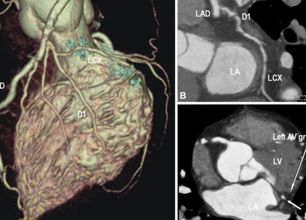
Coronary artery anomalies are diagnosed in 0.6 to 1.5% of patients who undergo coronary angiography (CAG). They may present with life threatening conditions but are generally asymptomatic. Recognition and adequate visualization of the anomaly is essential for correct management of the condition. However, in some cases the exact orifice and course of an anomalous coronary vessel cannot be selectively identified by CAG. In this report, a 54-year-old man was admitted to the hospital with acute inferior myocardial infarction and had an anomalous origin of the left circumflex coronary artery (LCX) from the first diagonal branch (D1). In CAG, the right CAG showed no significant stenosis and fortunately we found an anomalous origin of the LCX from the D1. The course of LCX was precisely established by 64-slice multi-detector computed tomography. (Korean Circ J 2011;41:612-614)

KEY WORD: Coronary vessel anomalies.

Received: May 31, 2010 Revision Received: July 19, 2010 Accepted: November 1, 2010

Correspondence: Young Soo Lee, MD, Division of Cardiology, Daegu Catholic University Medical Center, 3056-6 Daemyeong 4-dong, Nam- gu, Daegu 705-718, Korea

Tel: 82-53-650-3010, Fax: 82-53-621-3166 E-mail: [email protected]

• The authors have no financial conflicts of interest.

cc

This is an Open Access article distributed under the terms of the Cre- ative Commons Attribution Non-Commercial License (http://creativecom- mons.org/licenses/by-nc/3.0) which permits unrestricted non-commer- cial use, distribution, and reproduction in any medium, provided the origi- nal work is properly cited.

Introduction

The incidence of coronary artery anomalies in a routine co- ronary angiography (CAG) series is between 0.6% and 1.5%.

1)

The ostium of an aberrant vessel may be difficult to identify in the diagnostic procedure, but recognition and adequate visualization of the anomaly is essential for appropriate pa- tient management, especially in patients undergoing evalua- tion for percutaneous coronary intervention (PCI) and car- diac surgery.

2)

This report describes the anomalous origin of the left cir-

cumflex coronary artery (LCX) from the first diagonal br- anch in a 54-year-old man, which presented as an acute in- ferior myocardial infarction.

Case

A 54-year-old man with hypertension and 30 pack-years smoking history presented with chest pain. The resting elec- trocardiogram was demonstrating ST-segment elevation in leads II, III and aVF. CAG showed no critical stenosis in the right coronary artery (RCA) and no visualization of the LCX.

Initially, we considered total occlusion of ostium of the LCX

but the guide wire could not bypass the site which usually ex-

ists in an ostium of the LCX. We then identified the small ar-

tery originating from the first diagonal branch with throm-

bolysis in the myocardial infarction flow grade 1 (Fig. 1A and

B). After balloon angioplasty was accomplished for that ar-

tery and D1, its course was similar to that of the LCX (Fig. 1C

and D). To trace the exact anatomical course of coronary ar-

teries, 64-slice multi-detector computed tomography (MD-

CT) was performed. MDCT demonstrated an anomalous ori-

gin of the LCX coursed to the left atrioventricular groove

(2)

Jung Hyun Kim, et al. 613

from the first diagonal branch (Fig. 2).

Discussion

Coronary artery anomalies have been identified in 0.6 to 1.5% of coronary angiograms.

1)

Previous studies reported that LCX anomalies were the third most common coronary artery anomaly diagnosed by using CAG.

3)

A total of 71% of patients with LCX anomaly had significant stenosis of the proximal LCX and 11% had severe atherosclerosis in this vessel alone.

4)

The origin of the LCX from the right sinus of Valsalva or RCA is a relatively common anatomical varia- tion

1)5)

and some cases reported the origin of the LCX from the pulmonary artery.

6)

However, an anomalous origin of the LCX from the first diagonal branch is a very rare congenital anomaly.

Since the variable clinical presentation and prognosis of coronary anomalies depends on the proximal course of the

anomalous arising coronary artery in relation to the great ves- sels, early detection and exact delineation of their proximal course are crucial.

2)

Conventional CAG has traditionally been used to diagnose coronary anomalies but in some cases the exact origin of anomalous coronary vessels cannot be selec- tively identified by this technique.

6)7)

The three dimensional acquisitions of either MDCT or cardiovascular magnetic res- onance allow unambiguous interpretation of the locations of coronary origins.

6)7)

For our patient, initially we recognized an anomalous origin of the LCX from the first diagonal branch by conventional CAG and finally the origin of the LCX could be precisely established by MDCT.

REFERENCES

1) Yamanaka O, Hobbs RE. Coronary artery anomalies in 126,595 pa- tients undergoing coronary arteriography. Cathet Cardiovasc Diagn 1990;21:28-40.

2) Gavrielatos G, Letsas KP, Pappas LK, Antonellis I, Kardaras F. Anom-

A

C

B

D

Fig. 1. Coronary angiographic finding. A and B: coronary angiography shows the small artery (arrow) with TIMI flow grade 1 originating from

the D1 (arrowhead). C and D: after percutaneous coronary intervention for the small artery, the coronary angiography reveals an anoma-

lous origin of the LCX from the D1. A and C: left anterior oblique cranial view. B and D: left anterior oblique caudal views. TIMI: thrombolysis

in myocardial infarction, LAD: left anterior descending coronary artery, LCX: left circumflex coronary artery, D1: first diagonal branch.

(3)

614 Anomalous Origin of the Left Circumflex Coronary Artery From the Diagonal Branch

alous origin of the entire coronary system with separate ostia within the right sinus of valsalva: a rare congenital anomaly and a review of the literature. Cardiology 2007;107:209-12.

3) Aydinlar A, Cicek D, Senturk T, et al. Primary congenital anomalies of the coronary arteries: a coronary arteriographic study in Western Tur- key. Int Heart J 2005;46:97-103.

4) Wilkins CE, Betancourt B, Mathur VS, et al. Coronary artery anoma- lies: review of over 10,000 patients from the Clayton Cardiovascular Laboratories. Tex Heart Inst J 1988;15:166-73.

5) Lee YS, Lee JB, Kim KS. Anomalous origin of the left circumflex coro-

nary artery from the right sinus of valsalva identified by imaging with multidetector computed tomography. Korean Circ J 2006;36:823-5.

6) Korosoglou G, Ringwald G, Giannitsis E, Katus HA. Anomalous ori- gin of the left circumflex coronary artery from the pulmonary artery: a very rare congenital anomaly in an adult patient diagnosed by cardio- vascular magnetic resonance. J Cardiovasc Magn Reson 2008;10:4.

7) Post JC, van Rossum AC, Bronzwaer JG, et al. Magnetic resonance angiography of anomalous coronary arteries: a new gold standard for delineating the proximal course. Circulation 1995;92:3163-71.

A

B

C

Fig. 2. 64-slice multi-detector computed tomography demonstrates an anomalous origin of the LCX coursed to the left atrioventricular (AV)

groove from the D1. LAD: left anterior descending coronary artery, LCX: left circumflex coronary artery, D1: first diagonal branch, LA: left

atrium, LV: left ventricle.

수치

Fig. 1. Coronary angiographic finding. A and B: coronary angiography shows the small artery (arrow) with TIMI flow grade 1 originating from  the D1 (arrowhead)
Fig. 2. 64-slice multi-detector computed tomography demonstrates an anomalous origin of the LCX coursed to the left atrioventricular (AV)  groove from the D1

참조

관련 문서

Coronary artery fistula give rise to some clinical manifestations including myocardial ischemia, congestive heart failure, bacterial endocarditis, secondary valvular

nance imaging enabled noninvasive morphological assessment of the coronary arteries. 5) reported a 65-year-old wom- an with ALCAPA confirmed by coronary angiography with a clue

autopsy population of congenital heart disease. Sudden cardiac death asso- ciated with isolated congenital coronary artery anomalies. Clinical features of pro- gnosis of

demonstration of coronary steal phenomenon. 4) Takenaka T, Horimoto M, Igarashi K, Inoue H, Sakuragi H. Coronary artery-left ventricle fistula complicating balloon angioplasty: a

1) Yamanaka O, Hobbs RE. Coronary artery anomalies in 126, 595 patients undergoing coronary arteriography. Coronary angioplasty of anomalous cor- onary arteries: Notes on

We reported a middle aged man who developed an acute transmural inferior wall myocardial in- farction associated with coronary artery fistula secondary to blunt chest trauma in

We report a case surgically treated with neo-ostium formation in anomalous origin of the left coronary artery from the right cor- onary sinus.. Key

Management was coronary artery bypass grafting to the left anterior descending artery and obtuse marginal arteries, closure of the left main coronary artery