Case Report pISSN: 1011-8942 eISSN: 2092-9382
Korean J Ophthalmol 2013;27(5):372-375 http://dx.doi.org/10.3341/kjo.2013.27.5.372
© 2013 The Korean Ophthalmological Society
This is an Open Access article distributed under the terms of the Creative Commons Attribution Non-Commercial License (http://creativecommons.org/licenses /by-nc/3.0/) which permits unrestricted non-commercial use, distribution, and reproduction in any medium, provided the original work is properly cited.
372
Acute Bilateral Visual Loss Related to Orthostatic Hypotension
Jung-Yeul Kim, Kyoung-Nam Kim, Woo-Jin Kim, Yeon-Hee Lee
Department of Ophthalmology, Chungnam National University College of Medicine, Daejeon, Korea
Ischemic optic neuropathy (ION) may develop in set- tings of hemodynamic compromise, such as systemic hy- potension, blood loss and anemia [1,2]. Perioperative ION is a subgroup of ION and is associated with perioperative hypotension and anemia [3]. Another well known ION re- lated to hypotension is “uremic optic neuropathy” that presents in patients with chronic renal failure and dialysis [4-6].
Prolonged immobilization of the body has extensive del- eterious physiologic consequences. After a long period of immobilization, there is a marked pooling of blood in the lower extremities causing a decrease in the circulating blood volume. This causes orthostatic hypotension, if an individual attempts to sit or stand, depleting the brain of blood and oxygen and often leading to fainting [7].
We report an individual who developed acute bilateral visual loss with optic disc edema after standing up from a prolonged bed-ridden position. We believe it to be a case of ION related to orthostatic hypotension.
Case Report
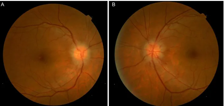
A 50-year-old man was referred to us due to decreased visual acuity in both eyes. The patient had spondylolisthe- sis and had undergone a lumbar laminectomy three months prior to these symptoms. After the operation, he had been bed-ridden in the supine position. The day before the visit, the patient had sat up for the first time and experienced momentary dizziness, nausea and facial pallor. Blood pres- sure decreased from 135 / 85 mmHg to 90 / 60 mmHg. A few hours later, the patient complained of blurred vision in both eyes. The next day, we measured a best-corrected vi- sual acuity (BCVA) of 0.3 in both eyes. The pupillary light reflex was decreased in both eyes. Fundus examination re- vealed optic disc edema in both eyes and a peripapillary flame-shaped hemorrhage in the left eye (Fig. 1). The Humphrey visual field test demonstrated generally de- creased sensitivity in both eyes. Fluorescein angiography showed definite delayed dye filling of both of the optic discs. Contrast-enhanced orbital magnetic resonance im- aging did not show remarkable findings, and there was no retrobulbar pain. The patient had iron deficiency anemia, and his hemoglobin level was 10.5 g/dL. He also had be- nign prostate hypertrophy and was prescribed tamsulosin (0.2 mg daily). Systemic evaluations including laboratory A 50-year-old man had undergone lumbar vertebral surgery and was confined to bed in the supine position for three months. When he sat up from the prolonged supine position, he showed clinical signs of orthostatic hypotension and reported decreased vision in both eyes. He also had underlying anemia.
Ophthalmologic findings suggested bilateral anterior ischemic optic neuropathy (ION) as the cause of the visual loss. Although there are numerous reports of ION in the setting of hemodynamic compromise, such as systemic hypotension, cases of ION-associated orthostatic hypotension are very rare.
Key Words: Ischemic optic neuropathy, Optic disc edema, Orthostatic hypotension, Visual loss
Received: May 16, 2011 Accepted: Dcember 8, 2011
Corresponding Author: Yeon-Hee Lee, MD. Department of Ophthalmol- ogy, Chungnam National University Hospital, #282 Munhwa-ro, Jung- gu, Daejeon 301-721, Korea. Tel: 82-42-280-8447, Fax: 82-42-255-3745, E-mail: [email protected]
373 JY Kim, et al. Visual Loss Related to Orthostatic Hypotension
and cardiovascular work-up showed no other abnormal findings. The cardiovascular work-up included echocardio- gram and magnetic resonance angiography of the carotid arteries. Methylprednisolone pulse therapy was initiated.
However, the next day, vision in the right eye further de- creased to 0.05. Vision stabilized for the next 10 days. Two weeks after the initial visit, fluorescein angiography showed improved dye filling on both optic discs. However, improvement was not complete in the right optic disc (Fig. 2). Vision slowly improved and, after six months, BCVA was 0.4 and 0.6 in the right and left eyes, respec- tively. Both optic discs were pale, and retinal nerve fiber layer thinning was prominent (Fig. 3). The visual field re- mained severely constricted.
Discussion
More than 300 cases of ION induced by shock have been reported and have established a firm link between hypo- tension and ION [1,2]. ION related to hypotension is most often observed in patients with chronic renal failure and on dialysis [4]. Haider et al. [5] observed four cases of ante- rior ION in a series of 60 patients undergoing dialysis over a two-year follow-up. Hypotension was acute and tempo- rary and associated with the dialysis procedure. Most pa- tients were also chronically anemic. Servilla and Groggel [8] reported a case of typical anterior ION in a uremic pa- tient, which occurred following an acute hypotensive epi- sode during hemodialysis. Jackson et al. [9], Michaelson et Fig. 1. Fundus photograph indicating features of optic disc edema in both eyes (A,B) and peripapillary flame-shaped hemorrhage in the left eye (B).
A B
A B C D
Fig. 2. Fluorescein angiography on the day of initial visit (A,C) and two weeks later (B,D). (A) Venous phase demonstrating definite de- layed dye filling on the right optic disc. (B) Arterio-venous phase two weeks later showing improved filling on the right optic disc. (C) Venous phase showing delayed dye filling on the left optic disc. (D) Venous phase two weeks later showing improved filling on the left optic disc.
374
Korean J Ophthalmol Vol.27, No.5, 2013
al. [10], and Basile et al. [11] also reported cases believed to be secondary to a hypotensive episode. Several cases of anterior ION resulting from rapid correction of malignant hypertension have also been reported [12-14]. The patho- genesis of anterior ION that occurs in this setting is likely to be related to impaired autoregulation of the vessels sup- plying the optic nerve head and to the reduction in perfu- sion pressure, leading to ischemia [4]. There are numerous reports of ION associated with spontaneous or traumatic blood loss [1,2,15,16]. Visual loss was usually bilateral in the reported cases. About 50% of patients experienced some recovery of vision, and 10% to 15% of patients recov- ered completely. Hayreh [1] reported that there is typically a time delay of up to 10 days between bleeding and the on- set of visual loss. Cases of perioperative ION are examples of ION associated with hemodynamic compromise. Most of the cases have bilateral simultaneous involvement.
Pathogenesis is believed be associated with perioperative hypotension and anemia [3].
Cases of ION induced by orthostatic hypotension are rare. We found only one case in MEDLINE. Connolly et al. [17] reported a case of anterior ION induced by a re- peated episode of orthostatic hypotension in patients who underwent simultaneous transplantation of the pancreas and kidney.
Painless acute visual loss accompanying optic edema is a characteristic clinical presentation of anterior ION. Bilat- eral presentation is unusual in ION, although it is a typical feature of ION in the setting of hemodynamic compromise [4]. Optic disc filling delay on fluorescein angiography is a common feature of non-arteritic anterior ION [18], and it is not a feature of non-ischemic optic disc edema [19]. In the present case, optic disc filling delay was definite in both eyes at the acute stage, and the filling delay had improved two weeks later. This observation gives strong evidence of ischemic optic disc edema. The substantial recovery of vi- sual acuity in the presenting case is not a typical feature of ordinary anterior ischemic optic neuropathy (AION).
However, this is common phenomenon in cases of ION as- sociated with hemorrhage. Many authors also have report- ed substantial visual improvement in cases of ION related to hypotension [6,14,17,20]. They have postulated that vol- ume replacement, correction of uremia with dialysis, or corticosteroid therapy might have been effective for vision recovery. These reports, however, are case reports or small case series, not controlled studies. Substantial visual im- provement might be a natural course of ION related to hy- potension. We believe that the presenting case is anterior ION related to orthostatic hypotension and anemia, consid- ering the numerous clinical settings and features.
We believe that the temporary ischemic insult by tempo- rary hypoperfusion and underlying anemia is a major causative mechanism of the case. Impaired autoregulation of vessels supplying the optic nerve head might be a fur- ther contributing mechanism, as in cases of anterior ION resulting from rapid correction of malignant hypertension.
Impaired autoregulation is also thought to be one of the causative mechanisms of conventional AION [18]. The re- duced fluctuation in systemic blood pressure over a long period might have caused blunting of the autoregulatory reflex.
Tamsulosin is a α1-blocker, which is reported to be asso- ciated with orthostatic hypotension that results from an impaired compensatory reflex. The compensatory reflex mechanism mediated by the sympathetic nervous system Fig. 3. (A) Optic nerve disc edema in both eyes and peripapillary
flame-shaped hemorrhage in the left eye were resolved. Both optic discs showed temporal pallor after six months of decreased vision. (B) Optical coherence tomography demonstrating prom- inent thinning in the retinal nerve fiber layer in both eyes six months after the initial event. TEMP = temporal; SUP = superior;
NAS = nasal; INF = inferior.
A
B
375 JY Kim, et al. Visual Loss Related to Orthostatic Hypotension
is normally brought into effect when standing [21]. There- fore, this might be an additional contributing factor to the orthostatic hypotension in this case.
Prolonged bed rest may cause deep vein thrombosis and resultant pulmonary embolism. In rare case of paradoxical embolism, venous thrombi may cause arterial thrombosis.
However, in the present case, there was no evidence of deep vein thrombosis, lateral opening of the heart, or arte- rial embolic phenomenon. Therefore, we do not consider deep vein thrombosis as a possible cause of the optic neu- ropathy.
Conflict of Interest
No potential conflict of interest relevant to this article was reported.
References
1. Hayreh SS. Anterior ischemic optic neuropathy. VIII. Clini- cal features and pathogenesis of post-hemorrhagic amauro- sis. Ophthalmology 1987;94:1488-502.
2. Johnson MW, Kincaid MC, Trobe JD. Bilateral retrobulbar optic nerve infarctions after blood loss and hypotension: a clinicopathologic case study. Ophthalmology 1987;94:1577- 84.
3. Katz DM, Trobe JD, Cornblath WT, Kline LB. Ischemic op- tic neuropathy after lumbar spine surgery. Arch Ophthalmol 1994;112:925-31.
4. Miller NR, Newman NJ, Biousse V, Kerrison JB. Walsh and Hoyt’s clinical neuro-ophthalmology. 6th ed. Philadelphia:
Lippincott Williams & Wilkins; 2005. p. 371-4.
5. Haider S, Astbury NJ, Hamilton DV. Optic neuropathy in uraemic patients on dialysis. Eye (Lond) 1993;7(Pt 1):148-51.
6. Knox DL, Hanneken AM, Hollows FC, et al. Uremic optic neuropathy. Arch Ophthalmol 1988;106:50-4.
7. Chobanian AV, Lille RD, Tercyak A, Blevins P. The meta-
bolic and hemodynamic effects of prolonged bed rest in normal subjects. Circulation 1974;49:551-9.
8. Servilla KS, Groggel GC. Anterior ischemic optic neuropa- thy as a complication of hemodialysis. Am J Kidney Dis 1986;8:61-3.
9. Jackson TL, Farmer CK, Kingswood C, Vickers S. Hypo- tensive ischemic optic neuropathy and peritoneal dialysis.
Am J Ophthalmol 1999;128:109-11.
10. Michaelson C, Behrens M, Odel J. Bilateral anterior isch- aemic optic neuropathy associated with optic disc drusen and systemic hypotension. Br J Ophthalmol 1989;73:762-4.
11. Basile C, Addabbo G, Montanaro A. Anterior ischemic optic neuropathy and dialysis: role of hypotension and anemia. J Nephrol 2001;14:420-3.
12. Cove DH, Seddon M, Fletcher RF, Dukes DC. Blindness af- ter treatment for malignant hypertension. Br Med J 1979;2:245-6.
13. Pryor JS, Davies PD, Hamilton DV. Blindness and malig- nant hypertension. Lancet 1979;2:803.
14. Taylor D, Ramsay J, Day S, Dillon M. Infarction of the op- tic nerve head in children with accelerated hypertension. Br J Ophthalmol 1981;65:153-60.
15. Chisholm IA. Optic neuropathy of recurrent blood loss. Br J Ophthalmol 1969;53:289-95.
16. Klewin KM, Appen RE, Kaufman PL. Amaurosis and blood loss. Am J Ophthalmol 1978;86:669-72.
17. Connolly SE, Gordon KB, Horton JC. Salvage of vision af- ter hypotension-induced ischemic optic neuropathy. Am J Ophthalmol 1994;117:235-42.
18. Hayreh SS. Ischemic optic neuropathy. Prog Retin Eye Res 2009;28:34-62.
19. Arnold AC, Badr MA, Hepler RS. Fluorescein angiography in nonischemic optic disc edema. Arch Ophthalmol 1996;114:293-8.
20. Saini JS, Jain IS, Dhar S, Mohan K. Uremic optic neuropa- thy. J Clin Neuroophthalmol 1989;9:131-3.
21. Nieminen T, Koobi T, Tammela TL, Kahonen M. Hypoten- sive potential of sildenafil and tamsulosin during orthostasis.
Clin Drug Investig 2006;26:667-71.