549
http://dx.doi.org/10.4196/kjpp.2015.19.6.549 eISSN 2093-3827
ABBREVIATIONS: AktiIV, Akt inhibitor IV; CCL11, C-C motif ligand 11; ERK, extracellular signal-regulated kinase; HAoSMCs, human aortic smooth muscle cells; MAPK, mitogen-activated protein kinase;
MEK, mitogen-activated protein/extracellular signal-regulated kinase kinase; PI3K, phosphoinositide 3 kinase; PTX, pertussis toxin; PKC, protein kinase C; VSMCs, vascular smooth muscle cells.
Received July 14, 2015, Revised August 19, 2015, Accepted September 11, 2015
Corresponding to: Koanhoi Kim, Department of Pharmacology, School of Medicine, Pusan National University, Busandaehak-ro 49, Mulgeum, Yangsan 50612, Korea. (Tel) 82-51-510-8064, (Fax) 82- 51-510-8068, (E-mail) [email protected]
This is an Open Access article distributed under the terms of the Creative Commons Attribution Non-Commercial License (http://
creativecommons.org/licenses/by-nc/4.0) which permits unrestricted non-commercial use, distribution, and reproduction in any medium, provided the original work is properly cited.
Copyright ⓒ Korean J Physiol Pharmacol
Multiple Signaling Pathways Contribute to the Thrombin-induced Secretory Phenotype in Vascular Smooth Muscle Cells
Ji Young Jeong
1, Younghae Son
1, Bo-Young Kim
1, Seong-Kug Eo
2, Byung-Yong Rhim
1, and Koanhoi Kim
11
Department of Pharmacology, School of Medicine, Pusan National University Yangsan 50612,
2College of Veterinary Medicine and Bio-Safety Research Institute, Chonbuk National University, Iksan 54596, Korea
We attempted to investigate molecular mechanisms underlying phenotypic change of vascular smooth muscle cells (VSMCs) by determining signaling molecules involved in chemokine production. Treatment of human aortic smooth muscle cells (HAoSMCs) with thrombin resulted not only in elevated tran- scription of the (C-C motif) ligand 11 (CCL11) gene but also in enhanced secretion of CCL11 protein.
Co-treatment of HAoSMCs with GF109230X, an inhibitor of protein kinase C, or GW5074, an inhibitor of Raf-1 kinase, caused inhibition of ERK1/2 phosphorylation and significantly attenuated expression of CCL11 at transcriptional and protein levels induced by thrombin. Both Akt phosphorylation and CCL11 expression induced by thrombin were attenuated in the presence of pertussis toxin (PTX), an inhibitor of Gi protein-coupled receptor, or LY294002, a PI3K inhibitor. In addition, thrombin-induced production of CCL11 was significantly attenuated by pharmacological inhibition of Akt or MEK which phosphorylates ERK1/2. These results indicate that thrombin is likely to promote expression of CCL11 via PKC/Raf-1/ERK1/2 and PTX-sensitive protease-activated receptors /PI3K/Akt pathways in HAoSMCs.
We propose that multiple signaling pathways are involved in change of VSMCs to a secretory phenotype.
Key Words: CCL11, Secretory phenotype, Signaling pathway, Thrombin, Vascular smooth muscle cell
INTRODUCTION
Thrombin, a serine protease, is released during tissue damage and converts fibrinogen to fibrin at the final step of the blood coagulation cascade [1]. The released thrombin molecules not only contribute to repair of damaged blood vessels but also are associated with progression of vascular diseases [2]. Interaction of thrombin molecules with plate- lets at sites of vascular injury ensures rapid formation of haemostatic plugs [2,3]. Binding of thrombin to its re- ceptors, protease-activated receptors (PARs), leads to molec- ular and cellular events that occur atherosclerosis. PARs, a subfamily of related seven transmembrane G-protein-coupled receptors (GPCRs), are expressed on surface of multiple vascular cells, including endothelial cells and vascular smooth muscle cells (VSMCs) [4,5], and activation of PARs cause
production of mediators, including platelet-derived growth factor and inflammatory chemokines [3], and migration and proliferation of the cells [6-8].
Among CC chemokines whose expression is elevated in atherosclerotic lesions and injured arteries, expression of C-C motif chemokines 11 (CCL11) is of interest because this chemokine is believed to be involved remodeling of blood vessels [9,10]. CCL11, which is also known as eosinophil chamotactic protein and eotaxin-1, is a protein of the CC family chemokines that in human is encoded by the CCL11 gene with a high degree of amino acid sequence homology with monocyte chemotactic proteins (MCPs) [11]. Overex- pression of CCL11 protein has been detected in smooth muscle cells (SMCs) of human atheroma, with negligible expression in normal vessels, and CCL11 mRNA is up-regu- lated in SMCs of rat aortic allografts exposed to prolonged ischemic storage [12,13]. However, it is not clear which fac- tors induce expression of CCL11 in VSMCs.
In the normal arteries expression of PARs is restricted
mainly to the endothelial layer whereas their expression
is significantly elevated in areas rich in SMCs within athero-
sclerotic lesions, indicating activation of seine protease-medi-
ated pathways in atherosclerosis [14]. As thrombin is re-
leased during injury or inflammation, subendothelial SMCs will be come into contact with thrombin after endothelial injury and thrombus formation [15]. Moreover, due to pro- tection from inactivation by circulating inhibitors, thrombin bound to the subendothelial extracellular matrix remains functionally active [16], and SMCs are likely to be exposed to thrombin in atherosclerotic arteries. Therefore, to better understand underlying mechanisms associated with changes in VSMCs in the injured artery, it is necessary to determine molecular pathways via which thrombin exerts its effects on VSMCs because activation of thrombin receptors cause proliferation, migration, and production of chemokines and extracellular matrix synthesis of VSMCs [7,17,18].
In the current study, we attempted to determine signal- ing molecules below PARs whose activity is necessary for thrombin to induce production of CCL11 using human aort- ic smooth muscle cells (HAoSMCs) in order to clarify molec- ular mechanisms involved in change of VSMCs to secretory phenotype in the artery.
METHODS Cell culture and reagents
HAoSMCs purchased from American Type Culture Collection were grown in ATCC-formulated F-12K medium supplemented with vascular smooth muscle growth kit, 50 units/ml penicillin and 50 μg/ml streptomycin, as pre- viously reported [8]. The cells in between passage 6 and 9 were used. Thrombin, pertussis toxin (PTX), GF109203X, LY294002, N-acetylcysteine (NAC) and diphenyleneiodo- nium (DPI) were purchased from Sigma-Aldrich Co. (St.
Louis, MO, USA). U0126, Akt Inhibitor IV (AktiIV), and anti-phosphorylated Akt antibody were purchased from Cell Signaling Technology (Danvers, MA, USA). GW5074 and anti-phosphorylated ERK1/2 antibody were purchased from Santa Cruz Biotechnology (Santa Cruz, CA, USA).
Enzyme-linked immunosorbent assay (ELISA) of CCL11 The amount of CCL11 protein released from HAoSMC was determined using commercially available ELISA kit ac- cording to the manufacturer’s instructions (R&D systems, Minneapolis, MN, USA). HAoSMCs were incubated for 12 h in the presence of 1% fetal bovine serum (FBS) and ex- posed to thrombin prior to isolation of culture medium. The isolated culture medium and standard dilutions of CCL11 protein were added to each well. After incubation for 2 h, each well was washed and the Conjugate was added. After incubation for 1 h, each well was washed and the Substrate Solution was added. After incubation for 30 min, the Stop Solution was added and color intensity was measured at 450 nm.
Reverse transcription (RT) - polymerase chain reaction (PCR)
Total RNAs were reverse-transcribed to complementary DNA (cDNA) for 1 h at 42
oC with Moloney Murine Leukemia Virus reverse transcriptase using the oligod(T)
15primer, followed by non-quantitative and quantitative real-time PCR. For non-quantitative PCR, transcripts of genes of in- terest were amplified using Hot Start Taq Polymerase. The cDNA was denatured at 90
oC for 5 min followed by 25 cycles
of PCR (95 C for 30 sec, 55 C for 30 sec, 72 C for 30 sec) in the presence of forward and reverse primers of the genes.
Glyceraldehyde-3-phosphate dehydrogenase (GAPDH) was amplified as an internal control. Real-time quantitative PCR was performed in triplicate; each 20-μl reaction con- sisted of 10 μl of SYBR Green Master Mix, 2 μl of forward and reverse primers (10 pM each) of genes to be analyzed, and cDNA template. Thermal cycling conditions were as fol- low: 95
oC for 10 min, and 45 cycles at 95
oC for 10 sec, 50
oC for 10 sec, and an elongation period for 10 sec at 72
oC. The relative expression of each gene was then calculated as a ratio to GAPDH. The primers were CCL11: 5’-aaccacctgct- gctttaacc-3’ (forward) and 5’-tggctttggagttggagatt-3’ (reverse);
and GAPDH: 5’-gagtcaacggatttggtcct-3’ (forward) and 5’-tg- tggtcatgagtccttcca-3’ (reverse).
Western blot analysis
Cells were lysed with the lysis buffer (1% SDS, 1 mM NaVO
3, 10 mM Tris-HCl, pH 7.4) containing protease in- hibitors and centrifuged (15,000 xg) for 5 min at 4
oC. Super- natants were isolated, and protein concentration of the lyaste was determined. An equal amount of protein was separated by 12% SDS-PAGE and transferred to polyvi- nylidene fluoride (PVDF) membranes. After blocking for an hour in 5% skim milk in 0.1% Tween 20/Tris-buffered saline (TBS) (pH 7.6), the membranes were incubated with in- dicated primary antibodies diluted in the blocking solution at 4
oC overnight. After washing three times with 0.1%
Tween 20/TBS for 10 min each, the membranes were in- cubated with horseradish peroxidase (HRP)-conjugated sec- ondary antibodies diluted in the blocking solution (1 : 5,000) for an hour at room temperature. After washing three times with washing buffer for 10 min each, bands were detected using the Pierce ECL Western Blotting Substrate (Thermo Fisher Scientific Inc., Rockford, IL, USA).
Statistics
Statistical analyses were performed using GraphPad PRISM (version 5.0) (GraphPad Software Inc., San Diego, CA, USA), and p<0.05 was considered statistically sig- nificant.
RESULTS
Up-regulated expression of CCL11 in human VSMCs by treatment with thrombin
We investigated the possibility of change of VSMCs to secretory phenotype in the presence of thrombin, a PARs ligand, by determining production of CCL11 chemokine.
Treatment of HAoSMCs with thrombin resulted in an in-
creased expression of the CCL11 gene. The increase was
observed at 6 h post-treatment and persisted up to 12 h
post-treatment with thrombin (Fig. 1A). We also examined
the effects of thrombin on CCL11 at protein level. Treatment
with thrombin resulted in significantly elevated secretion
of CCL11 from HAoSMC in proportion to the treatment du-
ration with thrombin up to 12 h (Fig. 1B).
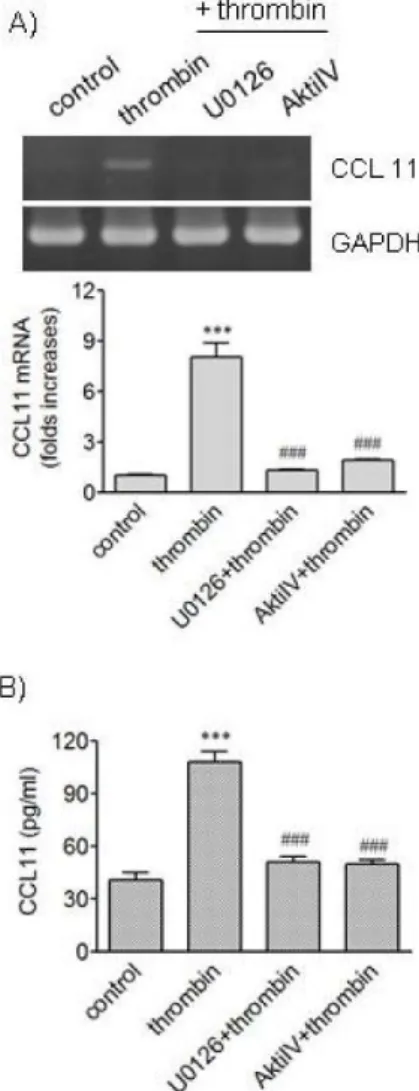
Fig. 2. Effects of inhibitors of MEK and Akt in thrombin-induced expression of CCL11. (A, B) HAoSMCs were incubated with or without U0126 and Akti IV (10 μM each) for 2 h followed by stimulation with thrombin for 9 h. (A) CCL11 transcripts were amplified by RT-PCR (upper panel) and the levels of CCL11 transcripts were determined by realtime PCR (lower panel). (B) The levels of CCL11 protein secreted into culture media were deter- mined by ELISA. Data are expressed as mean±SD (n=3 replicates for each group). ***p<0.001 vs. control;
###p<0.001 vs. thrombin.
Fig. 1. Effects of thrombin on expression of CCL11 in human VSMCs. (A, B) HAoSMCs were treated for the indicated time periods with thrombin (1 U/ml). (A) CCL11 transcripts were amplified by RT-PCR (upper panel) and the levels of CCL11 transcripts were determined by realtime PCR (lower panel). (B) The levels of CCL11 protein secreted into culture media were determined by ELISA.
Data are expressed as mean±SD (n=3 replicates for each group).
**p<0.01 vs. control cultured in the absence of thrombin (0 h);
***p<0.001 vs. 0 h.
Active roles of ERK and Akt pathways in CCL11 ex- pression induced by thrombin
Treatment of VSMCs with thrombin caused elevated phosphorylation of extracellular signal-regulated kinase 1/2 (ERK1/2) and Akt [8]. We investigated whether ERK and Akt pathways are involved in CCL11 production using U0126 and AktiIV, respectively (Fig. 2). U0126 inhibits mi- togen-activated protein/extracellular signal-regulated kin- ase kinase (MEK) which activates ERK1/2, and AktiIV con- trols activity of Akt. Thrombin increased transcription of the CCL11 gene, and pharmacological inhibition of ERK kinase and Akt pathways resulted in blockage of the CCL11 gene transcription induced by thrombin. Secretion of CCL11 increased by thrombin was also abrogated when ERK kinase or Akt pathway was inhibited using the inhibitors.
These results suggest participation of ERK and Akt path- ways in production of CCL11 in response to thrombin.
Involvement of PKC-mediated pathway in ERK1/ 2 phosphorylation and CCL11 expression induced by thrombin
Thrombin increases activity of protein kinase C (PKC),
and PKC can activate ERK pathway [8,19]. Therefore, it
was investigated whether PKC played roles in ERK1/2
phosphorylation and CCL11 production induced by thrombin
(Fig. 3). Consistent with the previous study, treatment of
HAoSMCs with thrombin resulted in increased phosphor-
ylation of ERK1/2. An addition of GF109230X, a potent in-
hibitor of PKC isoforms, resulted in abrogation of the
ERK1/2 phosphorylation. Treatment with GF109230X also
led to significantly attenuated transcription of the CCL11
gene and secretion of CCL11 protein induced by thrombin.
Fig. 3. Effects of inhibitors of PKC and Raf-1 on phosphorylation of ERK and expression of CCL11 induced by thrombin. (A) HAo- SMCs were stimulated with thrombin for 5 min after incubation with or without GF109203X (3 μM) and GW5074 (25 nM) for 2 h. An equal amount of protein was subjected to Western blot analysis with antibodies against α-tubulin and phosphorylated and unphosphorylated forms of ERK1/2. (B, C) HAoSMCs were incubated with or without GF109203X (3 μM) and GW5074 (25 nM) for 2 h followed by stimulation with thrombin for 9 h. The levels of CCL11 transcripts were determined by realtime PCR (B), and the levels of CCL11 protein secreted into culture media were determined by ELISA (C). Data are expressed as mean±SD (n=3 replicates for each group). ***p<0.001 vs. control;
###p<0.001 vs.
thrombin.
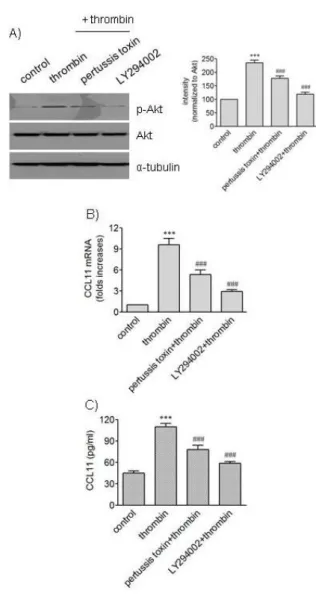
Fig. 4. Effects of pertussis toxin and LY294002 on phosphorylation of Akt and expression of CCL11 induced by thrombin. (A) HAo- SMCs were stimulated with thrombin for 5 min after incubation with or without pertussis toxin (100 ng/ml) and LY294002 (10 μM) for 2 h. An equal amount of protein was subjected to Western blot analysis with antibodies against α-tubulin and phosphorylated and unphosphorylated forms of Akt. The mean band intensity of p-Akt was normalized to that of Akt. (B, C) HAoSMCs were in- cubated with or without pertussis toxin (100 ng/ml) and LY294002 (10 μM) for 2 h followed by stimulation with thrombin for 9 h.
The levels of CCL11 transcripts were determined by realtime PCR (B), and the levels of CCL11 protein secreted into culture media were determined by ELISA (C). Data are expressed as mean±SD (n=3 replicates for each group). ***p<0.001 vs. control;
###p<0.001 vs. thrombin.
Because Raf-1 kinase participates in ERK cascade [19], it was investigated whether the kinase was involved in the effects of thrombin on VSMCs. Exposure of HAoSMCs to GW5074, an inhibitor of Raf-1 kinase, resulted in attenu- ated phosphorylation of ERK1/2 and reduced expression of CCL11 at transcriptional and protein levels. These results suggest requirement of PKC and Raf-1 kinase in throm- bin-induced phosphorylation of ERK1/2 and expression of
CCL11 in VSMCs.
Involvement of PTX-sensitive PARs in Akt phospho-
rylation and CCL11 expression induced by thrombin
Akt is activated via phosphorylation by phosphoinosi-
tide-3-kinase (PI3K) [20]. We investigated the possibility
that PI3K inhibition affected action of thrombin in VSMCs
(Fig. 4). Exposure of HAoSMCs to LY294002, an inhibitor
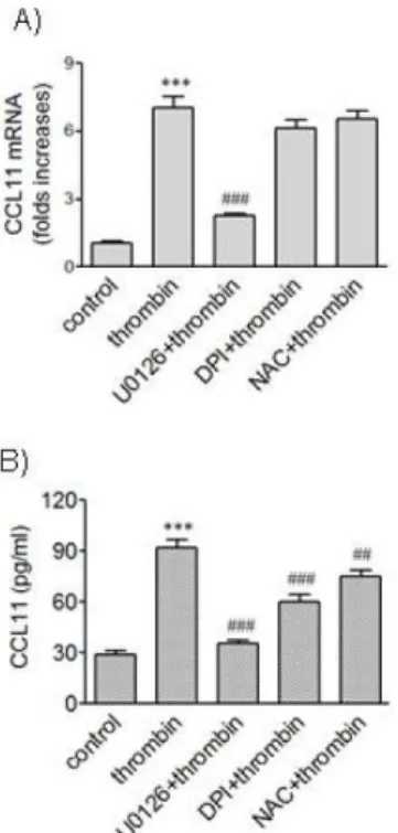
Fig. 5. Effects of ROS quenchers on expression of CCL11 induced by thrombin. (A, B) HAoSMCs were incubated with or without DPI (10 μM) and NAC (5 mM) for 2 h followed by stimulation with thrombin for 9 h. The levels of CCL11 transcripts were determined by realtime PCR (A), and the levels of CCL11 protein secreted into culture media were determined by ELISA (B). Data are expressed as mean±SD (n=3 replicates for each group). ***p<0.001 vs. control;
#