Fluvastatin inhibits AGE-induced cell
proliferation and migration via an
ERK5-dependent Nrf2 pathway in vascular smooth
muscle cells
Ae-Rang Hwang1, Jung-Hwa Han1,2, Jae Hyang Lim3, Young Jin Kang1, Chang-Hoon Woo1,2
*
1 Department of Pharmacology, Yeungnam University College of Medicine, Daegu, Republic of Korea, 2 Smart-Aging Convergence Research Center, Yeungnam University College of Medicine, Daegu, Republic
of Korea, 3 Department of Microbiology, Ewha Womans University School of Medicine, Seoul, Republic of Korea
Abstract
Advanced glycation endproduct (AGE)-induced vascular smooth muscle cell (VSMC) proliferation and reactive oxygen species (ROS) production are emerging as important mechanisms of diabetic vasculopathy, but little is known about the molecular mechanism responsible for the antioxidative effects of statins on AGEs. It has been reported that statins exert pleiotropic effects on the cardiovascular system due to decreases in AGE-induced cell proliferation, migration, and vascular inflammation. Thus, in the present study, the authors investigated the molecular mechanism by which statins decrease AGE-induced cell prolifer-ation and VSMC migrprolifer-ation. In cultured VSMCs, statins upregulated Nrf2-related antioxidant gene, NQO1 and HO-1, via an ERK5-dependent Nrf2 pathway. Inhibition of ERK5 by siRNA or BIX02189 (a specific ERK5 inhibitor) reduced the statin-induced upregulations of Nrf2, NQO1, and HO-1. Furthermore, fluvastatin was found to significantly increase ARE pro-moter activity through ERK5 signaling, and to inhibit AGE-induced VSMC proliferation and migration as determined by MTT assay, cell counting, FACS analysis, a wound scratch assay, and a migration chamber assay. In addition, AGE-induced proliferation was dimin-ished in the presence of Ad-CA-MEK5αencoding a constitutively active mutant form of MEK5α(an upstream kinase of ERK5), whereas depletion of Nrf2 restored statin-mediated reduction of AGE-induced cell proliferation. Moreover, fluvastatin suppressed the protein expressions of cyclin D1 and Cdk4, but induced p27, and blocked VSMC proliferation by regulating cell cycle. These results suggest statin-induced activation of an ERK5-dependent Nrf2 pathway reduces VSMC proliferation and migration induced by AGEs, and that the ERK5-Nrf2 signal module be viewed as a potential therapeutic target of vasculopathy in patients with diabetes and complications of the disease.
a1111111111 a1111111111 a1111111111 a1111111111 a1111111111 OPEN ACCESS
Citation: Hwang A-R, Han J-H, Lim JH, Kang YJ, Woo C-H (2017) Fluvastatin inhibits AGE-induced cell proliferation and migration via an ERK5-dependent Nrf2 pathway in vascular smooth muscle cells. PLoS ONE 12(5): e0178278.https:// doi.org/10.1371/journal.pone.0178278
Editor: Aamir Ahmad, University of South Alabama Mitchell Cancer Institute, UNITED STATES Received: September 1, 2016
Accepted: May 10, 2017 Published: May 22, 2017
Copyright:© 2017 Hwang et al. This is an open access article distributed under the terms of the Creative Commons Attribution License, which permits unrestricted use, distribution, and reproduction in any medium, provided the original author and source are credited.
Data Availability Statement: All relevant data are within the paper.
Funding: This research was supported by the Medical Research Center Program
(2015R1A5A2009124) and Basic Science Research Program (NRF-2015R1D1A1A01059398) through the National Research Foundation of Korea funded by the Ministry of Science, ICT, and Future Planning.
Competing interests: The authors have declared that no competing interests exist.
Introduction
Hydroxy-3-methylglutaryl coenzyme A reductase inhibitors (statins) are potent inhibitors of cholesterol biosynthesis and are widely used to reduce serum cholesterol levels in hyperlipidemic patients [1]. However, recent reports have shown statins also ameliorate cardiovascular disor-ders, and remarkably, have preventative effects on cardiovascular diseases [2]. In addition, statins have been reported to improve endothelial dysfunction by increasing nitric oxide availability, to inhibit proliferation and inflammatory responses, and to stabilize atherosclerotic plaque [3,4].
Extracellular signal-regulated kinase 5 (ERK5) is an atypical member of MAPK family, and reportedly, regulates endothelial integrity and protects against vascular dysfunction and car-diovascular diseases in rodent models. On the other hand, MEK5, an upstream kinase of ERK5, plays critical roles in cell proliferation, migration, and differentiation [5]. Transcription factor nuclear factor-erythroid 2-related factor 2 (Nrf2) is an important regulator of cellular oxidative stress [6,7], and under homeostatic conditions, binds to Kelch-like ECH-associated protein 1, and is subsequently degraded via the proteasome system or stored in cytoplasm [8]. In the presence of oxidative stress, Nrf2 translocates to the nucleus, where it forms Nrf2/small Maf heterodimer, which binds specifically to antioxidant response elements (AREs), and acti-vates the gene expressions of antioxidant proteins, such as, NAD(P)H:quinone oxidoreduc-tase-1 (NQO1), and heme oxygenase-1 (HO-1) [9–15]. Nrf2 has also been reported to play an atheroprotective roles by regulating antioxidant genes in the cardiovascular system [16]. Cellu-lar redox balance is tightly controlled by various antioxidant systems, and in VSMCs, ROS activates the Nrf2 signaling pathway, which in turn, induces anti-atherosclerotic gene expres-sion [17], and it has been reported Nrf2-activating drugs decrease VSMC proliferation and migration by antioxidant gene expression [18,19]. Furthermore, studies have demonstrated ERK5 is a molecular target for regulating laminar blood flow-mediating Nrf2-dependent gene expression and suggested it may have significant therapeutic potential for the treatment of ath-erosclerosis [20].
It was recently shown advanced glycation endproducts (AGEs) and their receptor-ligand interactions play key roles in neointimal formation after vascular injury [21,22]. AGEs are known to be formed in diabetes and to promote inflammation via specific receptors on endo-thelial cells [23,24], and to mediate pro-inflammatory responses and cell proliferation through the NFκB signaling pathway [25,26]. We hypothesized that statin might decrease AGE-induced proliferation and migration via ERK5-Nrf2-dependent gene regulation in VSMCs, and thus, we sought to identify the molecular mechanism responsible for the reductions in AGE-induced cell proliferation and VSMC migration by statins.
Materials and methods
Reagents and antibodies
Fluvastatin, pitavastatin, and BIX02189 (a specific inhibitor of ERK5) were purchased from Selleck Chemicals (Houston, TX) [27]. AGE-BSA was obtained from Calbiochem (Darmstadt, Germany), and MTT reagents were purchased from Amresco (Solon, Ohio). Antibodies were purchased from the following vendors: ERK1/2 (#9102, anti-rabbit), ERK5 (#3372, anti-rab-bit), phosphor-ERK1/2 (#9106, anti-rabbit) and phospho-ERK5 (#3371, anti-rabbit) from Cell Signaling Technology (Danvers, MA), Nrf2 (sc-13032, 200μg/ml, anti-rabbit), NQO1 (sc-32739,μg/ml, anti-mouse), CDK4 (sc-260 C-22, 100 μg/ml, anti-rabbit), p27 (sc-528 C-19, 100μg/ml, anti-rabbit) and HA (anti-rabbit) from Santa Cruz Biotechnology (Santa Cruz, CA), HO-1 (ADI-SPA-895, 1 mg/ml, anti-rabbit) from Enzo lifesciences, cyclin D (06–137, 1 mg/ml, anti-rabbit) from Millipore and tubulin (anti-mouse) from Sigma (St. Louis, MO).
Cell culture and treatment conditions
Sprague-Dawley rats were anesthetized with ketamine and xylazine. Primary rat vascular smooth muscle cells (VSMCs) were isolated from rat thoracic aorta. The cells were processed using a 1 mm chop setting in a 10 cm culture dish, and cultured with 50% FBS-DMEM with 1% antibiotics-antimycotics in a CO2 incubator. VSMCs were maintained in DMEM supple-mented with 10% fetal bovine serum (FBS), 50 U/mL penicillin, and 50μg/mL streptomycin at 37˚C in a 95% air-5% CO2atmosphere. VSMCs from passages 4–7 were cultured for 9–24 h in
the presence of 5μM fluvastatin with or without 10 μg/ml AGEs. Cells were harvested at indi-cated time points. All animal experiments were handled in accordance with the protocol approved by the Institutional Animal Care and Use Committee at Yeungnam University Col-lege of Medicine, Daegu,Republic of Korea.
Western blotting analysis
Cells were lysed with radioimmunoprecipitation assay (RIPA) lysis buffer supplemented with 1 mM phenylmethylsulfonyl fluoride (PMSF) and 0.01 mM protease inhibitor cocktail (PIC), and lysates were incubated on ice for 15 min and centrifuged at 15,000× g for 10 min at 4˚C. Protein concentrations were determined using a Bradford assay. Proteins were separated by SDS-PAGE and transferred to polyvinylidene difluoride (PVDF) membranes, which were then immunoblotted with primary antibodies (1:1,000 dilution) followed by corresponding second-ary antibodies (1:4,000 dilution). Signals were visualized using electrochemiluminescence (ECL) detection reagents (Millipore, Temecula, CA), according to the manufacturer’s instructions.
Quantitative real time RT-PCR (qRT-PCR)
The mRNA expressions of Nrf2 targeted genes were determined by qRT-PCR as described previously [7]. Briefly, total RNA was isolated with TRIzol, and reverse transcription was con-ducted using TaqMan reverse transcription reagents, according to the manufacturer’s instruc-tions. qRT-PCR was conducted using 1μL of template cDNA and Power SYBR Green in an ABI PRISM 7500. Quantification was performed using the efficiency-correctedΔΔCq method. The primers used to amplify DNA sequences were as follows: NQO1, forward 50
-TTACTATG GGATGGGGTCCA-30and reverse 50
-TGCCAAAACTGTTCACCAAA-30, Nrf2, forward 50
-AAA CCACCCTGAAAGCACAG-30and reverse 50-AGTGTTCTGGTGATGCCACA-30; and GAPDH,
forward 50
-GGAGCCAAAAGGGTCATCAT-30and reverse 50
-GTGATGGCATGGACTGTGGT-30. PCR conditions were as follows: preliminary denaturation at 50˚C for 2 min, followed by 95˚C for 10 min, 95˚C for 15 s, and 60˚C for 1 min.
Small interfering RNA (siRNA)
VSMCs were transiently transfected with 50 or 100 pM of control or specific siRNAs against Nrf2 or ERK5 using Lipofectamine 2000 reagent (Invitrogen) according to the manufacturer’s instructions. The targeting sequences of siRNAs were as follows: rat Nrf2 siRNA (5’-CAAAC AGAAUGGACCUAAAdTdT-3’); rat/mouse ERK5 siRNA(5’-GAAAGGGTGCGAGCCTATAU U-3’). Non-specific control siRNA was purchased from Bioneer and used as a negative con-trol. Cells were harvested 48–72 h after siRNA transfection, and mRNA expression levels were measured.
MTT assay
AGE-induced proliferation was quantified using a MTT assay. Briefly, VSMCs were cultured on 24-well plates and when ~80% confluent, medium was replaced with serum free DMEM.
Cells were then pretreated with BIX02189 (2μM) and stimulated with fluvastatin (5 μM) for 24 h. MTT reagents were added for 4 h at 37˚C the removed by washing with PBS, and eluted with DMSO. Proliferation was measured using a microplate reader (Biorad) at 570 nm.
Reporter gene assay
A luciferase assay was used to determine ARE promoter activity. The resultant construct was co-transfected with pRL-tk vector containing Renilla luciferase reporter gene into cultured VSMCs using the lipofectamine 2000 method and Plus transfection reagent. Cells were lysed at 24 h post-transfection, and Firefly-to-Renilla luciferase activity ratios in lysates were measured with dual-luciferase assay kit (Promega) to evaluate ARE promoter activity.
Flow cytometric analysis
Cells (1×10⁵) were trypsinized, fixed in 95% ethanol, and stained with propidium iodide (PI) (50μg/ml) for 30 min at 37˚C. PI stained cells were filtered using a 5 ml polystyrene round bottom tube fitted with a cell-strainer cap prior to flow cytometry. All flow cytometry measure-ments were obtained using a FACSCalibur (Becton Dickinson, San Jose, CA). Cell cycle analy-sis was performed using Cell-Quest pro-software.
Wound scratch assays and migration chamber assays
VSMC cells were cultured until >90% confluent in 6 well dishes, and then medium was replaced with serum free DMEM overnight. Wounds were made with a sterile 200μL pipet tip by drawing a line through cells perpendicular to the line above. Cells were then pretreated with BIX02189 (2μM) and stimulated with fluvastatin (5 μM) for 24 h.Finally, images were taken using a phase contrast at 40×. For selective migration assay, transwell system was used. Cells were seeded in the inner chamber and pretreated with BIX02189 (2μM) in the presence or absence of fluvastatin (5μM). AGE (10 μg/ml) were added to the lower for 12 h. After fixing, cells were visualized by crystal violet staining. Unmigrated cells were scraped off and then migrated cells were counted under a light microscope.
Statistics
Results shown in the bar graph are means± SDs. The significances of differences were deter-mined using ANOVA and Student’st test. P values of < 0.05 were considered significant.
Results
Inhibition of ERK5 reduced statin-induced expressions of Nrf2 and
NQO1 in VSMCs
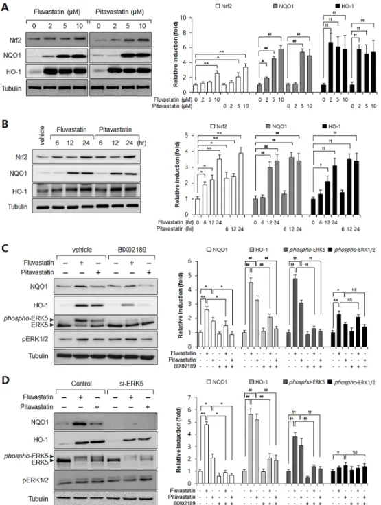
To investigate the effect of statins on the Nrf2 antioxidant system, we assessed Nrf2, NQO1, and HO-1 protein levels in VSMCs. As shown inFig 1, fluvastatin and pitavastatin strongly increased Nrf2 and NQO1 expressions in dose- and time-dependent manner (Fig 1A and 1B). To examine the role of ERK5 in Nrf2 antioxidant system induction by statins, we used an ERK5 inhibitor and ERK5 specific siRNA. Inhibition of ERK5 was found to markedly attenu-ate fluvastatin-induced NQO1 and HO-1 expression (Fig 1C and 1D). Interestingly, statin-induced phosphorylation levels of ERK1/2 were not affected by ERK5 inhibitor and siRNA
(Fig 1C and 1D), suggesting the specific role of ERK5 in statin-induced Nrf2 signaling in
vas-cular smooth muscle cells.
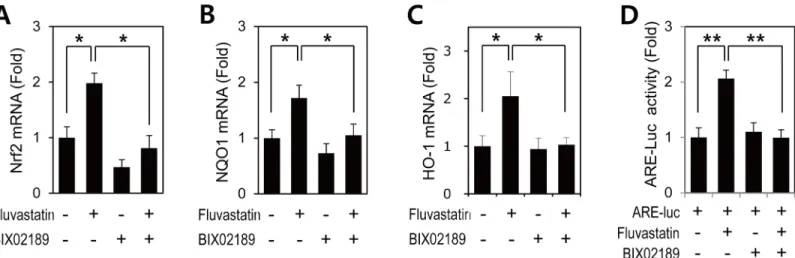
Next, we studied the effect of fluvastatin on the mRNA expressions of Nrf2, NQO1, and HO-1 in VSMCs. Consistent with protein levels, fluvastatin significantly induced the mRNA
Fig 1. The involvement of ERK5 in statin-induced Nrf2 signaling in VSMCs. (A-B) Western blot analysis
of Nrf2, NQO1, and HO-1 in statin-treated VSMCs. Cells were exposed to fluvastatin and pitavastatin for 24 h at the indicated dosages. In addition, protein samples wererefinedfrom cultured VSMCs treated with fluvastatin (5μM) or pitavastatin (5μM) for the indicated times. (C) Western blot analysis of ERK5, NQO1, HO-1, phospho-ERK1/2, and phospho-ERK5 in BIX02189 treated VSMCs. Protein samples were obtained from cultured VSMCs treated with 5μM fluvastatin or 5μM pitavastatin for 24 h. (D) VSMCs were transfected with control or ERK5 siRNA (50 pM) for 30 h and then treated with 5μM fluvastatin for 24 h. Protein levels of NQO1, HO-1, ERK5 and tubulin were determined by Western blotting with specific antibodies. In addition, phosphorylation levels of ERK1/2 and ERK5 were determined by immunoblotting with specific antibodies. Bar graphs present the densitometric quantification of Western blot bands. Results are representative of three independent experiments.*, #, †, p<0.05;**, ##, ††, p<0.01 compared with control. NS indicates not significant.
expressions of Nrf2, NQO1, and HO-1 (Fig 2), and ERK5 inhibition suppressed the fluvasta-tin-induced expressions Nrf2, NQO1, and HO-1 mRNA (Fig 2). These results indicated that ERK5 contributed to the fluvastatin-induced Nrf2 antioxidant genes in VSMCs.
Inhibition of ERK5 attenuated fluvastatin-induced ARE promoter activity
in VSMCs
Activated Nrf2 translocates to the nucleus and then binds specifically to ARE in the promoters of target genes, such as, NQO1 and HO-1 [8]. Thus, we investigated whether fluvastatin could promote Nrf2 to ARE promoter binding. VSMCs were transfected with the reporter ARE, and ARE promoter activity was measured using a reporter gene assay. ARE promoter activity was found to be enhanced in response to fluvastatin, and to be significantly inhibited by BIX02189
(Fig 2D). These observations suggest that ERK5 is an important regulator of
fluvastatin-induced Nrf2 activation and that it does so by regulating transcription.
Fluvastatin regulated AGE-induced cell proliferation through ERK5-Nrf2
signal module
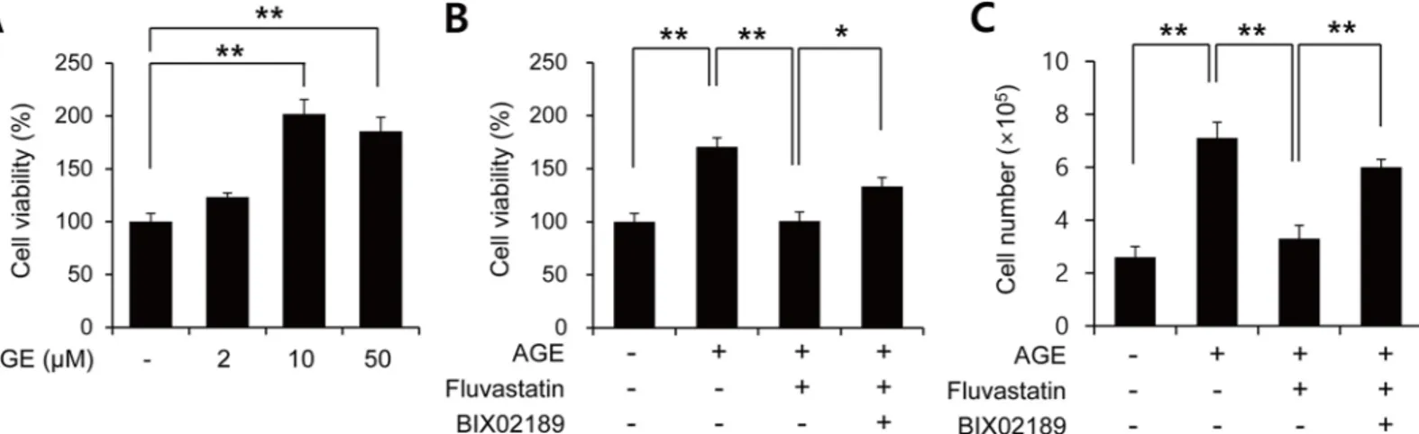
It has been reported that fluvastatin reduces AGE-induced VSMC proliferation [28]. To con-firm this effect, VSMCs were treated with AGEs in the presence or absence of fluvastatin and then subject to MTT assay. AGEs were found to dose-dependently induce cell proliferation
(Fig 3A), and this was significantly suppressed by fluvastatin (Fig 3B). In addition to MTT
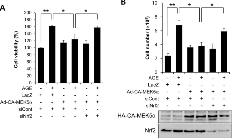
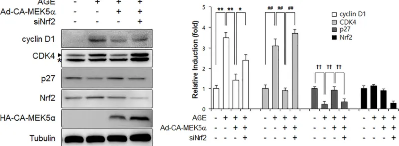
assay, we got the similar results with cell counting (Fig 3C). Interestingly, this suppressive effect of fluvastatin was prevented when VSMCs were pretreated with BIX02189, which sug-gested the involvement of ERK5 in the fluvastatin-mediated inhibition of AGE-induced cell proliferation (Fig 3B and 3C). We also examined whether ERK5 activation could reduce prolif-eration by using Ad-CA-MEK5α encoding a constitutively active mutant form of MEK5α (an upstream kinase of ERK5). As shown inFig 4, AGE-induced proliferation determined by both MTT assay and cell counting was significantly diminished in the presence of Ad-CA-MEK5α, and Nrf2 depletion using siRNA restored AGE-induced proliferation. These results indicate fluvastatin inhibited AGE-induced VSMC proliferation through ERK5-Nrf2 signal module.
Fig 2. The involvement of ERK5 in fluvastatin-induced Nrf2 signaling in VSMCs. (A-C) Quantitative RT-PCR analysis of the mRNA expressions of
Nrf2, NQO1, and HO-1 in VSMCs treated with fluvastatin. VSMCs were treated with BIX02189 (2μM) for 1 h and then incubated with 5μM fluvastatin for 6 h. qRT-PCR analysis was performed in triplicate. Results are representative of three independent experiments.*, p<0.05. (D) VSMCs were co-transfected with pARE and pRL-tk and then stimulated with 5μM fluvastatin for 24 h in the presence or absence of BIX02189 (2μM). Promoter activity was measured by using a Dual-Luciferase reporter assay kit and a GloMax 20/20 luminometer. Transfection efficiency was normalized versus Renilla luciferase activity derived from pRL-tk construct. Reporter assay was performed in triplicate. Results are presented as the means±SDs of three independent experiments.**p<0.01. https://doi.org/10.1371/journal.pone.0178278.g002
Fluvastatin regulated AGEs-induced cell cycle progression through the
ERK5-Nrf2 pathway
The cell cycle is controlled by activators (cyclins) and inhibitors (Rb, p16, p21, p27). Among them, cyclin D is a major cell cycle associated cyclin and interacts with four cyclin-dependent kinases (Cdks; Cdk2, 4, 5 and 6). Cyclin D-Cdk4/6 complex accumulation is required for cell cycle progression [29,30]. On the other hand, p27 inhibits Cdk by binding directly to Cdk-cyclin complex and blocking its protein kinase activity. Statins have been shown to induce cell cycle arrest in various cancer cells, including those of prostate [31] and breast cancer [32], and p53, p21, and p27 proteins are known to regulate VSMC proliferation [33]. Furthermore, it has been demonstrated that statins cause G1 arrest in VSMCs by up-regulating p27 and down-regulating cyclin E [34,35].
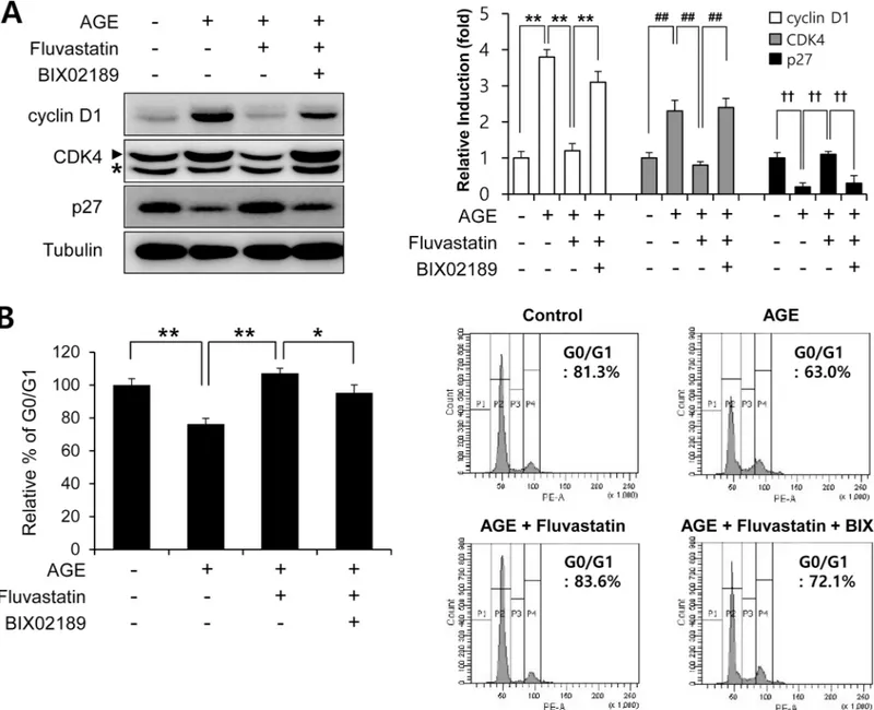
In order to examine the effects of fluvastatin on cell cycle progression at the cell cycle regu-latory gene level. We first checked that AGEs increased cyclin D1 and Cdk4 protein levels, but reduced p27 level. Fluvastatin was found to suppress cyclin D1 and Cdk4 protein levels, but to enhance p27 level in VSMCs (Fig 5A), and these effects were prevented by BIX02189 pretreat-ment. As compared with cells treated with fluvastatin (5μM) in the presence of AGEs (10 μg/ ml), cells in G0/G1phase was significantly reduced by BIX02189 pretreatment (Fig 5B). To investigate whether ERK5 activation induced cell cycle arrest, we pretreated cells with Ad-CA-MEK5α. It was found that the AGE-induced protein expressions of cyclin D1 and CDK4 were markedly reduced in the presence of Ad-CA-MEK5α, whereas depletion of Nrf2 pre-vented these reductions (Fig 6). These results show fluvastatin blocks VSMC proliferation via an ERK5-Nrf2 pathway and that this leads to cell cycle arrest.
Fluvastatin regulated AGE-induced cell migration through an
ERK5-dependent pathway
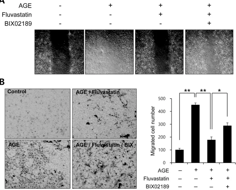
To determine the functional significance of the fluvastatin-mediated ERK5 activation pathway, we examined the effect of fluvastatin on AGE-induced VSMC migration using both a scratch assay (Fig 7A) and a migration chamber assay (Fig 7B). Fluvastatin significantly suppressed AGE-induced cell migration in response to wound injury, and BIX02189 blocked this inhibi-tory effect of fluvastatin (Fig 7). Taken together, our results suggest that fluvastatin suppresses
Fig 3. Fluvastatin inhibited AGEs-induced cell proliferation through the ERK5-Nrf2 pathway in VSMCs. (A) Serum starved VSMCs were exposed to
the indicated concentrations of AGEs for 24 h. (B) Serum starved VSMCs were pretreated with 5μM fluvastatin in the presence or absence of BIX02189 (2μM) for 1 h and then exposed to AGEs (10μg/ml) for 24 h. Cell viability was determined using a MTT assay. (C) Cell proliferation was determined by using cell counting with same condition. Results are representative of three independent experiments.*, p<0.05;**, p<0.01.
AGE-induced cell proliferation and migration by inducing antioxidant genes through the ERK5-dependent activation of the Nrf2 pathway.
Discussion
Antioxidant defense mechanisms protect cells from excessive oxidative stress [16], and the transcription factor Nrf2 contributes to these processes by regulating the expressions of antiox-idant genes. The present study provides evidence that in VSMCs, ERK5 activation increases the expressions of Nrf2 targeted genes involved in cytoprotective responses, and that Nrf2 transcriptional activity is lost when ERK5 expression is depleted or inhibited by ERK5 siRNA or BIX02189 (a biochemical inhibitor of ERK5). To the best of our knowledge, these findings provide first evidence fluvastatin exerts its antioxidant effects through the ERK5-dependent activation of the Nrf2 pathway in VSMCs. Furthermore, the study indicates statins exert their antioxidant effects by increasing the expressions of antioxidant genes in VSMCs, by showing fluvastatin and pitavastatin significantly induce the expressions of antioxidant enzymes (ERK5, Nrf2, NQO1, and HO-1) (Fig 1). Interestingly, we found the up-regulations Nrf2-regu-lated genes by statin was diminished by ERK5 siRNA or BIX02189 (Fig 1andFig 2). It has been reported that statins reduce intracellular ROS in endothelial cells by S-nitrosylating thior-edoxin as well as other various effects [18,24,36].
Fig 4. The effect of Nrf2 siRNA on the ERK5 activation-mediated inhibition of AGE-induced VSMC proliferation. (A) VSMC cells were infected with
adenovirus encoding LacZ or CA-MEK5αfor 48 h, and then exposed to AGEs for 24 h. Cell viability was determined using a MTT assay. Results are presented as the means±SEs of three independent experiments.*, p<0.05;**, p<0.01. (B) Cell proliferation was determined by using cell counting with same condition. Results are representative of three independent experiments.**, p<0.01. Amounts of protein expression were determined by immunoblotting with specific antibodies for HA and Nrf2.
A number of recent reports have shown that statins may also have important anti-in-flammatory effects, in addition to their effects on lowering plasma lipids [37,38]. Since in-flammation is closely linked to ROS production, the molecular basis of their observed anti-inflammatory effects might be related to their abilities to block ROS production [18,37,39]. It has been well established that ROS are related to the development of cardiovascular disease, and many studies support the notion that ROS released from a dysfunctional mitochondrial respiratory chain other sources plays a role in the development of atherosclerosis and its com-plications [40–42], which have been attributed to ROS-mediated vascular signaling pathways
Fig 5. Fluvastatin regulated AGE-induced cell cycle progression through an ERK5 activating pathway in VSMCs. (A) Serum starved VSMCs were
pretreated with 5μM fluvasatin in the presence or absence of BIX02189 (2μM), and then exposed to AGEs 10μg/ml AGEs for 24 h. Protein expressions were determined by immunoblotting with anti-cyclin D1, anti-CDK4, anti-p27, and anti-tubulin. Results are representative of three independent experiments. Asterisk indicates a nonspecific band. Bar graphs present the densitometric quantification of Western blot bands. (B) VSMCs were seeded onto 6 well plates at a density of 1×10⁴cells/ml, pretreated with fluvastatin (5μM) in the presence or absence of BIX02189 (2μM), and then exposed to AGEs (10μg/ml) for 24
h. For cell cycle analysis, cells were then detached, stained with PI, and subjected to flow cytometry.*, p<0.05;**, p<0.01 (n = 5). https://doi.org/10.1371/journal.pone.0178278.g005
[17,18]. In the present study, fluvastatin activated Nrf2 in VSMCs and this markedly increased ARE-driven promoter activity (Fig 2C) and the upregulation of ARE-targeted antioxidant genes (NQO1 and HO-1). These findings provide first evidence fluvastatin exerts its antioxidant effects via the ERK5-dependent activation of the Nrf2 pathway in vascular cells [17–19,43].
It has been recently demonstrated AGEs and their receptor-ligand interactions play key roles in neointimal formation after vascular injury [28]. In fact, AGE-induced VSMC prolifer-ation and ROS production are emerging as important mechanisms of atherosclerosis. Before commencing the present study, we hypothesized fluvastatin might reduce AGE-induced pro-liferation, and thus, we sought to identify the mechanism underlying the involvement of flu-vastatin in AGE-induced cellular signaling. Treatment with AGEs increased the protein levels of the proliferation markers, cyclin D1 and CDK4, and markedly decreased p27 level, and these effects of AGEs were lost when ERK5 expression was depleted or inhibited by BIX02189
(Fig 5A). Our MTT assay and cell counting results show the antioxidant effects of fluvastatin
are largely dependent on the ERK5-dependent Nrf2 pathway, because Nrf2 siRNA abolished their antioxidant cytoprotective responses. In addition, the overexpression of CA-MEK5α using an adenoviral system inhibited AGE-induced cell proliferation and cell cycle progres-sion, and these effects were prevented by Nrf2 depletion (Fig 4andFig 6), which indicates ERK5 activation regulates cell cycle arrest via a Nrf2-dependent pathway.
NQO1 and HO-1 have been suggested as a downstream target of Nrf2 in inhibition of vas-cular smooth muscle cell proliferation through cell cycle arrest [44–46]. In this study, our data showed that statin induced Nrf2 and its target genes including NQO1 and HO-1 in an ERK5-dependent manner. In addition, inhibition of ERK5 and Nrf2 failed to inhibit fluvastatin-induced protein regulation of cell cycle-related genes including cyclin D1, CDK4, and p27 sug-gesting that ERK5-Nrf2 signal nodule regulates G1 cell cycle arrest in vascular smooth muscle cells.
A growing body of evidence suggested that ERK5 has cytoprotective effects via multiple pathways. Based on the genetic approach with gene knock out, it has been established that ERK5 is essential for endothelial viability and protects vascular leakage [47]. In addition,
Fig 6. The effect of Nrf2 siRNA on the ERK5 activation-mediated inhibition of AGE-induced cell cycle progression. VSMCs infected with Ad-LacZ or
Ad-CA-MEK5αwere transfected with siNrf2 for 48 h and then immunoblotted using anti-cyclin D1, anti-CDK4, anti-p27, anti-Nrf2, anti-HA, and anti-tubulin. Results are representative of three independent experiments. Asterisk indicates a nonspecific band. Bar graphs present the densitometric quantification of Western blot bands.
laminar shear stress protects endothelial cells from oxidative stress and inflammatory re-sponses via ERK5 activation. However, laminar shear stress reduces endothelial cell prolifera-tion an ERK5-dependent manner. On the other hand, ERK5 can be activated by many growth factors that are important in vascular smooth muscle cell proliferation. It is not clear whether growth factor-mediated ERK5 activation could regulate Nrf2 or not. ERK5 is not only a kinase but also a transcriptional coactivator with C-terminal transactivation domain, which is a uni-que characteristics compared to ERK1/2. It has been reported that statin induces both phos-phorylation and transcriptional activation of ERK5 [48]. Therefore it is reasonable to pursue
Fig 7. Fluvastatin regulated AGE-induced cell migration through an ERK5-dependent pathway in VSMCs. (A) VSMC cells were cultured until near
confluent in 6 well dishes. Cell migration was assessed using a modified scratch assay. VSMCs were pretreated with 5μM fluvastatin in the presence or absence of BIX02189 (2μM), and then exposed to 10μg/ml AGEs for 24 h. Wound areas were visualized using a phase contrast microscope. Results are representative of three independent experiments. (B) For selective migration assay, transwell system was used. Cells were seeded in the inner chamber and pretreated with BIX02189 (2μM) in the presence or absence of fluvastatin (5μM). AGE (10μg/ml) were added to the lower for 12 h. After fixing, cells were visualized by crystal violet staining. Unmigrated cells were scraped off and then migrated cells were counted under a light microscope. Results are representative of three independent experiments.*, p<0.05;**, p<0.01.
the molecular mechanism of ERK5-Nrf2 signal nodule in aspect of transcriptional regulation regarding the ambiguity of ERK5.
This study provides a molecular basis for the protective effects for statins. In particular, our finding regarding their induction of the ERK5-dependent regulation of cell proliferation and cell cycle arrest in VSMCs could eventually contribute to the development of a treatment for cardiovascular diseases. This study shows fluvastatin exerts induces antioxidant enzymes via an ERK5-dependent Nrf2 pathway.
Author Contributions
Conceptualization: ARH CHW. Data curation: ARH JHH YJK CHW. Formal analysis: ARH JHH CHW. Funding acquisition: CHW. Investigation: ARH JHH CHW.Methodology: ARH JHH JHL YJK CHW. Project administration: CHW.
Resources: CHW. Supervision: CHW.
Validation: ARH JHH JHL YJK CHW. Visualization: ARH CHW.
Writing – original draft: ARH JHH JHL YJK CHW. Writing – review & editing: ARH JHH JHL YJK CHW.
References
1. Maron DJ. The epidemiology of low levels of high-density lipoprotein cholesterol in patients with and without coronary artery disease. Am J Cardiol. 2000; 86(12A):11L–4L. PMID:11374848
2. Pedersen TR, Wilhelmsen L, Faergeman O, Strandberg TE, Thorgeirsson G, Troedsson L, et al. Fol-low-up study of patients randomized in the Scandinavian simvastatin survival study (4S) of cholesterol lowering. Am J Cardiol. 2000; 86(3):257–62. PMID:10922429
3. Zhou Z, Wang K, Penn MS, Marso SP, Lauer MA, Forudi F, et al. Receptor for AGE (RAGE) mediates neointimal formation in response to arterial injury. Circulation. 2003; 107(17):2238–43.https://doi.org/ 10.1161/01.CIR.0000063577.32819.23PMID:12719284
4. Duckers HJ, Boehm M, True AL, Yet SF, San H, Park JL, et al. Heme oxygenase-1 protects against vascular constriction and proliferation. Nat Med. 2001; 7(6):693–8.https://doi.org/10.1038/89068
PMID:11385506
5. Wang X, Tournier C. Regulation of cellular functions by the ERK5 signalling pathway. Cell Signal. 2006; 18(6):753–60.https://doi.org/10.1016/j.cellsig.2005.11.003PMID:16376520
6. Motohashi H, Yamamoto M. Nrf2-Keap1 defines a physiologically important stress response mecha-nism. Trends Mol Med. 2004; 10(11):549–57.https://doi.org/10.1016/j.molmed.2004.09.003PMID:
15519281
7. Nigro P, Abe J, Woo CH, Satoh K, McClain C, O’Dell MR, et al. PKCzeta decreases eNOS protein sta-bility via inhibitory phosphorylation of ERK5. Blood. 2010; 116(11):1971–9.https://doi.org/10.1182/ blood-2010-02-269134PMID:20538799
8. Itoh K, Wakabayashi N, Katoh Y, Ishii T, Igarashi K, Engel JD, et al. Keap1 represses nuclear activation of antioxidant responsive elements by Nrf2 through binding to the amino-terminal Neh2 domain. Genes Dev. 1999; 13(1):76–86. PMID:9887101
9. Forman HJ, Ruden D. Introduction to serial reviews on EpRE and its signaling pathway. Free Radic Biol Med. 2004; 36(10):1197–8.https://doi.org/10.1016/j.freeradbiomed.2004.03.004PMID:15110383 10. Itoh K, Tong KI, Yamamoto M. Molecular mechanism activating Nrf2-Keap1 pathway in regulation of
adaptive response to electrophiles. Free Radic Biol Med. 2004; 36(10):1208–13.https://doi.org/10. 1016/j.freeradbiomed.2004.02.075PMID:15110385
11. Chan K, Lu R, Chang JC, Kan YW. NRF2, a member of the NFE2 family of transcription factors, is not essential for murine erythropoiesis, growth, and development. Proc Natl Acad Sci U S A. 1996; 93 (24):13943–8. PMID:8943040
12. Ishii T, Itoh K, Takahashi S, Sato H, Yanagawa T, Katoh Y, et al. Transcription factor Nrf2 coordinately regulates a group of oxidative stress-inducible genes in macrophages. J Biol Chem. 2000; 275 (21):16023–9. PMID:10821856
13. Li J, Ichikawa T, Janicki JS, Cui T. Targeting the Nrf2 pathway against cardiovascular disease. Expert Opin Ther Targets. 2009; 13(7):785–94.https://doi.org/10.1517/14728220903025762PMID:19530984 14. Mann GE, Bonacasa B, Ishii T, Siow RC. Targeting the redox sensitive Nrf2-Keap1 defense pathway in
cardiovascular disease: protection afforded by dietary isoflavones. Curr Opin Pharmacol. 2009; 9 (2):139–45.https://doi.org/10.1016/j.coph.2008.12.012PMID:19157984
15. Itoh K, Chiba T, Takahashi S, Ishii T, Igarashi K, Katoh Y, et al. An Nrf2/small Maf heterodimer mediates the induction of phase II detoxifying enzyme genes through antioxidant response elements. Biochem Biophys Res Commun. 1997; 236(2):313–22. PMID:9240432
16. Makabe S, Takahashi Y, Watanabe H, Murakami M, Ohba T, Ito H. Fluvastatin protects vascular smooth muscle cells against oxidative stress through the Nrf2-dependent antioxidant pathway. Athero-sclerosis. 2010; 213(2):377–84.https://doi.org/10.1016/j.atherosclerosis.2010.07.059PMID:20864107 17. Wassmann S, Laufs U, Muller K, Konkol C, Ahlbory K, Baumer AT, et al. Cellular antioxidant effects of
atorvastatin in vitro and in vivo. Arterioscler Thromb Vasc Biol. 2002; 22(2):300–5. PMID:11834532 18. Haendeler J, Hoffmann J, Zeiher AM, Dimmeler S. Antioxidant effects of statins via S-nitrosylation and
activation of thioredoxin in endothelial cells: a novel vasculoprotective function of statins. Circulation. 2004; 110(7):856–61.https://doi.org/10.1161/01.CIR.0000138743.09012.93PMID:15289372 19. Takemoto M, Liao JK. Pleiotropic effects of 3-hydroxy-3-methylglutaryl coenzyme a reductase
inhibi-tors. Arterioscler Thromb Vasc Biol. 2001; 21(11):1712–9. PMID:11701455
20. Kim M, Kim S, Lim JH, Lee C, Choi HC, Woo CH. Laminar flow activation of ERK5 protein in vascular endothelium leads to atheroprotective effect via NF-E2-related factor 2 (Nrf2) activation. J Biol Chem. 2012; 287(48):40722–31.https://doi.org/10.1074/jbc.M112.381509PMID:23043106
21. Brownlee M, Cerami A, Vlassara H. Advanced glycosylation end products in tissue and the biochemical basis of diabetic complications. N Engl J Med. 1988; 318(20):1315–21.https://doi.org/10.1056/ NEJM198805193182007PMID:3283558
22. Sakaguchi T, Yan SF, Yan SD, Belov D, Rong LL, Sousa M, et al. Central role of RAGE-dependent neointimal expansion in arterial restenosis. J Clin Invest. 2003; 111(7):959–72.https://doi.org/10.1172/ JCI17115PMID:12671045
23. Hofmann MA, Drury S, Fu C, Qu W, Taguchi A, Lu Y, et al. RAGE mediates a novel proinflammatory axis: a central cell surface receptor for S100/calgranulin polypeptides. Cell. 1999; 97(7):889–901. PMID:10399917
24. Surapisitchat J, Hoefen RJ, Pi X, Yoshizumi M, Yan C, Berk BC. Fluid shear stress inhibits TNF-alpha activation of JNK but not ERK1/2 or p38 in human umbilical vein endothelial cells: Inhibitory crosstalk among MAPK family members. Proc Natl Acad Sci U S A. 2001; 98(11):6476–81.https://doi.org/10. 1073/pnas.101134098PMID:11353829
25. Bierhaus A, Chevion S, Chevion M, Hofmann M, Quehenberger P, Illmer T, et al. Advanced glycation end product-induced activation of NF-kappaB is suppressed by alpha-lipoic acid in cultured endothelial cells. Diabetes. 1997; 46(9):1481–90. PMID:9287050
26. Stephenson K, Tunstead J, Tsai A, Gordon R, Henderson S, Dansky HM. Neointimal formation after endovascular arterial injury is markedly attenuated in db/db mice. Arterioscler Thromb Vasc Biol. 2003; 23(11):2027–33.https://doi.org/10.1161/01.ATV.0000096394.32433.E9PMID:14500292
27. Tatake RJ, O’Neill MM, Kennedy CA, Wayne AL, Jakes S, Wu D, et al. Identification of pharmacological inhibitors of the MEK5/ERK5 pathway. Biochem Biophys Res Commun. 2008; 377(1):120–5.https:// doi.org/10.1016/j.bbrc.2008.09.087PMID:18834865
28. Yoon SJ, Yoon YW, Lee BK, Kwon HM, Hwang KC, Kim M, et al. Potential role of HMG CoA reductase inhibitor on oxidative stress induced by advanced glycation endproducts in vascular smooth muscle cells of diabetic vasculopathy. Exp Mol Med. 2009; 41(11):802–11.https://doi.org/10.3858/emm.2009. 41.11.086PMID:19641377
29. Hall C, Nelson DM, Ye X, Baker K, DeCaprio JA, Seeholzer S, et al. HIRA, the human homologue of yeast Hir1p and Hir2p, is a novel cyclin-cdk2 substrate whose expression blocks S-phase progression. Mol Cell Biol. 2001; 21(5):1854–65.https://doi.org/10.1128/MCB.21.5.1854-1865.2001PMID:11238922
30. Adams PD. Regulation of the retinoblastoma tumor suppressor protein by cyclin/cdks. Biochim Biophys Acta. 2001; 21(3):M123–33.
31. Hoque A, Chen H, Xu XC. Statin induces apoptosis and cell growth arrest in prostate cancer cells. Can-cer Epidemiol Biomarkers Prev. 2008; 17(1):88–94.https://doi.org/10.1158/1055-9965.EPI-07-0531
PMID:18199714
32. Sanchez CA, Rodriguez E, Varela E, Zapata E, Paez A, Masso FA, et al. Statin-induced inhibition of MCF-7 breast cancer cell proliferation is related to cell cycle arrest and apoptotic and necrotic cell death mediated by an enhanced oxidative stress. Cancer Invest. 2008; 26(7):698–707.https://doi.org/10. 1080/07357900701874658PMID:18608208
33. Zhu P, Chen JM, Chen SZ, Zhang C, Zheng SY, Long G, et al. Matrine inhibits vascular smooth muscle cell proliferation by modulating the expression of cell cycle regulatory genes. Acta Pharmacol Sin. 2010; 31(10):1329–35.https://doi.org/10.1038/aps.2010.145PMID:20835268
34. Fouty BW, Rodman DM. Mevastatin can cause G1 arrest and induce apoptosis in pulmonary artery smooth muscle cells through a p27Kip1-independent pathway. Circ Res. 2003; 92(5):501–9.https://doi. org/10.1161/01.RES.0000061180.03813.0FPMID:12600884
35. Rao S, Gray-Bablin J, Herliczek TW, Keyomarsi K. The biphasic induction of p21 and p27 in breast can-cer cells by modulators of cAMP is posttranscriptionally regulated and independent of the PKA pathway. Exp Cell Res. 1999; 252(1):211–23.https://doi.org/10.1006/excr.1999.4620PMID:10502413
36. Kim SE, Thanh Thuy TT, Lee JH, Ro JY, Bae YA, Kong Y, et al. Simvastatin inhibits induction of matrix metalloproteinase-9 in rat alveolar macrophages exposed to cigarette smoke extract. Exp Mol Med. 2009; 41(4):277–87.https://doi.org/10.3858/emm.2009.41.4.031PMID:19299917
37. Antonopoulos AS, Margaritis M, Lee R, Channon K, Antoniades C. Statins as anti-inflammatory agents in atherogenesis: molecular mechanisms and lessons from the recent clinical trials. Curr Pharm Des. 2012; 18(11):1519–30.https://doi.org/10.2174/138161212799504803PMID:22364136
38. Jain MK, Ridker PM. Anti-inflammatory effects of statins: clinical evidence and basic mechanisms. Nat Rev Drug Discov. 2005; 4(12):977–87.https://doi.org/10.1038/nrd1901PMID:16341063
39. Alegret M, Silvestre JS. Pleiotropic effects of statins and related pharmacological experimental approaches. Timely Top Med Cardiovasc Dis. 2007; 18(11).
40. De Rosa S, Cirillo P, Paglia A, Sasso L, Di Palma V, Chiariello M. Reactive oxygen species and antioxi-dants in the pathophysiology of cardiovascular disease: does the actual knowledge justify a clinical approach? Curr Vasc Pharmacol. 2010; 8(2):259–75. PMID:19758111
41. Madamanchi NR, Runge MS. Mitochondrial dysfunction in atherosclerosis. Circ Res. 2007; 100 (4):460–73.https://doi.org/10.1161/01.RES.0000258450.44413.96PMID:17332437
42. Goncalves DO, Calou IB, Siqueira RP, Lopes AA, Leal LK, Brito GA, et al. In vivo and in vitro anti-inflam-matory and anti-nociceptive activities of lovastatin in rodents. Braz J Med Biol Res. 2011; 44(2):173–81. PMID:21243316
43. Lee HH, Park SA, Almazari I, Kim EH, Na HK, Surh YJ. Piceatannol induces heme oxygenase-1 expression in human mammary epithelial cells through activation of ARE-driven Nrf2 signaling. Arch Biochem Biophys. 2010; 501(1):142–50.https://doi.org/10.1016/j.abb.2010.06.011PMID:20558128 44. Choi HC, Lee KY, Lee DH, Kang YJ. Heme oxygenase-1 induced by aprotinin inhibits vascular smooth
muscle cell proliferation through cell cycle arrest in hypertensive rats. Korean J Physiol Pharmacol. 2009; 13(4):309–13.https://doi.org/10.4196/kjpp.2009.13.4.309PMID:19885015
45. Kim JE, Kang YJ, Lee KY, Choi HC. Isoproterenol inhibits angiotensin II-stimulated proliferation and reactive oxygen species production in vascular smooth muscle cells through heme oxygenase-1. Biol Pharm Bull. 2009; 32(6):1047–52. PMID:19483313
46. Oh CJ, Park S, Kim JY, Kim HJ, Jeoung NH, Choi YK, et al. Dimethylfumarate attenuates restenosis after acute vascular injury by cell-specific and Nrf2-dependent mechanisms. Redox Biol. 2014; 2:855– 64.https://doi.org/10.1016/j.redox.2014.06.003PMID:25009787
47. Hayashi M, Kim SW, Imanaka-Yoshida K, Yoshida T, Abel ED, Eliceiri B, et al. Targeted deletion of BMK1/ERK5 in adult mice perturbs vascular integrity and leads to endothelial failure. J Clin Invest. 2004; 113(8):1138–48.https://doi.org/10.1172/JCI19890PMID:15085193
48. Akaike M, Che W, Marmarosh NL, Ohta S, Osawa M, Ding B, et al. The hinge-helix 1 region of peroxi-some proliferator-activated receptor gamma1 (PPARgamma1) mediates interaction with extracellular signal-regulated kinase 5 and PPARgamma1 transcriptional activation: involvement in flow-induced PPARgamma activation in endothelial cells. Mol Cell Biol. 2004; 24(19):8691–704.https://doi.org/10. 1128/MCB.24.19.8691-8704.2004PMID:15367687