INTRODUCTION
Asthma is a chronic bronchial inflammatory disease associat- ed with several processes, including hyper-reactivity of the bron- chi and local inflammation/thickening of the bronchial walls leading to mucus formation and bronchoconstriction.1-3 Accord- ing to the World Health Organization (WHO) estimates, nearly 235 million individuals suffer from asthma worldwide and its incidence, constantly increasing, is often underrated due to misdiagnosis. Though some cases of asthma, especially devel- oping in childhood period, can be remitted and disappear, the disease is not usually curable for life because about 10% of asth- ma patients do not respond to currently available therapies4 Asthma control is based on a daily background treatment (anti-
inflammatory drugs, inhaled corticosteroids, or anti-leukotri- enes) and an emergency treatment (fast-acting bronchodila- tors).5-7 Difficulty in developing efficient therapeutic strategies may reflect the fact that the pathogenesis of asthma is poorly understood.
Allergy Asthma Immunol Res. 2018 May;10(3):260-267.
https://doi.org/10.4168/aair.2018.10.3.260 pISSN 2092-7355 • eISSN 2092-7363
Overexpression of miR-155-5p Inhibits the Proliferation and Migration of IL-13-Induced Human Bronchial Smooth Muscle Cells by Suppressing TGF-β-Activated Kinase 1/MAP3K7-Binding Protein 2
Yujia Shi,
1Xingli Fu,
2Qi Cao,
1Zhengdao Mao,
1Yi Chen,
1Yun Sun,
1Zhiguang Liu,
1Qian Zhang
1*
1Department of Respiratory Medicine, The Affiliated Changzhou No. 2 People’s Hospital of Nanjing Medical University, Changzhou, China
2Health Science Center, Jiangsu University, Zhenjiang, China
This is an Open Access article distributed under the terms of the Creative Commons Attribution Non-Commercial License (http://creativecommons.org/licenses/by-nc/4.0/) which permits unrestricted non-commercial use, distribution, and reproduction in any medium, provided the original work is properly cited.
Purpose: Molecular mechanisms leading to asthma is still ill-defined. Though the function of microRNAs (miRNAs) in asthma was previously re- ported, the involvement of miR-155 in important features of this disease remains unknown. The present study was designed to uncover the proba- ble involvement of miR-155-5p in the proliferation and migration of IL-13-induced human bronchial smooth muscle cells (BSMCs) and the intrinsic regulatory mechanism. Methods: The effects of different concentrations of IL-13 on the proliferation and migration of BSMCs as well as the ex- pression of miR-155-5p and its predicted target transforming growth factor (TGF)-β-activated kinase 1/MAP3K7-binding protein 2 (TAB2) were in- vestigated. The effects of miR-155-5p on the proliferation and migration of interleukin (IL)-13-induced BSMCs was determined in vitro using BSMCs transfected with miR-155 mimic/inhibitor and induced by a high concentration of IL-13. The quantitative real-time polymerase chain reaction (qRT- PCR) was employed for determining the expression of miR-155-5p and TAB2. Western blotting was applied to analyze the expression of TAB2 at the protein level. Cell proliferation and migration were assessed using the 3-(4,5-dimethylthiazol-2-yl)-2,5-diphenyltetrazolium bromide (MTT) and Tran- swell assays, respectively. Results: The proliferation and migration of BSMCs were dose-dependently increased with IL-13 treatment. Contrari- wise, IL-13 dose-dependently inhibited the expression of miR-155-5p in BSMCs. Mechanistic studies showed that inhibition of miR-155-5p further promoted the stimulatory effects of IL-13, whereas overexpression of miR-155 significantly inhibited these effects. In silico studies and luciferase re- porter assays indicated that TAB2 was a negatively regulated miR-155-5p target. Conclusions: These results suggested that miR-155-5p-inhibit the IL-13-induced proliferation and migration of BSMCs by targeting TAB2 and that the IL-13/miR-155/TAB2 pathway could serve as a therapeutic target for pulmonary diseases, especially asthma.
Key Words: miR-155-5; asthma; bronchial smooth muscle cells; TAB2; proliferation; migration
Correspondence to: Qian Zhang, PhD, Department of Respiratory Medicine, Affiliated Changzhou No. 2 People’s Hospital, Nanjing Medical University, 29 Xinlong Road, Changzhou 213003, China.
Tel: +86-519-81087391; Fax: +86-519-88115560;
E-mail: [email protected]
Received: March 8, 2017; Revised: August 16, 2017;
Accepted: December 13, 2017
•Yujia Shi and Xingli Fu contributed equally to this paper.
•There are no financial or other issues that might lead to conflict of interest.
There are several cytokines and chemokines that are involved in the pathophysiology of asthma. Cytokines bind to membrane- specific receptors of their target cells and trigger signal transduc- tion pathways that alter gene expression in these target cells.8-10 Currently, cytokines are considered therapeutic targets, and even some specific anticytokines, such as interleukin (IL)-5 an- tibodies (mepolizumab or reslizumab), have already been ap- proved as a treatment option for severe asthma.11,12
The role of IL-13, particularly in severe asthma, has been con- firmed. In bronchial biopsy specimens of severe asthmatic pa- tients, there is a large number of inflammatory cells with increased expression of mRNAs encoding IL-13.13,14 In vivo animal experi- ments have shown the critical role of IL-13 in the development of asthma.15,16 In vitro, IL-13 showed a significant effect on various cellular processes within the airway and correlated with the clin- ical aspects of airway diseases.17 Specifically, IL-13 is found to in- duce apoptosis and fibrosis in lung fibroblasts18 and cooperates with alveolar macrophages and dendritic cells to promote in- flammation and chemokine release.19 In addition, IL-13 positive- ly regulates collagen type-1 in airway fibroblasts in asthma.20 Therefore, establishing innovative therapeutic tactics to target this critical cytokine will be vital for therapeutic purposes.21,22
The detection of key microRNAs (miRNAs), a class of small, noncoding, single-stranded RNAs that regulate gene expression, may be useful for setting up therapeutic strategies for asthma, via probable therapeutic modulation using miRNA mimics or inhibitors. In previous studies, miR-155 was found to be down- regulated in many pathological conditions, including cancer and cardiovascular disease, and decreased miR-155 expression was involved in the pathogenesis of asthma.23,24 Mechanistic studies about the role of miR-155 in asthma pathophysiology have previously been reported using in vitro and in vivo mod- els. For example, miR-155 was found to be essential for T(H)2- mediated allergen-induced eosinophilic inflammation in the lung. In addition, miR-155 is a critical regulator of type 2 innate lymphoid cells and IL-33 signaling in experimental models of allergic airway inflammation. However, the potential role of miR- 155 in asthma pathophysiology and its underlying mechanism remain to be determined. In addition, to the best of our knowl- edge, studies investigating the regulatory effect of IL-13 on miR- NAs in asthma pathogenesis are scarce and ill-defined, espe- cially regarding miR-155 and its functional role.
Therefore, the aim of the present study was to determine the regulatory effect of IL-13 on the expression of miR-155 and to explore the role of miR-155 in the proliferation and migration of IL-13-induced bronchial smooth muscle cells (BSMCs) and the underlying molecular mechanism involved.
MATERIALS AND METHODS Cell culture
Human BSMCs were obtained from Lonza (Walkersville, MD,
USA). The M199 culture medium added with 10% new born calf serum (ThermoFisher Scientific, Carlsbad, CA, USA), 0.5 μg/L epidermal growth factor, and 2 μg/L fibroblast growth fac- tor was employed for cell cultivation in a humidified 5% CO2 at- mosphere at 37°C. Once cell growth reached 70%-80% conflu- ence, it was stopped for 48 hours by transfer in serum-free me- dia containing a 50:50 mix of F12/DMEM enhanced with 10 mL/L ITS Premix (BD Bioscience, Bedford, MA, USA). Cells were then treated with different concentrations of IL-13 for 24 hours or left untreated. IL-13 was purchased from Sigma-Al- drich (St. Louis, MO, USA).
Transfection
The MISSION® hsa-miR-155 mimic (HMI0254) or hsa-miR-155 hairpin inhibitor (HSTUD0254) were obtained from Sigma-Al- drich corporation. The human transforming growth factor (TGF)- β-activated kinase 1/MAP3K7-binding protein 2 (TAB2) small interfering RNA (siRNA) (h) was purchased from Santa Cruz Biotechnology. BSMCs were seeded onto 96-well plates, and once cells reached 50%-80% confluence, they were transfected with hsa-miR-155 mimic (10 nM)/hsa-miR-155 inhibitor or TAB2 siRNA following the manufacturer’s instructions. Cells were har- vested after 72 hours for subsequent analyses.
In silico analysis of predicted miRNA targets
The online bioinformatics’ tool TargetScan (Whitehead Insti- tute for Biomedical Research, Cambridge, MA, USA; www.tar- getscan.org) was exploited to uncover candidate binding sites of miR-155 and the corresponding seed sequences of target tran- scripts.
Luciferase reporter assay
The miR-155 binding sites from the 3′-untranslated region (UTR) of TAB2 or mutant 3′-UTR of TAB2 were cloned into the control pGL3 downstream of the open reading frame (ORF) en- coding the Firefly luciferase at the XbaI restriction site (Gene- Chem, Montreal, QC, Canada). Recombinant clones with the 3′-UTR regions inserted in the sense orientation were selected.
BSMCs were cultured in 96-well plates. Aliquots of 10 nM of miR-155 mimic or control miRNA were cotransfected with 100 ng of the pGL3-TAB2 3′-UTR wildtype or mutant TAB2 plasmid DNA into BSMCs using Lipofectamine 2000. After transfection for 48 hours, luciferase assays were performed using the Dual Glo Luciferase Assay SystemTM (Promega, Madison, WI, USA) kit. The cells of each well were lysed using the lysis buffer (PLB 5X) for 15 minutes with stirring. The cell extracts were then trans- ferred to a 96-well microplate and the bioluminescence intensi- ties were determined using the Xenius XLTM luminometer (SA- FAS corporation, Clifton, NJ, USA). The relative luciferase activ- ity was standardized against Renilla data (ratio luciferase activi- ty/renilla activity). The values were then quantitated from the control sample considered to be 100%. The experiments were
conducted in triplicate.
Quantitative polymerase chain reaction (PCR)
Total RNA containing miRNA was extracted and enriched for the using miRVana isolation protocols (Ambion; ThermoFisher Scientific). The miScript II RT Kit was used for reverse transcrip- tion of total RNA, including miRNA into cDNA. The extracted RNA was quantified with a NanoDrop device (ThermoFisher Scientific) and kept in a refrigerator at -70°C pending subsequent analysis. The expression level of miR-155-5p was assessed us- ing the Taqman MicroRNA Reverse Transcription Kit (Applied Biosystems, Foster City, CA, USA). The quantitative real-time polymerase chain reaction (qRT-PCR) experiments were per- formed using the hsa-miR-155 Primers (QIAGEN, Germantown, MD, USA), TAB2 Sense (5′-CGATCAGCTGTTGCGAGCGCTG- CAC-3′) and anti-sense (5′-GTGCAGCGCTCGCAACAGCTGA- TCG-3′) primers on the CFX96 TouchTM Real-Time PCR system (Bio-Rad, Hercules, CA, USA). The small nuclear RNA U6 was used as endogenous control for miR-155 while glyceraldehyde- 3-phosphate dehydrogenase (GAPDH) was the housekeeping gene used for TAB2. The ∆∆Ct approach was applied to estimate the relative gene expression levels. The experiments were per- formed in triplicates. The following settings were used for PCR amplification experiments: 30 seconds at 95°C, followed by 45 cycles at 95°C for 5 seconds and 58°C for 34 seconds.
Cell proliferation assays
Three-(4,5-dimethylthiazol-2-yl)-2,5-diphenyltetrazolium bro- mide (MTT) assay was used to measure the effect of miR-155 on the proliferation of human BSMCs. Briefly, cells transfected with miR-155-5p mimic/inhibitor, negative controls, or untrans- fected cells were stimulated with IL-13 (80 ng/mL) and cultured for 72 hours. Next, 20 µL of 5 mg/mL MTT reagent was added to the culture, followed by additional incubation for 4 hours. After replacement of culture supernatant by 150 µL of dimethyl sulf- oxide (DMSO) for solubilization of the formazan crystal, the ab- sorbance at 570 nm was measured by using spectrophotometry.
Cell migration assays
The migration of BSMCs was evaluated by the Boyden cham- ber migration assay using 8-micron Transwell filters. BSMCs transfected with miR-155 mimic/inhibitor, negative controls, or untransfected cells were placed in the upper chamber at a den- sity of 4,000 cells/well. Migration of BSMCs was stimulated by addition of different concentrations of IL-13 to the lower com- partment. Next, cells were left in the chamber for 6 hours in terms of which cells that had migrated through the membrane were dyed, photographed, and counted with the help of the Axiovert 135 microscope. Assays were performed in triplicate.
Western blot analysis
Cells were treated with RIPA buffer at 4°C for 15 minutes for
cell lysis. Then, BCA Protein Assay kit was used to determine protein concentration of cell lysates. After protein purification on 10% sodium dodecyl sulfate polyacrylamide gel electropho- resis (SDS-PAGE), aliquots were transferred on the polyvinyli- dene difluoride (PVDF) membrane and blocked with 5% bovine serum albumin (BSA) in tris-buffered saline containing 0.1%
Tween 20 (TBST) at ambient temperature for 1 hour. The mem- branes were incubated at 4°C overnight with anti-TAB2 antibody (ab172412; Abcam, Cambridge, MA, USA) or anti-GAPDH anti- body (ab9483, Abcam) as a loading control. After incubation, the blots were washed 3 times with TBST and incubated in pres- ence of horseradish peroxidase (HRP)-conjugated secondary antibodies prior to visualization with the Pierce ECL Western Blotting Substrate (ThermoFisher Scientific). Densitometry anal- ysis was performed using the Image J software (National Insti- tutes of Health, Bethesda, MD, USA).
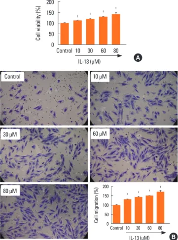
Fig. 1. Treatment with IL-13 induces proliferation and migration of BSMCs. (A) MTT assay was performed to evaluate the effects of IL-13 on the proliferation of BSMCs. Treatment with IL-13 significantly induced cell proliferation in a dose- dependent manner. (B) Transwell migration assay was performed to evaluate the effects of IL-13 on the migration of BSMCs. Treatment with IL-13 signifi- cantly induced cell migration in a dose-dependent manner. BSMC, bronchial smooth muscle cell; MTT, 3-(4,5-dimethylthiazol-2-yl)-2,5-diphenyltetrazolium bromide; IL, interleukin. *P<0.01, †P<0.001, and ‡P<0.0001 when compared with the control group.
Control 10 μM
60 μM
80 μM 30 μM
Cell viability (%)
Control 10 30 60 80 IL-13 (μM) 200
150 100 50 0
† ‡ ‡ ‡
A
B
Cell migration (%)
Control 10 30 60 80 IL-13 (μM) 200
150 100 50 0
‡
‡ ‡
‡
Statistical analysis
All statistical analyses were conducted using Graphpad Prism software version 6.01 for windows (GraphPad Software, La Jol- la, CA, USA). The data are expressed as mean±standard devia- tion. Differences between the groups were assessed by one-way analysis of variance (ANOVA) followed by the Bonferroni mul- tiple comparison tests or two-way ANOVA, with Bonferroni mul- tiple comparison posttests. A P value of <0.05 was considered statistically significant.
RESULTS
Treatment with IL-13 induces proliferation and migration of BSMCs
In order to reliably explore the effects of IL-13 related pathways on the proliferation and migration of BSMCs, IL-13-treated cells were subjected to MTT cell proliferation and Transwell migra- tion assays. The results indicated that IL-13 treatment signifi- cantly induced the proliferation of BSMCs in a dose-dependent manner (Fig. 1A). Similarly, in Transwell assay, the migration of BSMCs was significantly increased following IL-13 treatment, dose-dependently (Fig. 1B).
Expression of miR-155-5p is down-regulated in IL-13-induced BSMCs
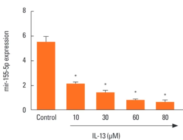
To identify the role of miR-155-5p in the regulation of BSMC phenotype, miR-155-5p expression was examined in BSMCs treated with different concentrations of IL-13 which is known as a key cytokine driving asthma pathogenesis. The qRT-PCR experiments indicated that miR-155 expression was significant- ly and dose-dependently down-regulated in IL-13-induced BS- MCs compared with the control untreated cells (Fig. 2).
Overexpression of miR-155-5p inhibits proliferation and migration of IL-13-induced BSMCs
The transfection of BSMCs with miR-155-5p mimic or inhibi- tor significantly promoted or inhibited the expression of miR- 155. Similarly, overexpression of miR-155-5p inhibited the pro- liferation of BSMCs, while the inverse effect was found for miR- 155-5p inhibitor (Fig. 3A). Meanwhile, during the migration as- say, we observed that inhibition of miR-155-5p was accompa- nied by increased number of migrated cells, whereas transfec- tion with miR-155-5p mimic showed a remarkable hindrance of BSMC migration ability (Fig. 3B).
mir-155-5p expression
Control 10 30 60 80
IL-13 (μM) 8
6
4
2
0
*
*
* *
Fig. 2. Treatment with IL-13 inhibits the expression of miR-155-5p in BSMCs.
qRT-PCR was performed to evaluate the effects of IL-13 on the expression of miR-155-5p in BSMCs. Treatment with IL-13 significantly inhibited miR-155-5p expression in a dose-dependent manner. IL, interleukin; BSMC, bronchial smooth muscle cell; qRT-PCR, quantitative real-time polymerase chain reaction. *P<0.0001 when compared with the control group.
Fig. 3. Overexpression of miR-155-5p inhibits proliferation and migration of IL-13-induced BSMCs. (A) MTT assay was performed to evaluate the effects of miR-155- 5p on the proliferation of IL-13-induced BSMCs. Treatment with miR-155-5p inhibitor significantly induced cell proliferation while miR-155-5p mimics significantly in- hibited cell proliferation. (B) Transwell migration assay was performed to evaluate the effects of miR-155-5p on the migration of IL-13-induced BSMCs. Treatment with miR-155-5p inhibitor significantly induced cell migration, while miR-155-5p mimics significantly inhibited cell migration. IL, interleukin; BSMC, bronchial smooth muscle cell; MTT, 3-(4,5-dimethylthiazol-2-yl)-2,5-diphenyltetrazolium bromide; ns, not significant. *P<0.0001 when compared with inhibitor control (Inh Ctrl) or mim- ic control (Mi Ctrl).
Cell proliferation (%)
Inh ctrl mir-155-5p Inh
mir-155-5p Mi Mi Ctrl 200
150 100 50 0
*
ns
*
Cell migration (%)
Inh ctrl mir-155-5p Inh
mir-155-5p Mi Mi Ctrl 150
100
50
0
*
ns
*
Inh Ctrl Mi Ctrl
mir-155 Inh mir-155-5p Mi
A B
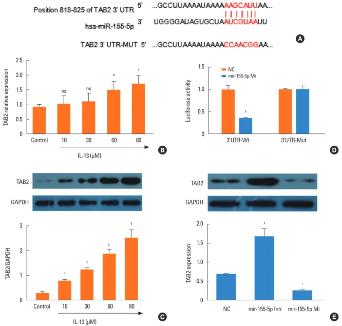
TAB2 is a direct target of miR-155
To elucidate the mechanisms responsible for the regulation of BSMC phenotype by miR-155, we used bioinformatics to iden- tify the target transcripts of miR-155. TAB2 was predicted as a potential miR-155 target (Fig. 4A). Therefore, we assessed TAB2 expression at protein and mRNA levels in BSMCs induced by different concentrations of IL-13. The mRNA expression of TAB2 was significantly and dose-dependently increased in IL-13-in- duced BSMCs compared with untreated cells (Fig. 4B). Western blot analysis showed that TAB2 protein level was equally signif- icantly and dose-dependently increased in IL-13-induced BSMCs
compared with untreated cells (Fig. 4C). Furthermore, using the luciferase reporter assay, we found that miR-155-5p signifi- cantly inhibited luciferase activity in BSMCs transfected with the vector harboring the wild-type 3′-UTR TAB2, while no sig- nificant difference was recorded with cells transfected with mu- tant 3′-UTR vector (Fig. 4D). These observations indicated that TAB2 was a direct target of miR-155-5p. Inhibition of miR-155- 5p with the inhibitor resulted in the up-regulation of TAB2 pro- tein expression in IL-13-induced BSMCs (Fig. 4E). On the con- trary, miR-155 mimic led to decreased TAB2 protein levels. Nei- ther miR-155 inhibitor nor mimic affected TAB2 mRNA expres- Fig. 4. TAB2 is a direct target of miR-155-5p. (A) Search in the online bioinformatics tool Targetscan indicated that 3′-UTR of TAB2 mRNA is a potential target of miR- 155-5p. (B) qRT-PCR experiments indicated that IL-13 dose-dependently induced the expression of TAB2 mRNA in BSMCs. (C) Western blot analysis indicated that IL- 13 dose-dependently induced the expression of TAB2 protein in BSMCs. (D) Luciferase reporter assay indicated TAB2 is a direct target of miR-155-5p. (E) miR-155-5p mimic significantly inhibited the expression of TAB2. TAB2, transforming growth factor (TGF)-β-activated kinase 1/MAP3K7-binding protein 2; UTR, untranslated re- gion; qRT-PCR, quantitative real-time polymerase chain reaction; IL, interleukin; BSMC, bronchial smooth muscle cell; NC, negative control; ns, not significant. *P<0.05,
†P<0.01, ‡P<0.001, and §P<0.0001 when compared with control or NC.
TAB2 relative expression
Control 10 30 60 80
IL-13 (μM) 2.5
2.0 1.5 1.0 0.5 0
ns ns
*
†
Luciferase activity
3’UTR-Wt 3’UTR-Mut
1.5
1.0
0.5
0
§
NCmir-155-5p Mi
TAB2/GAPDH
Control 10 30 60 80
IL-13 (μM) 3
2
1
0
†
§
§
§
TAB2
GAPDH
TAB2 expression
NC mir-155-5p Inh mir-155-5p Mi 2.0
1.5
1.0
0.5
0
§
†
TAB2
GAPDH
A
B
C
D
E
sion (Fig. 4E). These results indicated that miR-155-5p is a post- transcriptional regulator of TAB2 and that miR-155-5p negative- ly regulated TAB2. These results suggested that overexpression of miR-155-5p inhibits the proliferation and migration of IL-13- induced BSMCs by suppressing TAB2.
Silencing of TAB2 mimics the effects of miR-155-5p on BSMCs
To evaluate possible involvement of miR-155-5p/TAB2 in IL- 13-induced proliferation and migration of BSMCs, we determined the effects of TAB2 siRNA on the effects of BSMCs transfected with miR-155 mimic. MTT assay showed that TAB2 siRNA mim- icked the inhibitory effects of miR-155 mimic on IL-13-induced proliferation of BSMCs (Fig. 5A). Similar effects were recorded on cell migration in Transwell migration assay (Fig. 5B). The present data indicated that miR-155-5p-mediated inhibition of TAB2 is involved in the inhibitory effect of mir-155-5p on the proliferation and migration of BSMCs. The results equally indi- cated that the IL-13/miR-155-5p/TAB2 pathways could be in- volved in the proliferation and migration of BSMCs, which can probably be implicated in the pathogenesis of asthma and oth- er pulmonary diseases.
DISCUSSION
Cytokines are known as the main drivers of inflammatory dis- eases, such as asthma. TH2 cytokines (IL-4, IL-5, and IL-13) are related to disease activity, symptom score, eosinophilia of the airways, and bronchial hyper-reactivity. They increase after an allergenic stimulation and decrease with the corticoid treatment.
Similar to IL-4, IL-13 stimulates the production of immunoglob- ulin E (IgE) by B lymphocytes, activates monocytes and macro- phages, and increases mucus secretion by goblet cell hyperpla- sia.25,26 Recent publications have highlighted the involvement of
IL-13 in asthma,27,28 but the molecular mechanism involved in this process is poorly studied. In asthma, IL-13 is reported to in- duce mucus production.29-31 Additional studies also indicate im- portant functions of IL-13 in airway smooth muscle cells, such as gene regulation, cell proliferation, and migration.32-34 Specifical- ly, IL-13 was found to promote contractility of BSMCs by up-reg- ulating RhoA protein.35 In addition, IL-13 was found to induce BSMC proliferation by upregulating CysLT1 receptor IL-13 in response to LTD4.36 In the present study, we observed that IL- 13 dose-dependently induced the proliferation and migration of BSMCs, thus confirming its role in bronchial asthma and the necessity to further explore its underlying mechanism.
Studies on the cross-talk between IL-13 and miRs in the patho- physiology of asthma, especially the phenotypic changes of BS- MCs, are scarce and the underlying mechanism is ill-defined.
In mouse in vivo studies, a set of miRs that are involved in the response to allergen attacks have been discovered and some of them, involved in the regulation of IL-13 and TH2 which are key components in asthmatic reactions, have been proved promis- ing for pre-miR and anti-miR manipulations.37,38 Other studies have identified miRs that are implicated in bronchial smooth muscle hyperresponsiveness and proliferation.40-42 Up to now, the most frequently studied miR in the regulation of asthma mo- lecular and cellular processes is miR-155, which is found to be up-regulated in asthma.39,43 Previous studies have demonstrated that miR-155 participates in allergic airway inflammation through regulating the transcription factor PU.1.44 In vivo experiments indicated that miR-155 deficiency contributes to allergic airway inflammation improvement by modulating Th2 responses and ATP-/P2R-induced activation of dendritic cells in mice.45 An ad- ditional study documented that miR-155 promotes the prolifer- ation of Th cells in allergic asthma by down-regulating cytotox- ic T-lymphocyte-associated protein 4 (CTLA-4).46 All these find- ings suggested miR-155 to be a potential therapeutic target for Fig. 5. Silencing of TAB2 mimics the effects of miR-155-5p on BSMCs. (A) MTT assay was performed to evaluate the effects of TAB2 siRNA and miR-155-5p mimic on the proliferation of IL-13-induced BSMCs. Transfection with miR-155-5p mimic significantly inhibited cell proliferation. The same effect was found with TAB2 siR- NA. (B) Transwell migration assay was performed to evaluate the effects of TAB2 siRNA and miR-155-5p mimic on the proliferation of IL-13-induced BSMCs. Trans- fection with miR-155-5p mimic significantly inhibited cell migration. The same effect was found with TAB2 siRNA. TAB2, transforming growth factor (TGF)-β-activated kinase 1/MAP3K7-binding protein 2; BSMC, bronchial smooth muscle cell; MTT, 3-(4,5-dimethylthiazol-2-yl)-2,5-diphenyltetrazolium bromide; siRNA, small interfer- ing RNA; IL, interleukin; ns, not significant. *P<0.05, †P<0.01, ‡P<0.001, §P<0.0001; llP<0.05 and ¶P<0.001 when compared with the control siRNA+IL-13 group.
Cell proliferation (%)
IL-13 (80 μM) - + + +
ctrl siRNA + + - -
TAB2 siRNA - - + -
mir-155-5p Mi - - - +
200 150 100 50 0
§
‡, ll
*, ¶
Cell migration (%)
IL-13 (80 μM) - + + +
ctrl siRNA + + - -
TAB2 siRNA - - + -
mir-155-5p Mi - - - +
200 150 100 50 0
§
†, ll
ns¶
Ctrl siRNA
TAB2 siRNA+IL-13
Ctrl siRNA+IL-13
mir-155-5p Mi+IL-13
A B
allergic asthma. However, the molecular and cellular mecha- nism via which miR-155 exerts its action in asthma is not fully elucidated and needs further clarification. The present study was to scrutinize the mechanisms involved in the cross-talk be- tween miR-155-5p and IL-13 and their action on the prolifera- tion and migration of BSMCs, which could have beneficial im- plications in the understanding and treatment of pulmonary diseases, especially asthma. The main results were as follows: 1) IL-13 induced proliferation and migration of BSMCs dose-depen- dently; 2) IL-13 dose-dependently decreased the expression of miR-155-5p in IL-13-induced BSMCs; 3) Overexpression of miR- 155-5p inhibited the proliferation and migration of IL-13-induced BSMCs; 4) TAB2 was a direct target of miR-155-5p and silencing of TAB2 mimicked the effects of miR-155-5p. Previous studies have shown that in human macrophages, miR-155 directly tar- gets IL13Ralpha1 and reduces the levels of IL13Ralpha1 protein, leading to diminished activation of signal transducer and acti- vator of transcription 6 (STAT6) and demonstrated that miR-155 affects the IL-13-dependent regulation of several genes (SOCS1, DC-SIGN, CCL18, CD23, and SERPINE) involved in the estab- lishment of an M2/pro-Th(2) phenotype in macrophages. Our study, to the best of our knowledge, is the first to report the in- volvement of the IL-13/mir-155/TAB2 axis in the proliferation and migration of BSMCs. These findings corroborated, along with a previous study, that miR-155 is a negative regulator of TAB2 in mesenchymal stem cells (MSCs).47 A direct functional role of TAB2 in asthma has not been reported previously, but previous studies demonstrated its involvement in the activation of inflammatory pathways.47-49 Our findings also provided evi- dence that IL-13-inhibits TAB2 expression via up-regulating miR- 155-5p in BSMCs, which suggest its role in asthma inflammation.
Our work presents some limitations. IL-13-induced asthma hy- perresponsiveness may be one of the key features in asthmatics, in which airway smooth muscle contraction plays a critical role.
Nevertheless, in the present study, we did not check incre ased Ca++ oscillations or cell contraction in IL-13-treated BSMCs; thus, it is hard to precisely define the function of miR-155-overexpressed BSMCs. Further studies are needed to clarify this aspect.
We conclude that overexpression of miR-155-5p may inhibit the proliferation and migration of IL-13-induced human BSMCs by suppressing TAB2. In addition, the results suggest that mod- ulation of the IL-13/mir-155/TAB2 pathway could be a poten- tial strategy in the treatment of allergic asthma.
ACKOWLEDGMENTS
This study was supported by the National Natural Science Foundation of China (81270077), Jiangsu province “six big tal- ent peak” project (WSN092), Changzhou High-Level Medical Talents Training Project (2016CZLJ017), Jiangsu province “333 talents” project (BRA2016119) and the Changzhou Sci & Tech Program (CJ20179031).
ORCID
Yujia Shi https://orcid.org/0000-0001-6683-2932 Xingli Fu https://orcid.org/0000-0002-9199-7525 Qi Cao https://orcid.org/0000-0001-6901-8928 Zhengdao Mao https://orcid.org/0000-0003-0225-7056 Yi Chen https://orcid.org/0000-0001-9611-8393 Yun Sun https://orcid.org/0000-0002-7393-6630 Zhiguang Liu https://orcid.org/0000-0001-6982-5164 Qian Zhang https://orcid.org/0000-0003-4928-3366
REFERENCES
1. Ariel D, Upadhyay D. The role and regulation of microRNAs in as- thma. Curr Opin Allergy Clin Immunol 2012;12:49-52.
2. Ghosh S, Erzurum SC. Nitric oxide metabolism in asthma patho- physiology. Biochim Biophys Acta 2011;1810:1008-16.
3. Gras D, Bourdin A, Chanez P, Vachier I. Airway remodeling in asth- ma: clinical and functional correlates. Med Sci (Paris) 2011;27:959- 65.
4. Chung KF. Asthma phenotyping: a necessity for improved therapeu- tic precision and new targeted therapies. J Intern Med 2016;279:192- 204.
5. Lubret M, Bervar JF, Thumerelle C, Deschildre A, Tillie-Leblond I.
Asthma: treatment of exacerbations. Rev Mal Respir 2012;29:245- 53.
6. Moss RB. Treatment options in severe fungal asthma and allergic bronchopulmonary aspergillosis. Eur Respir J 2014;43:1487-500.
7. Ricciardolo FL, Blasi F, Centanni S, Rogliani P. Therapeutic novel- ties of inhaled corticosteroids and bronchodilators in asthma. Pulm Pharmacol Ther 2015;33:1-10.
8. Dunican EM, Fahy JV. The role of type 2 inflammation in the patho- genesis of asthma exacerbations. Ann Am Thorac Soc 2015;12 Suppl 2:S144-9.
9. Hu D. Role of anti-inflammatory cytokines IL-35 and IL-37 in asth- ma. Inflammation 2017;40:697-707.
10. Walsh GM. Anti-IL-4/-13 based therapy in asthma. Expert Opin Emerg Drugs 2015;20:349-52.
11. Albers FC, Price RG, Smith SG, Yancey SW. Mepolizumab efficacy in patients with severe eosinophilic asthma receiving different con- troller therapies. J Allergy Clin Immunol 2017;140:1464-1466.e4.
12. Máspero J. Reslizumab in the treatment of inadequately controlled asthma in adults and adolescents with elevated blood eosinophils:
clinical trial evidence and future prospects. Ther Adv Respir Dis 2017;11:311-25.
13. Saha SK, Berry MA, Parker D, Siddiqui S, Morgan A, May R, et al.
Increased sputum and bronchial biopsy IL-13 expression in severe asthma. J Allergy Clin Immunol 2008;121:685-91.
14. Cai F, Hornauer H, Peng K, Schofield CA, Scheerens H, Morimoto AM. Bioanalytical challenges and improved detection of circulat- ing levels of IL-13. Bioanalysis 2016;8:323-32.
15. Chapman AM, Malkin DJ, Camacho J, Schiestl RH. IL-13 overex- pression in mouse lungs triggers systemic genotoxicity in peripher- al blood. Mutat Res 2014;769:100-7.
16. Ferreira CM, Pereira AT, de Souza RS, Coelho FM, Poole S, Teixeira MM, et al. Role of IL-13 in a model of Strongyloides venezuelensis infection in rats. Microbes Infect 2010;12:409-14.
17. Brightling CE, Saha S, Hollins F. Interleukin-13: prospects for new treatments. Clin Exp Allergy 2010;40:42-9.
18. Borowski A, Kuepper M, Horn U, Knupfer U, Zissel G, Hohne K, et al. Interleukin-13 acts as an apoptotic effector on lung epithelial cells and induces pro-fibrotic gene expression in lung fibroblasts.
Clin Exp Allergy 2008;38:619-28.
19. Crapster-Pregont M, Yeo J, Sanchez RL, Kuperman DA. Dendritic cells and alveolar macrophages mediate IL-13-induced airway in- flammation and chemokine production. J Allergy Clin Immunol 2012;129:1621-1627.e3.
20. Firszt R, Francisco D, Church TD, Thomas JM, Ingram JL, Kraft M.
Interleukin-13 induces collagen type-1 expression through matrix metalloproteinase-2 and transforming growth factor-beta1 in air- way fibroblasts in asthma. Eur Respir J 2014;43:464-73.
21. Kasaian MT, Miller DK. IL-13 as a therapeutic target for respiratory disease. Biochem Pharmacol 2008;76:147-55.
22. Mitchell J, Dimov V, Townley RG. IL-13 and the IL-13 receptor as therapeutic targets for asthma and allergic disease. Curr Opin In- vestig Drugs 2010;11:527-34.
23. Zhang YY, Zhong M, Zhang MY, Lv K. Expression and clinical sig- nificance of miR-155 in peripheral blood CD4(+);T cells of patients with allergic asthma. Xi Bao Yu Fen Zi Mian Yi Xue Za Zhi 2012;28:
540-3.
24. Zhou H, Li J, Gao P, Wang Q, Zhang J. miR-155: a novel target in al- lergic asthma. Int J Mol Sci 2016;17:E1773.
25. Vock C, Yildirim AO, Wagner C, Schlick S, Lunding LP, Lee CG, et al. Distal airways are protected from goblet cell metaplasia by di- minished expression of IL-13 signalling components. Clin Exp Al- lergy 2015;45:1447-58.
26. Parker JC, Thavagnanam S, Skibinski G, Lyons J, Bell J, Heaney LG, et al. Chronic IL9 and IL-13 exposure leads to an altered differenti- ation of ciliated cells in a well-differentiated paediatric bronchial epithelial cell model. PLoS One 2013;8:e61023.
27. Qaseem AS, Sonar S, Mahajan L, Madan T, Sorensen GL, Shamji MH, et al. Linking surfactant protein SP-D and IL-13: implications in asthma and allergy. Mol Immunol 2013;54:98-107.
28. Tomasiak-Łozowska MM, Bodzenta-Łukaszyk A, Tomasiak M, Ski- epko R, Zietkowski Z. The role of interleukin 13 and interleukin 5 in asthma. Postepy Hig Med Dosw (Online) 2010;64:146-55.
29. Jakiela B, Gielicz A, Plutecka H, Hubalewska-Mazgaj M, Mastalerz L, Bochenek G, et al. Th2-type cytokine-induced mucus metapla- sia decreases susceptibility of human bronchial epithelium to rhi- novirus infection. Am J Respir Cell Mol Biol 2014;51:229-41.
30. Lai HY, Rogers DF. Mucus hypersecretion in asthma: intracellular signalling pathways as targets for pharmacotherapy. Curr Opin Al- lergy Clin Immunol 2010;10:67-76.
31. Tanabe T, Fujimoto K, Yasuo M, Tsushima K, Yoshida K, Ise H, et al. Modulation of mucus production by interleukin-13 receptor al- pha2 in the human airway epithelium. Clin Exp Allergy 2008;38:122- 34.
32. Cleary RA, Wang R, Wang T, Tang DD. Role of Abl in airway hyper- responsiveness and airway remodeling. Respir Res 2013;14:105.
33. Jiang H, Xie Y, Abel PW, Toews ML, Townley RG, Casale TB, et al.
Targeting phosphoinositide 3-kinase gamma in airway smooth mus- cle cells to suppress interleukin-13-induced mouse airway hyper- responsiveness. J Pharmacol Exp Ther 2012;342:305-11.
34. Moynihan B, Tolloczko B, Michoud MC, Tamaoka M, Ferraro P, Martin JG. MAP kinases mediate interleukin-13 effects on calcium signaling in human airway smooth muscle cells. Am J Physiol Lung Cell Mol Physiol 2008;295:L171-7.
35. Chiba Y, Nakazawa S, Todoroki M, Shinozaki K, Sakai H, Misawa M.
Interleukin-13 augments bronchial smooth muscle contractility with an up-regulation of RhoA protein. Am J Respir Cell Mol Biol 2009;40:159-67.
36. Espinosa K, Bosse Y, Stankova J, Rola-Pleszczynski M. CysLT1 re- ceptor upregulation by TGF-beta and IL-13 is associated with bron- chial smooth muscle cell proliferation in response to LTD4. J Aller- gy Clin Immunol 2003;111:1032-40.
37. Wang Y, Yang F, Xue J, Zhou X, Luo L, Ma Q, et al. Antischistosomi- asis liver fibrosis effects of chlorogenic acid through IL-13/miR-21/
Smad7 signaling interactions in vivo and in vitro. Antimicrob Agents Chemother 2017;61:e01347-16.
38. Liu Y, Yang K, Shi H, Xu J, Zhang D, Wu Y, et al. MiR-21 modulates human airway smooth muscle cell proliferation and migration in asthma through regulation of PTEN expression. Exp Lung Res 2015;
41:535-45.
39. Comer BS, Camoretti-Mercado B, Kogut PC, Halayko AJ, Solway J, Gerthoffer WT. Cyclooxygenase-2 and microRNA-155 expression are elevated in asthmatic airway smooth muscle cells. Am J Respir Cell Mol Biol 2015;52:438-47.
40. Greene CM, Gaughan KP. microRNAs in asthma: potential thera- peutic targets. Curr Opin Pulm Med 2013;19:66-72.
41. Chiba Y, Misawa M. MicroRNAs and their therapeutic potential for human diseases: MiR-133a and bronchial smooth muscle hyper- responsiveness in asthma. J Pharmacol Sci 2010;114:264-8.
42. Chiba Y, Tanabe M, Goto K, Sakai H, Misawa M. Down-regulation of miR-133a contributes to up-regulation of Rhoa in bronchial smooth muscle cells. Am J Respir Crit Care Med 2009;180:713-9.
43. Malmhäll C, Johansson K, Winkler C, Alawieh S, Ekerljung L, Rådin- ger M. Altered miR-155 expression in allergic asthmatic airways.
Scand J Immunol 2017;85:300-7.
44. Malmhäll C, Alawieh S, Lu Y, Sjöstrand M, Bossios A, Eldh M, et al.
MicroRNA-155 is essential for T(H)2-mediated allergen-induced eosinophilic inflammation in the lung. J Allergy Clin Immunol 2014;
133:1429-38, 1438.e1-7.
45. Zech A, Ayata CK, Pankratz F, Meyer A, Baudiss K, Cicko S, et al.
MicroRNA-155 modulates P2R signaling and Th2 priming of den- dritic cells during allergic airway inflammation in mice. Allergy 2015;
70:1121-9.
46. Zhang Y, Sun E, Li X, Zhang M, Tang Z, He L, et al. miR-155 contrib- utes to Df1-induced asthma by increasing the proliferative response of Th cells via CTLA-4 downregulation. Cell Immunol 2017;314:1-9.
47. Xu C, Ren G, Cao G, Chen Q, Shou P, Zheng C, et al. miR-155 regu- lates immune modulatory properties of mesenchymal stem cells by targeting TAK1-binding protein 2. J Biol Chem 2013;288:11074-9.
48. Tan B, Mu R, Chang Y, Wang YB, Wu M, Tu HQ, et al. RNF4 negative- ly regulates NF-kappaB signaling by down-regulating TAB2. FEBS Lett 2015;589:2850-8.
49. Zhu S, Pan W, Song X, Liu Y, Shao X, Tang Y, et al. The microRNA miR-23b suppresses IL-17-associated autoimmune inflammation by targeting TAB2, TAB3 and IKK-alpha. Nat Med 2012;18:1077-86.