Sphingosine-1-Phosphate-Induced Migration and Differentiation of Human Mesenchymal Stem Cells to Smooth Muscle Cells
Hae Young Song
1, Sang Hun Shin
1,2, Min Young Kim
1,2and Jae Ho Kim
1,2*
1
Department of Physiology, School of Medicine,
2Medical Research Center for Ischemic Tissue Regeneration, Medical Research Institute, Pusan National University, Yangsan 626-870, Republic of Korea
Received November 8, 2010 /Accepted January 21, 2011
Migration and differentiation of mesenchymal stem cells are crucial for tissue regeneration in response to injury. Sphingosine-1-phosphate (S1P) is a bioactive lipid that regulates a variety of biological proc- esses, including proliferation, survival, differentiation and motility. In the present study, we de- termined the role of S1P in migration and differentiation of human bone marrow-derived mesen- chymal stem cells (BMSCs). S1P stimulated migration of BMSCs in a dose- and time-dependent man- ner, and pre-incubation of the cells with pertussis toxin completely abrogated S1P-induced migration, suggesting involvement of G
i-coupled receptors in S1P-induced cell migration. S1P elicited elevation of intracellular concentration of Ca
2+([Ca
2+]
i) and pretreatment with VPC23019, an antagonist of S1P
1/S1P
3, blocked S1P-induced migration and increase of [Ca
2+]
i. Small interfering RNA-mediated knockdown of endogenous S1P
1attenuated S1P-induced migration of BMSCs. Furthermore, S1P treat- ment induced expression of a-smooth muscle actin (a-SMA), a smooth muscle marker, and pretreat- ment with VPC23019 abrogated S1P-induced a-SMA expression. S1P induced phosphorylation of p38 mitogen-activated protein kinase (MAPK), and pretreatment of cells with SB202190, an inhibitor of p38 MAPK, or adenoviral overexpression of a dominant-negative mutant of the p38 MAPK blocked S1P-induced cell migration and a-SMA expression. Taken together, these results suggest that S1P stim- ulates migration and smooth muscle differentiation of BMSCs through an S1P
1-p38 MAPK-dependent mechanism.
Key words : Sphingosine-1-phosphate, mesenchymal stem cells, migration, p38 MAPK, S1P receptor
*Corresponding author
*Tel:+82-51-510-8073, Fax:+82-51-510-8076
*E-mail : [email protected]
Introduction
Mesenchymal stem cells (MSCs) possess self-renewal ca- pacity, long-term viability, and differentiation potential to- ward diverse cell types, such as adipogenic, osteogenic, chondrogenic, and myogenic lineages. They can be isolated from a variety of tissues, including bone marrow and adi- pose tissues [2,31,34,36], suggesting that MSCs are highly useful for clinical applications in regenerative medicine.
Intravenously-transplanted MSCs can home to injured and inflamed sites in animal models of myocardial infarction and cerebral ischemia [3]. Inflamed tissues produce a variety of inflammatory mediators, including chemokines, cyto- kines, and prostanoids [43], and several chemokines have been reported to attract MSCs in vitro [4,8,33,42]. MSCs have been shown to migrate in response to various growth fac- tors, including platelet-derived growth factor, insulin-like growth factor, epidermal growth factor and hepatocyte
growth factor [6,7,17,32,40] and chemokines, such as stro- mal-derived factor-1, fractalkine and monocyte chemo- attractant protein-1 [5,16,21]. However, the molecular mech- anisms involved in migration of MSCs are still elusive.
Sphingosine-1-phosphate (S1P) is a bioactive lysophos- pholipid that is present in human plasma and serum [45]
and released in large amounts (0.5 μM in serum) from acti-
vated platelets [10]. As a factor released from activated pla-
telets in inflamed or injured tissues, S1P regulates a variety
of cellular responses, including cell growth, survival, differ-
entiation and motility [14,35,37,39]. The effects of ex-
tracellular S1P are mediated by G protein-coupled receptors,
termed S1P
1-5, which regulate diverse intracellular pathways
[35,37,39]. Activation of S1P receptors has been shown to
stimulate diverse signaling pathways involving ERK, JNK,
p38 mitogen-activated protein kinase (MAPK), phospholi-
pase C, phosphoinositide-3-kinase, and RhoA [11,39]. S1P
has been reported to inhibit or stimulate cellular motility,
depending on the cell type, differences in S1P receptor ex-
pression, and S1P concentration [35]. Activation of S1P
1and
S1P
3stimulates migration through G
i-mediated activation
of phosphatidylinositol-3-kinase, Akt and Rac [35,39], whereas S1P
2signals through G
12/13for activation of RhoA and inhibition of Rac-dependent signaling, which is respon- sible for the inhibitory effect of S1P on cell migration [23,38,44]. S1P
4is mainly expressed in immune cells and stimulates migration of transfected Chinese-hamster ovary cells by activation of cdc42 through a pertussis toxin (PTX)-sensitive manner [18]. S1P has been reported to stim- ulate migration of mouse bone marrow-derived MSCs (BMSCs) [27]. In addition, S1P mediated homing of BMSCs into injured liver and stimulated differentiation of cells into α -SMA-positive myofibroblasts [24]. However, S1P treatment has been shown to have no significant effect on migration of human BMSCs [15]. Therefore, it is still unclear whether S1P can regulate migration and differentiation of BMSCs.
In the present study, we explored the effects of S1P on migration and α-SMA expression of human BMSCs and characterized the signaling pathways associated with S1P-induced cellular responses.
Materials and Methods Materials
Phosphate-buffered saline, α-minimum essential medium (α-MEM), trypsin, fetal bovine serum, and Lipofectamine
Plusreagent were purchased from Invitrogen (Carlsbad, CA).
S1P and PTX were purchased from BIOMOL (Plymputh Meeting, PA). VPC23019 was purchased from Avanti Polar Lipids, Inc. (Alabaster, AL). 1-Oleoyl-sn-glycero-3-phos- phate (1-oleoyl-LPA), fatty acid-free bovine serum albumin, and Ki16425 were purchased from Sigma–Aldrich (St. Louis, MO). LY294002, Y27632, U0126, SP600125, and SB202190 were purchased from EMD Biosciences (San Diego, CA).
Anti-phospho-p38 and anti-p38 MAPK antibodies were from Cell Signaling Technology (Beverly, MA). Fluo-4-AM was from Molecular Probes, Inc. (Eugene, OR). Culture plates were purchased from Nunc (Roskilde, Denmark).
Horseradish peroxidase-labeled secondary antibodies and the enhanced chemiluminescence Western blotting system were from Amersham Biosciences (Pittsburgh, PA).
Cell culture
After informed consent, heparinized bone marrow was obtained from patients undergoing total hip arthroplasty, as approved by the Institutional Review Board of Pusan National University Hospital. BMSCs were isolated from
bone marrow as previously described [22]. For isolation of BMSCs, mononuclear cells from bone marrow were sepa- rated by centrifugation in a Ficoll-Hypaque gradient (density=1.077 g/cm
3; Sigma, USA), and seeded at a density of 1×10
6cells/cm
2. Cultures were maintained at 37
oC in a humidified atmosphere containing 5% CO
2in growth me- dium (α-MEM, 10% fetal bovine serum, 100 units/ml of penicillin, 100 μg/ml of streptomycin) until they reached confluence. Primary BMSCs were subcultured in tissue cul- ture dishes at a density of 2,000 cells/cm
2. Cells expressed CD29, CD44, CD90, CD105 and did not express HLA-DR and c-kit
22.
Cell migration assay
BMSCs migration was assayed using a Boyden chamber apparatus, as previously described [19]. Briefly, BMSCs were harvested with 0.05% trypsin containing 0.02% EDTA, washed once, and suspended in α-MEM at a concentration of 2×10
5cells/ml. Membrane filters (8-μm pore size) in dis- posable 96-well chemotaxis chambers (Neuro Probe, Inc.;
Gaithersburg, MD) were precoated overnight with 20 μg/ml rat-tail collagen at room temperature. Aliquots (50 μl per well) of the cell suspension were loaded into the upper chambers, and test reagents were placed in the lower cham- ber, unless otherwise specified. To elucidate the signaling pathways involved in S1P-induced migration, cells were preincubated with pharmacological inhibitors for 15 min prior to loading. Following incubation of cells with S1P in the absence or presence of inhibitors for 12 hr at 37
oC, filters were disassembled, and the upper surface of each filter was scraped free of cells by wiping with a cotton swab. The numbers of cells that had migrated to the lower surfaces of each filter were determined by microscopic counting of cells in four places (×100 magnification) after staining with hematoxylin and eosin.
Measurement of intracellular calcium concentration
Spatially averaged photometric [Ca
2+] measurements from single cells were performed with the fluorescent Ca
2+indicator fluo-4-AM. In brief, BMSCs cells grown on 32 mm
dish were incubated with serum-free α-MEM for 24 hr, load-
ed with 5 µM fluo-4-AM for 40 min at 37
oC in buffer A
[135 mM NaCl, 4 mM KCl, 1 mM MgCl
2, 2 mM CaCl
2,
10 mM glucose, and 20 mM N-2-hydroxyethylpiper-
azine-N'-2-ethanesulfonic acid (HEPES), pH 7.3], and wash-
ed twice with Hanks’ balanced salt solution without phenol
red and Ca
2+. Fluo-4-AM-loaded BMSCs were treated with S1P. A Leica TCS-SP2 laser scanning confocal microscope (Leica Microsystems, Germany) was used for visualization of Ca
2+-mediated fluorescence in cells. Fluo-4 was excited with a 488-nm line of an argon laser, and fluo-4 fluorescence was collected between 510 and 525 nm. Scanning was per- formed every 1 sec for the indicated times, and the ratio of fluorescence intensity to initial fluorescence intensity (F/F
0) was calculated at each point for quantitative measurement.
Western blotting
Confluent, serum-starved BMSCs were treated with the indicated conditions, washed with ice-cold phosphate-buf- fered saline, and lysed in lysis buffer (20 mM Tris-HCl, 1 mM EGTA, 1 mM EDTA, 10 mM NaCl, 0.1 mM phenyl- methylsulfonyl fluoride, 1 mM Na
3VO
4, 30 mM sodium py- rophosphate, 25 mM b-glycerophosphate, 1% Triton X-100, pH 7.4). Lysates were resolved by sodium dodecyl sul- fate-polyacrylamide gel electrophoresis (SDS-PAGE), trans- ferred onto nitrocellulose membranes, and stained with 0.1% Ponceau S solution. After blocking with 5% nonfat milk, membranes were immunoblotted with appropriate primary antibodies, and bound antibodies were visualized with horseradish peroxidase-conjugated secondary anti- bodies and an enhanced chemiluminescence system.
Reverse transcription-polymerase chain reaction analysis
Total cellular RNA was extracted by the Trizol method (Invitrogen, Carlsbad, CA). For reverse transcription-poly- merase chain reaction (RT-PCR) analysis, aliquots of 2 μg each of RNA were subjected to cDNA synthesis with 200 U of M-MLV reverse transcriptase and 0.5 μg of oligo (dT) 15 primer (Promega, Madison, WI). cDNA in 2 μl of the reaction mixture was amplified with 0.5 U of GoTaq DNA polymerase (Promega, Madison, WI) and 10 pmol each of sense and antisense primers as follows: S1P
1receptor (429-bp product), sense, 5'-TATCAGCGCGGACAAGGA GAACAG-3' and antisense, 5'-ATAGGCAGGCCACCCAG GATGAG-3'; S1P
2receptor (220-bp product), sense, 5'-TCGGCCTTCATCGTCATCCTCT-3' and antisense, 5'-CC TCCCGGGCAAACCACTG-3'; S1P
3receptor (394-bp prod- uct), sense, 5'-CTGCCTGCACAATCTCCCTGACTG-3' and antisense, 5'-GGCCCGCCGCATCTCCT-3'; S1P
4receptor (454-bp product), sense, 5'-GAGAGCGGGGCCACCA
AGAC-3' and antisense, 5'-GGTTGACCGCCGAGTTGAGG AC-3'; GAPDH 5′-TCCATGACAACTTTGGTATCG-3′, 5′
-TGTAGCCAAATTCGTTGTCA-3′. The thermal cycle pro- file was as follows: denaturation at 95
oC for 30 sec, anneal- ing at 52-58
oC for 30 sec, depending on the primers used, and extension at 72
oC for 30 sec. Each PCR reaction was carried out for 30 cycles, and PCR products were size frac- tionated on 1.2% ethidium bromide/agarose gel and photo- graphed under UV transillumination.
Transfection with small interfering RNA (siRNA)
siRNA duplexes were synthesized, desalted, and purified by Samchully Pharm. Co. Ltd. (Siheung, GyeongGi, Korea) as follows: S1P
15′-GCU GCU CAA GAC CGU AAU UTT-3′
(sense) and 5′-AAU UAC GGU CUU GAG CAG CTT-3′
(antisense). Nonspecific control siRNA (D-001206-13-05) was purchased from Dharmacon, Inc. (Chicago, IL). For siRNA experiments, BMSCs were seeded on 60-mm dishes at 70%
confluence, and were then transfected with siRNAs using the Lipofectamine plus
TMreagent, according to manu- facturer’s instructions. Briefly, Lipofectamine plus
TMreagent was incubated with serum-free medium for 15 min, and respective siRNAs were then added to the mixtures.
Following incubation for 15 min at room temperature, the mixtures were diluted with serum free medium and added to each well. The final concentration of siRNAs in each well was 100 nM. Following incubation of BMSCs with se- rum-free medium containing siRNAs for 4 hr, cells were cultured in growth medium for 24 hr and the expression levels of S1P
1and GAPDH were then determined by RT-PCR analysis.
Adenoviral expression of a dominant negative p38 MAPK mutant
The gene encoding the dominant-negative mutant of p38 MAPK (TY>AF) was kindly provided by Dr. J. Han [13]
and used for production of recombinant adenoviruses using
the AdEasy system (Stratagene, La Jolla, CA). Following the
manufacturer’s manual, recombinant adenoviruses were
amplified and purified by density gradient ultra-
centrifugation according to manufacturer’s manual. For ad-
enoviral infection, exponentially growing BMSCs were in-
fected with the appropriate amount of adenoviruses and
incubated at 37
oC for 2 hr with gentle shaking. Afterwards,
fresh growth medium was added to each dish and further
incubated for 48 hr. Expression levels of the dominant neg-
A B
C D
Fig. 1. Role of Gi in S1P-induced migration of BMSCs. (A) BMSCs were loaded onto the upper chambers of the Boyden apparatus and serum-free media containing the indicated concentrations of S1P were placed in the lower chambers. The numbers of migratory BMSCs were determined after 12 hr, as described under “Materials and Methods”. (B) Serum-free media containing 0.1 μM S1P or vehicles (control) were added to the lower chambers and the number of BMSCs that migrated to the lower surface of filters was determined after the indicated time periods. (C) Checkerboard analyses of S1P-induced migration. 0.1 μM S1P or vehicles was placed in either the bottom, top, or both chambers of the Boyden apparatus, as noted, and cells were then allowed to migrate for 12 hr. Data represent average values±S.D. (n=4). (D) BMSCs were pretreated with serum-free medium containing vehicles or 100 ng/ml PTX for 24 hr, loaded into the upper chamber, and serum-free media containing vehicles (w/o), 0.5 μM 1-oleoyl-LPA or 0.1 μM S1P were added into the lower chambers. The number of cells migrated to lower surface of filters was determined after 12 hr. Data represent means±S.D. (n=4). *,
p
<0.05; **,p
<0.01 by two-way ANOVA and Scheffe’spost hoc
test.ative mutant of p38 MAPK (DN-p38 MAPK) were de- termined by Western blot analysis.
Statistical analysis
Results of multiple observations are presented as means±SD. Statistical significance was assessed using Student’s t-test or two-way ANOVA test, where indicated in the figure legends.
Results
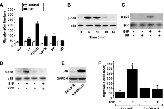
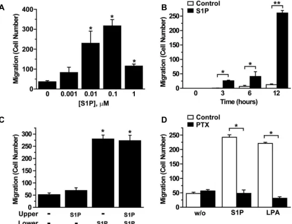
S1P stimulates migration of human BMSCs through a Gi-dependent mechanism
In order to explore the question of whether S1P can regu-
late migration of human BMSCs, we examined the effect of S1P on migration of BMSCs using a Boyden chamber apparatus. S1P dose-dependently increased the migration of BMSCs with a maximal stimulation at 0.1 μM (Fig. 1A) and S1P treatment stimulated migration of BMSCs in a time-dependent manner (Fig. 1B).
To examine the directional component of migration, we
examined S1P-induced migration of BMSCs using a checker-
board analysis. As shown in Fig. 1C, addition of S1P in
the bottom chamber induced migration of BMSCs into the
lower surfaces of the filters. S1P enhanced migration in the
absence of a gradient (equal concentrations in the top and
bottom), suggesting that S1P stimulates random migration
(chemokinesis) as well as directional migration (chemotaxis)
A B
Fig. 2. S1P induces elevation of [Ca2+]i in BMSCs. Serum-starved BMSCs were loaded with 5 μM Fluo-4-AM for 40 min at 37oC in the absence or presence of 10 μM of VPC23019 and then treated with serum-free medium containing 0.1 μM S1P in the absence or presence of 10 μM of VPC23019. Ca2+-dependent fluorescence was measured every second for the indicated time periods, and fluorescence intensities of more than 20 different cells from time-lapse images were quantified over time. Results are expressed as percentage of the control (0 sec) and expressed as means±S.D.
of BMSCs.
Involvement of G
i, a pertussis toxin-sensitive G protein, in S1P-induced migration has been reported in a variety of cell types [35,39]. We have demonstrated that LPA in- duced migration of human adipose tissue-derived MSCs through a Gi-dependent mechanism [20]. For assessment of whether the S1P-stimulated migration was mediated by G
iprotein, we examined the effect of PTX on migration of human BMSCs. As shown in Fig. 1D, PTX treatment com- pletely abrogated migration of BMSCs induced by not only LPA, but also S1P. These results indicate that S1P stimulates the migration of BMSCs through a G
i-dependent pathway.
Involvement of S1P
1receptor in S1P-induced increase of [Ca
2+]
iS1P has been reported to elevate intracellular concen- tration of calcium ([Ca
2+]
i) through activation of S1P receptors. Therefore, we next examined the effect of S1P on [Ca
2+]
iof BMSCs. As shown in Fig. 2A, S1P rapidly in- creased [Ca
2+]
iin BMSCs, and pretreatment of BMSCs with VPC23019, an antagonist specific for S1P
1and S1P
3, com- pletely inhibited S1P-induced elevation of [Ca
2+]
i(Fig. 2B), suggesting that S1P receptors, S1P
1/3, play a key role in S1P-induced elevation of [Ca
2+]
i.
Involvement of S1P1 receptor in S1P-induced migration
To further explore the question of whether S1P receptors are involved in S1P-induced migration, we examined the effects of VPC23019 on migration stimulated by S1P. As shown in Fig. 3A, VPC23019 completely inhibited S1P-in-
duced migration of BMSCs. In contrast, LPA-stimulated mi- gration of BMSCs was not affected by pretreatment with VPC23019, suggesting that S1P
1/3receptors may play a key role in S1P-induced migration of BMSCs.
To clarify the molecular identities of S1P receptors in- volved in S1P-stimulated migration, we examined the ex- pression levels of S1P receptors in BMSCs by RT-PCR analysis. As shown in Fig. 3B, S1P
1, S1P
2, and S1P
3, but not S1P
4, were expressed in BMSCs and S1P
1is a S1P re- ceptor isoform that is predominantly expressed in BMSCs.
To explore involvement of S1P
1in S1P-induced migration of BMSCs, we examined the effects of siRNA-mediated de- pletion of S1P
1receptor on S1P-induced migration. As shown in Fig. 3C, the mRNA level of S1P
1, but neither S1P
2nor S1P
3, in BMSCs was specifically down-regulated by transfection with siRNAs specific for S1P
1. We next exam- ined the effects of knockdown of S1P
1expression on S1P-stimulated migration. Depletion of endogenous S1P
1completely abrogated S1P-induced migration, whereas LPA-induced migration was not affected by knockdown of S1P
1expression (Fig. 3D). These results clearly indicate that S1P
1plays a key role in S1P-stimulated migration of BMSCs.
S1P induces migration of BMSCs through multiple signaling pathways
S1P reportedly activates multiple signaling pathways, in-
cluding ERK, p38 MAPK, RhoA, and phosphoinosi-
tide-3-kinase (PI3K) [35,39]. In order to explore the involve-
ment of these signaling enzymes in S1P-induced migration
of BMSCs, we examined the effects of pharmacological in-
hibitors of signaling enzymes on cell migration. As shown
A B
C D
Fig. 3. S1P stimulates migration of BMSCs through an S1P1-dependent mechanism. (A) BMSCs were pretreated with serum-free medium containing 10 μM of VPC23019 or 1 μM Ki16425 for 15 min, loaded into the upper chamber, and serum-free media containing vehicles (control), 0.5 μM 1-oleoyl-LPA or 0.1 μM S1P were added to the lower chambers. The number of cells that migrated to the lower surface of filters was determined after 12 hr. Data represent means±S.D. (n=4). *,
p
<0.01 by two-way ANOVA and Scheffe’spost hoc
test. (B) mRNA levels of S1P1, S1P2, S1P3and S1P4in BMSCs were determined by RT-PCR. (C) BMSCs were transfected with either control siRNA (si-control) or S1P1-specific siRNAs (si-S1P1) and the mRNA levels of S1P1, S1P2, S1P3, and GAPDH were determined by RT-PCR. (D) siRNA-transfected BMSCs were exposed to vehicles (w/o), 0.5 μM 1-oleoyl-LPA or 0.1 μM S1P, respectively, for 12 hr, and the number of migrated cells was determined. Data represent means±S.D. (n=4). * indicatesp
<0.01 by two-way ANOVA and Scheffe’spost hoc
test.in Fig. 4A, S1P-induced migration of BMSCs was markedly abrogated by pretreatment of cells with the PI3K inhibitor LY294002, the MEK/ERK pathway inhibitor U0126, the JNK inhibitor SP600125, or the p38 MAPK inhibitor SB202190.
However, the Rho kinase inhibitor Y27632 had no sig- nificant impact on S1P-induced cell migration. While the role of the ERK and PI3K pathways in cell migration has been evidently reported in other cell types [35,39], involve- ment of p38 MAPK in S1P-induced cell migration is largely elusive. To explore the question of whether S1P can activate p38 MAPK in BMSCs, we examined the effects of S1P on phosphorylation of p38 MAPK in these cells. S1P treatment resulted in time-dependent increase of the phosphorylation levels of p38 MAPK with a maximal stimulation at 5 min (Fig. 4B). Pretreatment of cells with the p38 MAPK inhibitor SB202190 blocked S1P-induced phosphorylation of p38 MAPK (Fig. 4C), supporting an implication of p38 MAPK
in S1P-induced cell migration. S1P-induced phosphorylation of p38 MAPK was blocked by pretreatment of cells with VPC23019, suggesting involvement of S1P
1in S1P-induced activation of p38 MAPK (Fig. 4D).
To confirm involvement of p38 MAPK in S1P-induced migration of BMSCs, cells were infected with an adenovirus bearing a kinase-deficient dominant negative mutant of p38 MAPK (DN-p38 MAPK). DN-p38 MAPK was highly ex- pressed in DN-p38 MAPK adenovirus-infected cells (Fig. 4E) and overexpression of DN-p38 MAPK abrogated S1P-in- duced migration of BMSCs (Fig. 4F). Taken together, these results suggest that p38 MAPK plays a key role in S1P-in- duced migration of BMSCs.
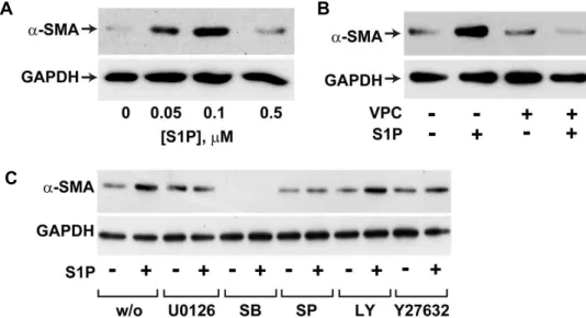
S1P induces α-SMA expression in BMSCs
S1P has been reported to induce differentiation of mouse
BMSCs to α-SMA-positive myofibroblasts in vivo, which
A B C
D E F
Fig. 4. Signaling pathways involved in S1P-induced migration of BMSCs. (A) BMSCs were pretreated with vehicles, 10 μM LY294002, 10 μM Y27632, 10 μM U0126, 10 μM SP600125 or 10 μM SB202190 for 15 min, and then exposed to serum-free medium containing 0.1 μM S1P or vehicles in the absence or presence of pharmacological inhibitors for 12 hr. The number of migrated cells was counted and data represent means±S.D. (n=4). (B) BMSCs were stimulated with 1 μM of S1P for the indicated time periods. (C) BMSCs, which were preincubated in the absence or presence of 10 μM of SB202190 for 15 min, were stimulated with 1 μM of S1P or vehicles for 5 min. (D) BMSCs were pretreated with serum-free medium containing 10 μM of VPC23019 or vehicles for 15 min and then treated with 1 μM S1P or vehicles for 5 min. Phosphorylation and expression levels of p38 MAPK were analyzed by Western blotting with phospho-p38 and p38 MAPK antibodies, respectively.
(E) BMSCs were infected with adenoviruses bearing DN-p38 MAPK or LacZ at MOI of 100 pfu for 48 h and then exposed to 1 μM S1P or vehicles for 5 min. Expression levels of p38 MAPK were determined by Western blotting. To ensure equal loading of proteins, the expression levels of GAPDH were determined. (F) Adenovirus-infected cells were exposed to serum-free media containing 1 μM S1P or vehicles for 12 hr. The number of migrated cells was counted and data represent means±S.D. (n=4). * indicates
p
<0.01 by two-way ANOVA and Scheffe’spost hoc
test.play a key role in tissue injury [24]. In order to explore the question of whether S1P treatment can directly induce differentiation of BMSCs to α-SMA-positive cells, we exam- ined the effect of S1P on the expression level of α-SMA in human BMSCs. As shown in Fig. 5A, S1P treatment in- creased the expression levels of α-SMA at 0.1 μM concen- tration, whereas the concentrations of S1P higher than 0.1 μ M inhibited α-SMA expression. S1P-induced α-SMA ex- pression was abrogated by VPC23019, suggesting that S1P induced expression of α-SMA in human BMSCs through an S1P
1-dependent pathway (Fig. 5B). To elucidate the sig- naling pathways involved in S1P-induced α-SMA ex- pression, we examined the effects of pharmacological in- hibitors in S1P-induced α-SMA expression. As shown in Fig.
5C, S1P-induced α-SMA expression was largely attenuated by pretreatment of cells with U0126, SP600125, SB202190,
or Y27632, suggesting involvement of ERK, JNK, p38 MAPKs, and Rho kinase in S1P-stimulated α-SMA ex- pression, whereas LY294002 had no significant impact on S1P-induced α-SMA expression, indicating that PI3K is not involved in the S1P induced α-SMA expression.
Discussion
S1P is released from activated platelets in inflamed or
injured tissues and regulates a variety of cellular responses,
including cell growth, survival, differentiation, and motility
[14,35,37,39]. S1P has been shown to regulate diverse cel-
lular responses by binding to its specific G protein-coupled
receptors [35,37,39]. In the present study, we demonstrated
that S1P induced migration of human BMSCs through an
S1P
1-dependent mechanism. Accumulating evidence con-
A B
C
Fig. 5. S1P-induced α-SMA expression in human BMSCs. (A) BMSCs were treated with the indicated concentrations of S1P for 4 days. (B) BMSCs were treated with 0.1 μM S1P or vehicles in the absence or presence of 10 μM of VPC23019 for 4 days. (C) BMSCs were pretreated with vehicles, 10 μM LY294002, 10 μM Y27632, 10 μM U0126, 10 μM SP600125, or 10 μM SB202190 for 15 min, and then exposed to serum-free medium containing 0.1 μM S1P or vehicles in the absence or presence of pharmacological inhibitors for 4 days. Expression levels of α-SMA and GAPDH were determined by Western blotting.
sistently suggests a pivotal role of S1P
1in S1P-induced mi- gration of various cell types [35,37,39]. Furthermore, S1P stimulated migration of mouse BMSCs through an S1P
1-mediated mechanism [1,27]. These results support the notion that S1P
1is involved in S1P-induced migration of human BMSCs.
In the present study, we demonstrated that S1P induced expression of α-SMA in human BMSCs through an S1P
1-dependent pathway. S1P
1knockout mice exhibited em- bryonic hemorrhage, leading to intrauterine death owing to a defective migration of vascular smooth muscle cells/pericytes and incomplete vascular maturation [25]. S1P has been reported to induce expression of smooth muscle markers in cultured smooth muscle cells, suggesting a piv- otal role of smooth muscle differentiation and vascular de- velopment [26]. α-SMA is expressed in not only contractile smooth muscle cells (SMCs) but also in myofibroblasts [9,29]. Myofibroblasts arise as a consequence of tissue injury, and they exhibit a contractile force that is required for wound closure [9,41]. S1P mediated homing of BMSCs in a mouse liver injury model and infiltrated BMSCs have re- cently been reported to contribute to formation of α -SMA-positive myofibroblasts within injured tissues [24].
Furthermore, S1P treatment induced expression of α-SMA in adipose tissue-derived MSCs [28]. These results suggest
that S1P plays a key role in differentiation of BMSCs to smooth muscle cells or myofibroblasts, which contribute to vascular development and regeneration of injured tissues.
Coupling of S1P
1via G
ifor activation of diverse signaling pathways, including Ras-MAP kinase, phosphoinosi- tide-3-kinase and the phospholipase C pathway, as well as coupling of S1P
2and S1P
3to multiple G proteins, i.e. G
q, G
12/13and G
ifor stimulation of the phospholipase C path- way and Rho pathway, as well as the G
i-dependent path- ways, have been reported [11,14,37]. We demonstrated here that S1P-induced migration was prevented by pretreatment of cells with PTX, suggesting involvement of G
iin S1P-in- duced cell migration. An increasing body of evidence sug- gests involvement of p38 MAPK in migration of diverse cell types, including smooth muscle cells, endothelial cells, and epithelial cells [12]. However, the role of p38 MAPK in S1P-induced cell migration has not been clearly understood. We demonstrated that pharmacological in- hibition of p38 MAPK or adenoviral expression of a domi- nant negative mutant of p38 MAPK abrogated S1P-stimu- lated cell migration. In support of the present study, the p38 MAPK inhibitor SB203580 completely abrogated the S1P-induced migration of OVCAR3 ovarian cancer cells [30].
Therefore, these results suggest that p38 MAPK plays a key
role in S1P-induced cell migration. In addition, we demon-
strated here that S1P-induced migration of BMSCs was com- pletely abrogated by either the MEK-ERK inhibitor U0126, JNK inhibitor, or the PI3K inhibitor LY294002, whereas the Rho kinase inhibitor Y27632 had only a marginal impact on S1P-induced cell migration. In addition, we showed that S1P induced α-SMA expression in BMSCs through mecha- nisms involving p38 MAPK, ERK, and JNK. In contrast to S1P-induced migration, S1P-induced α-SMA expression was abrogated by Y27632 but not by LY294002.These results led us to suggest that S1P-induced migration and differentiation of BMSCs to smooth muscle-like cells are differentially mediated by multiple signaling pathways involving p38 MAPK, JNK, ERK, Rho kinase and phosphoinosi- tide-3-kinase.
Acknowledgement
This work was supported for two years by a Pusan National University Research Grant.
References
1. Annabi, B., S. Thibeault, Y. T. Lee, N. Bousquet-Gagnon, N. Eliopoulos, S. Barrette, J. Galipeau, and R. Beliveau.
2003. Matrix metalloproteinase regulation of sphingo- sine-1-phosphate-induced angiogenic properties of bone marrow stromal cells.
Exp. Hematol.
31, 640-649.2. Barry, F. P. and J. M. Murphy. 2004. Mesenchymal stem cells: clinical applications and biological characterization.
Int. J. Biochem. Cell Biol.
36, 568-584.3. Chamberlain, G., J. Fox, B. Ashton, and J. Middleton. 2007.
Concise review: mesenchymal stem cells: their phenotype, differentiation capacity, immunological features, and poten- tial for homing.
Stem Cells
25, 2739-2749.4. Chavakis, E., C. Urbich, and S. Dimmeler. 2008. Homing and engraftment of progenitor cells: A prerequisite for cell therapy.
J. Mol. Cell Cardiol.
45, 514-522.5. Dwyer, R. M., S. M. Potter-Beirne, K. A. Harrington, A.
J. Lowery, E. Hennessy, J. M. Murphy, F. P. Barry, T.
O'Brien, and M. J. Kerin. 2007. Monocyte chemotactic pro- tein-1 secreted by primary breast tumors stimulates migra- tion of mesenchymal stem cells.
Clin. Cancer Res.
13, 5020-5027.6. Fiedler, J., G. Roderer, K. P. Gunther, and R. E. Brenner.
2002. BMP-2, BMP-4, and PDGF-bb stimulate chemotactic migration of primary human mesenchymal progenitor cells.
J. Cell Biochem.
87, 305-312.7. Forte, G., M. Minieri, P. Cossa, D. Antenucci, M. Sala, V.
Gnocchi, R. Fiaccavento, F. Carotenuto, P. De Vito, P. M.
Baldini, M. Prat, and P. Di Nardo. 2006. Hepatocyte growth factor effects on mesenchymal stem cells: proliferation, mi-
gration, and differentiation.
Stem Cells
24, 23-33.8. Fox, J. M., G. Chamberlain, B. A. Ashton, and J. Middleton.
2007. Recent advances into the understanding of mesen- chymal stem cell trafficking.
Br. J. Haematol.
137, 491-502.9. Gabbiani, G. 2003. The myofibroblast in wound healing and fibrocontractive diseases.
J. Pathol.
200, 500-503.10. Goetzl, E. J. and S. An. 1998. Diversity of cellular receptors and functions for the lysophospholipid growth factors lyso- phosphatidic acid and sphingosine 1-phosphate.
FASEB J.
12, 1589-1598.
11. Hla, T. 2004. Physiological and pathological actions of sphingosine 1-phosphate.
Semin. Cell Dev. Biol.
15, 513-520.12. Huang, C., K. Jacobson, and M. D. Schaller. 2004. MAP kinases and cell migration.
J. Cell Sci.
117, 4619-4628.13. Huang, S., Y. Jiang, Z. Li, E. Nishida, P. Mathias, S. Lin, R. J. Ulevitch, G. R. Nemerow, and J. Han. 1997. Apoptosis signaling pathway in T cells is composed of ICE/Ced-3 family proteases and MAP kinase kinase 6b.
Immunity
6, 739-749.14. Ishii, I., N. Fukushima, X. Ye, and J. Chun. 2004.
Lysophospholipid receptors: signaling and biology.
Annu.
Rev. Biochem.
73, 321-354.15. Jaganathan, B. G., B. Ruester, L. Dressel, S. Stein, M. Grez, E. Seifried, and R. Henschler. 2007. Rho inhibition induces migration of mesenchymal stromal cells.
Stem Cells
25, 1966-1974.16. Ji, J. F., B. P. He, S. T. Dheen, and S. S. Tay. 2004.
Interactions of chemokines and chemokine receptors medi- ate the migration of mesenchymal stem cells to the im- paired site in the brain after hypoglossal nerve injury.
Stem Cells
22, 415-427.17. Kang, Y. J., E. S. Jeon, H. Y. Song, J. S. Woo, J. S. Jung, Y. K. Kim, and J. H. Kim. 2005. Role of c-Jun N-terminal kinase in the PDGF-induced proliferation and migration of human adipose tissue-derived mesenchymal stem cells.
J. Cell Biochem.
95, 1135-1145.18. Kohno, T., H. Matsuyuki, Y. Inagaki, and Y. Igarashi. 2003.
Sphingosine 1-phosphate promotes cell migration through the activation of Cdc42 in Edg-6/S1P4-expressing cells.
Genes Cells
8, 685-697.19. Law, R. E., W. P. Meehan, X. P. Xi, K. Graf, D. A. Wuthrich, W. Coats, D. Faxon, and W. A. Hsueh. 1996. Troglitazone inhibits vascular smooth muscle cell growth and intimal hyperplasia.
J. Clin. Invest.
98, 1897-1905.20. Lee, M. J., E. S. Jeon, J. S. Lee, M. Cho, D. S. Suh, C. L.
Chang, and J. H. Kim. 2008. Lysophosphatidic acid in ma- lignant ascites stimulates migration of human mesen- chymal stem cells.
J. Cell Biochem.
104, 499-510.21. Lee, R. H., S. C. Hsu, J. Munoz, J. S. Jung, N. R. Lee, R.
Pochampally, and D. J. Prockop. 2006. A subset of human rapidly self-renewing marrow stromal cells preferentially engraft in mice.
Blood
107, 2153-2161.22. Lee, R. H., B. Kim, I. Choi, H. Kim, H. S. Choi, K. Suh, Y. C. Bae, and J. S. Jung. 2004. Characterization and ex- pression analysis of mesenchymal stem cells from human bone marrow and adipose tissue.
Cell Physiol. Biochem.
14,311-324.
23. Lepley, D., J. H. Paik, T. Hla, and F. Ferrer. 2005. The G protein-coupled receptor S1P2 regulates Rho/Rho kinase pathway to inhibit tumor cell migration.
Cancer Res.
65, 3788-3795.24. Li, C., Y. Kong, H. Wang, S. Wang, H. Yu, X. Liu, L. Yang, X. Jiang, L. Li, and L. Li. 2009. Homing of bone marrow mesenchymal stem cells mediated by sphingosine 1-phos- phate contributes to liver fibrosis.
J. Hepatol.
50, 1174-1183.25. Liu, Y., R. Wada, T. Yamashita, Y. Mi, C. X. Deng, J. P.
Hobson, H. M. Rosenfeldt, V. E. Nava, S. S. Chae, M. J.
Lee, C. H. Liu, T. Hla, S. Spiegel, and R. L. Proia. 2000.
Edg-1, the G protein-coupled receptor for sphingo- sine-1-phosphate, is essential for vascular maturation.
J.
Clin. Invest.
106, 951-961.26. Lockman, K., J. S. Hinson, M. D. Medlin, D. Morris, J. M.
Taylor, and C. P. Mack. 2004. Sphingosine 1-phosphate stimulates smooth muscle cell differentiation and pro- liferation by activating separate serum response factor co-factors.
J. Biol. Chem.
279, 42422-42430.27. Meriane, M., S. Duhamel, L. Lejeune, J. Galipeau, and B.
Annabi. 2006. Cooperation of matrix metalloproteinases with the RhoA/Rho kinase and mitogen-activated protein kinase kinase-1/extracellular signal-regulated kinase sig- naling pathways is required for the sphingosine-1-phos- phate-induced mobilization of marrow-derived stromal cells.
Stem Cells
24, 2557-2565.28. Nincheri, P., P. Luciani, R. Squecco, C. Donati, C.
Bernacchioni, L. Borgognoni, G. Luciani, S. Benvenuti, F.
Francini, and P. Bruni. 2009. Sphingosine 1-phosphate in- duces differentiation of adipose tissue-derived mesen- chymal stem cells towards smooth muscle cells.
Cell Mol.
Life Sci.
66, 1741-1754.29. Owens, G. K., M. S. Kumar, and B. R. Wamhoff. 2004.
Molecular regulation of vascular smooth muscle cell differ- entiation in development and disease.
Physiol Rev.
84, 767-801.30. Park, K. S., M. K. Kim, H. Y. Lee, S. D. Kim, S. Y. Lee, J. M. Kim, S. H. Ryu, and Y. S. Bae. 2007. S1P stimulates chemotactic migration and invasion in OVCAR3 ovarian cancer cells.
Biochem. Biophys. Res. Commun.
356, 239-244.31. Pittenger, M. F., A. M. Mackay, S. C. Beck, R. K. Jaiswal, R. Douglas, J. D. Mosca, M. A. Moorman, D. W. Simonetti, S. Craig, and D. R. Marshak. 1999. Multilineage potential of adult human mesenchymal stem cells.
Science
284, 143-147.32. Ponte, A. L., E. Marais, N. Gallay, A. Langonne, B.
Delorme, O. Herault, P. Charbord, and J. Domenech. 2007.
The
in vitro
migration capacity of human bone marrow mesenchymal stem cells: comparison of chemokine and growth factor chemotactic activities.Stem Cells
25,1737-1745.
33. Ponte, A. L., E. Marais, N. Gallay, A. Langonne, B.
Delorme, O. Herault, P. Charbord, and J. Domenech. 2007.
The
in vitro
migration capacity of human bone marrow mesenchymal stem cells: comparison of chemokine and growth factor chemotactic activities.Stem Cells
25, 1737-1745.34. Prockop, D. J. 1997. Marrow stromal cells as stem cells for nonhematopoietic tissues.
Science
276, 71-74.35. Sanchez, T. and T. Hla. 2004. Structural and functional characteristics of S1P receptors.
J. Cell Biochem.
92, 913-922.36. Short, B., N. Brouard, T. Occhiodoro-Scott, A.
Ramakrishnan, and P. J. Simmons. 2003. Mesenchymal stem cells.
Arch. Med. Res.
34, 565-571.37. Spiegel, S. and S. Milstien. 2003. Sphingosine-1-phosphate:
an enigmatic signalling lipid.
Nat. Rev. Mol. Cell Biol.
4, 397-407.38. Sugimoto, N., N. Takuwa, H. Okamoto, S. Sakurada, and Y. Takuwa. 2003. Inhibitory and stimulatory regulation of Rac and cell motility by the G12/13-Rho and Gi pathways integrated downstream of a single G protein-coupled sphingosine-1-phosphate receptor isoform.
Mol. Cell Biol.
23, 1534-1545.
39. Taha, T. A., K. M. Argraves, and L. M. Obeid. 2004.
Sphingosine-1-phosphate receptors: receptor specificity ver- sus functional redundancy.
Biochim. Biophys. Acta
1682, 48-55.40. Tamama, K., V. H. Fan, L. G. Griffith, H. C. Blair, and A. Wells. 2006. Epidermal growth factor as a candidate for ex vivo expansion of bone marrow-derived mesenchymal stem cells.
Stem Cells
24, 686-695.41. Tomasek, J. J., G. Gabbiani, B. Hinz, C. Chaponnier, and R. A. Brown. 2002. Myofibroblasts and mechano-regulation of connective tissue remodelling.
Nat. Rev. Mol. Cell Biol.
3, 349-363.
42. Wang, L., Y. Li, X. Chen, J. Chen, S. C. Gautam, Y. Xu, and M. Chopp. 2002. MCP-1, MIP-1, IL-8 and ischemic cere- bral tissue enhance human bone marrow stromal cell mi- gration in interface culture.
Hematology
7, 113-117.43. Wymann, M. P. and R. Schneiter. 2008. Lipid signalling in disease.
Nat. Rev. Mol. Cell Biol.
9, 162-176.44. Yamaguchi, H., J. Kitayama, N. Takuwa, K. Arikawa, I.
Inoki, K. Takehara, H. Nagawa, and Y. Takuwa. 2003.
Sphingosine-1-phosphate receptor subtype-specific positive and negative regulation of Rac and haematogenous meta- stasis of melanoma cells.
Biochem. J.
374, 715-722.45. Yatomi, Y., Y. Igarashi, L. Yang, N. Hisano, R. Qi, N.
Asazuma, K. Satoh, Y. Ozaki, and S. Kume. 1997.
Sphingosine 1-phosphate, a bioactive sphingolipid abun- dantly stored in platelets, is a normal constituent of human plasma and serum.
![Fig. 2. S1P induces elevation of [Ca 2+ ] i in BMSCs. Serum-starved BMSCs were loaded with 5 μM Fluo-4-AM for 40 min at 37 o C in the absence or presence of 10 μM of VPC23019 and then treated with serum-free medium containing 0.1 μM S1P in the absence or p](https://thumb-ap.123doks.com/thumbv2/123dokinfo/5022950.551080/5.892.160.754.846.1044/induces-elevation-starved-absence-presence-treated-containing-absence.webp)