ABSTRACT
Purpose: A relatively low response to chemotherapy has been reported for hormone receptor (HR)-positive breast cancer. In this study, we investigated the role of tryptophanyl-transfer RNA synthetase (WARS) in the chemotherapeutic response of HR-positive breast cancer.
Methods: Pre-chemotherapeutic needle biopsy samples of 45 HR-positive breast cancer patients undergoing the same chemotherapeutic regimen were subjected to immunohistochemistry. To investigate the biological functions of WARS in HR-positive breast cancer, we conducted cell viability assay, flow cytometry analysis, caspase activity assay, Quantitative real-time polymerase chain reaction, and western blotting using WARS gene-modulated HR-positive breast cancer cells (T47D, ZR-75-1, and MCF7).
Results: WARS overexpression in HR-positive breast cancer patients showed a significant correlation with favorable chemotherapy response. Downregulation of WARS increased cell viability following docetaxel treatment in tumor cell lines. On the other hand, WARS overexpression sensitized the therapeutic response to docetaxel. Additionally, downregulation of WARS caused a decrease in the number of apoptotic cell populations by docetaxel. Poly (ADP-ribose) polymerase cleavage and caspase 3/7 activity were increased in docetaxel-treated tumor cells with WARS overexpression.
Conclusion: Our results suggest that WARS might be a potential predictor for chemotherapy response in patients with HR-positive breast cancer as well as a novel molecular target to improve chemosensitivity.
Keywords: Apoptosis; Breast neoplasms; Drug resistance; Neoadjuvant therapy;
WARS1 protein, human
Original Article
Received: Sep 16, 2020 Accepted: Dec 6, 2020 Correspondence to Han Suk Ryu
Department of Pathology and Center for Medical Innovation, Biomedical Research Institute, Seoul National University Hospital, Seoul National University College of Medicine, 101 Daehak-ro, Jongno-gu, Seoul 03080, Korea.
E-mail: [email protected]
© 2020 Korean Breast Cancer Society This is an Open Access article distributed under the terms of the Creative Commons Attribution Non-Commercial License (https://
creativecommons.org/licenses/by-nc/4.0/) which permits unrestricted non-commercial use, distribution, and reproduction in any medium, provided the original work is properly cited.
ORCID iDs Kyung-Min Lee
https://orcid.org/0000-0002-1359-7382 Eun Hye Hwang
https://orcid.org/0000-0001-5414-821X Seong Eun Kang
https://orcid.org/0000-0003-4119-7607 Cheng Hyun Lee
https://orcid.org/0000-0003-0714-8473 Hyebin Lee
https://orcid.org/0000-0001-8442-6479 Hyeon Jeong Oh
https://orcid.org/0000-0002-9998-3988 Kwangsoo Kim
https://orcid.org/0000-0002-4586-5062 Jiwon Koh
https://orcid.org/0000-0002-7687-6477
Kyung-Min Lee 1, Eun Hye Hwang 2, Seong Eun Kang 2, Cheng Hyun Lee 2, Hyebin Lee 3, Hyeon Jeong Oh 4, Kwangsoo Kim 5, Jiwon Koh 2,
Han Suk Ryu 1,2
1 Center for Medical Innovation, Biomedical Research Institute, Seoul National University Hospital, Seoul, Korea
2 Department of Pathology, Seoul National University Hospital, Seoul National University College of Medicine, Seoul, Korea
3 Department of Radiation Oncology, Kangbuk Samsung Hospital, Sungkyunkwan University School of Medicine, Korea
4 Department of Pathology, Seoul National University Bundang Hospital, 82 Gumi-ro 173, Bundang-gu, Seongnam, Korea
5 Transdisciplinary Department of Medicine & Advanced Technology, Seoul National University Hospital, Seoul, Korea
Tryptophanyl-tRNA Synthetase
Sensitizes Hormone Receptor-Positive Breast Cancer to Docetaxel-Based
Chemotherapy
Han Suk Ryu
https://orcid.org/0000-0003-2508-9796 Funding
This research was supported by the National Research Foundation of Korea (NRF), funded by the Ministry of Science, ICT &
Future Planning (2018R1A1A1A05077484, 2019R1C1C1006465), the Basic Science Research Program through the Seoul National University Hospital Research Fund (26-2018- 0010), and a grant from the Korea Health Technology R&D Project through the Korea Health Industry Development Institute (KHIDI), funded by the Ministry of Health &
Welfare, Republic of Korea (grant number:
HI17C0048).
Conflict of Interest
The authors declare that they have no competing interests.
Author Contributions
Conceptualization: Ryu HS; Data curation:
Lee KM, Hwang EH, Kang SE, Lee CH, Ryu HS;
Formal analysis: Lee KM, Hwang EH, Kang SE, Lee CH, Kim K, Ryu HS; Funding acquisition:
Lee KM, Ryu HS; Investigation: Lee KM, Hwang EH, Kang SE, Lee CH, Ryu HS; Methodology:
Lee KM, Hwang EH, Kang SE, Lee CH, Oh HJ, Kim K; Resources: Lee H, Oh HJ, Koh J, Ryu HS; Validation: Koh J, Ryu HS; Visualization:
Lee KM; Writing - original draft: Lee KM;
Writing - review & editing: Lee KM, Ryu HS.
INTRODUCTION
Although approximately 70% of breast cancer patients currently receive standard chemotherapeutic regimens, the pathologic complete response (pCR) rate is still low [1]. Among the four intrinsic subtypes of breast cancer, the pCR rate for neoadjuvant chemotherapy has been reported to be the lowest in hormone receptor (HR)-positive breast cancer, despite the better overall prognosis [2,3]. Therefore, the need for a new therapeutic strategy that promotes chemotherapy responses is required to optimize treatment in HR- positive breast cancer.
Recently, transfer RNAs (tRNAs) have been proposed as biomarkers to overcome clinical chemoresistance [4,5]. The role of tRNA is to specify the sequence of the genetic code with the corresponding amino acid, and covalent attachment of the specific amino acid to the 3′ end of tRNA is catalyzed by an enzyme called aminoacyl-tRNA synthetase (ARS) [6,7].
Among these enzymes, tryptophanyl-tRNA synthetase (WARS) has recently been reported as a biomarker for unnecessary adjuvant chemotherapy after surgery for gastric cancer [8].
WARS exhibits anti-inflammatory properties through diverse mechanisms such as alternative splicing and proteolytic cleavage [9]. It also plays a central regulatory role in interferon (IFN)- γ-induced anti-angiogenesis and cell death [10].
Here, we evaluated the relationship between WARS expression and chemotherapy response in HR-positive breast cancer patients. In addition, we confirmed the mechanism by which WARS sensitized docetaxel-based chemotherapy through programmed cell death. Our findings suggest that WARS overexpression may be used as a potential biomarker for the prediction of pCR to chemotherapy, as well as a novel target to overcome resistance to docetaxel in HR-positive breast cancer.
METHODS
Patient and clinical tissue sample selection
Pre-chemotherapeutic needle biopsy samples from 45 patients with HR-positive invasive ductal carcinoma with available post-chemotherapeutic surgical specimens for microscopic assessment of therapeutic effectiveness were enrolled under the approval of the Institutional Review Board (IRB) at Seoul National University Hospital (IRB No. 1809-120-975). The requirement for informed consent from the patients was waived by the ethics committee.
The inclusion criteria for this study were female patients who received chemotherapeutic medication of adriamycin, cyclophosphamide, and docetaxel as preoperative therapy.
Histologically confirmed T2 tumors with positive axillary nodes (N1) were included in the study. Immunohistochemistry (IHC) for estrogen receptor (ER), progesterone receptor (PR), and human epidermal growth factor receptor 2 (HER2) was routinely performed on resected specimens according to the guidelines of the American Society of Clinical Oncology/College of American Pathologists (ASCO/CAP) [11]. All cases were enrolled and tissue samples were divided into 2 response groups, including 31 patients with non-complete remission (nCR) and 14 with pathologic complete remission (pCR), based on sequential surgical samples where the pathologists had judged the presence of residual tumor cells. The presence of residual tumor cells was evaluated by examination of hematoxylin and eosin-stained histological sections by experienced pathologists (HJO, JK, and HSR).
IHC
IHC staining was performed on formalin-fixed, paraffin-embedded sections (FFPE) of 3 μm thickness. Staining was performed on the Ventana Medical Systems BenchMark XT automated IHC platform using the ultraView Universal DAB Detection Kit (Ventana Medical Systems Inc., Tucson, USA), standard antigen retrieval, antibody incubation (anti- WARS, 1:300, PA5-29102, Thermo Fisher Scientific, Waltham, USA), washing procedure, counterstaining with hematoxylin and bluing reagent. The immunohistochemistry results were analyzed by employing a semi-quantitative approach using an ‘H-score’ [11] in a blind and independent manner by 2 pathologists (HJO and HSR).
Public data
To evaluate the clinical significance of WARS in predicting the chemotherapeutic response, we also employed a publicly available dataset (GSE25066) [12], which is a microarray-based detailed clinical dataset of HR-positive breast cancer patients who received anthracycline/
taxane-based neoadjuvant chemotherapy. The disease-free survival (DFS) and overall survival (OS) of breast cancer patients in the GEO datasets were analyzed using Cancer Target Gene Screening (CTGS; http://ctgs.biohackers.net), a recently developed web application tool [13].
Cell culture and chemicals
T47D, MCF7, and ZR-75-1 cell lines were obtained from the Korea Cell Line Bank (KCLB, Seoul, Korea). The T47D and ZR-75-1 cells were cultured in RPMI (Gibco, Carlsbad, USA) containing 10% fetal bovine serum (FBS; Invitrogen, Carlsbad, USA) and 1% penicillin/
streptomycin (PS; Gibco). MCF7 cells were cultured in DMEM (Gibco) containing 10% FBS and 1% PS. Cells were maintained at 37°C in a humidified atmosphere of 95% air and 5%
CO2 and periodically screened for mycoplasma contamination. All cell lines were confirmed by short tandem repeat (STR) DNA profiling test performed at the Korean Cell Line Bank (KCLB). Docetaxel, adriamycin, and cyclophosphamide were purchased from Sigma-Aldrich (St. Louis, USA).
Small interfering RNA (siRNA) transfection
siRNAs targeting WARS and an AccuTarget Negative Control siRNA were purchased from Bioneer (Daejeon, Korea). Cells were transfected using Lipofectamine RNAiMAX (Invitrogen) following the manufacturer's instructions. After incubation for 48 hours, WARS gene silencing was confirmed by assessing the mRNA expression levels.
Generation of lentiviral WARS overexpression cells
Lentiviral vectors encoding human WARS cDNA (Precision LentiORF, LOHS_100009313) and control vector encoding green fluorescent protein (GFP) were used for WARS overexpression, and were purchased from Thermo Fisher Scientific (Loughborough, UK).
For generation of the lentivirus, the lentiviral vectors were co-transfected with pdPAX2 and pMD2.G (Addgene, Watertown, USA) into HEK293T cells (ATCC) using Lipofectamine 2000 (Life Technologies, Carlsbad, USA). Supernatants were collected at 24 and 48 hours and filtered through 0.45 μm pore syringes. ZR-75-1 cells were infected with the viral supernatant along with 8 μg/mL polybrene, and stable cell lines were selected using blasticidin
(concentration range of 1–10 μg/mL).
Cell viability assay
Cytotoxicity effects of the chemotherapeutic drugs were determined (docetaxel, doxorubicin, and cyclophosphamide, respectively). Cells were plated in triplicate (3,000
cells/well) and incubated in medium containing 10% FBS. After 24 hours, the complete medium was replaced with the test medium containing the vehicle control and various doses of drugs for 48 hours at 37°C. Cell viability was assessed by measuring intracellular levels of ATP using the Cell Titer-Glo luminescent cell viability assay kit (Promega, Madison, USA).
Luminescence was measured using a luminometer (Glomax® Explore Multimode Microplate Reader; Promega).
Quantitative real-time polymerase chain reaction (PCR)
Total RNA was isolated from cells using the AccuPrep® Universal RNA Extraction Kit (Bioneer) according to the manufacturer's protocol. Genomic DNA was removed by DNase treatment using RNase-Free-DNase Set (Qiagen, Hilden, Germany). cDNA was synthesized using AccuPower® RocketScript Cycle RT PreMix (Bioneer). The sequences of the primers used for amplifying WARS were 5′-TTCACTGACAGCGACTGCAT-3′ and 5′-GGATCCTGGTCAATGGCACA-3′. Data analysis was based on the relative quantification method and ΔΔCT value was used to determine the relative fold change in expression. All data were normalized to the expression levels of glyceraldehyde 3-phosphate dehydrogenase (GAPDH), which was used as the reference gene.
Western blotting
Cells were collected and homogenized using RIPA lysis buffer (Thermo Fisher Scientific) on ice. Subsequently, the cell lysates were centrifuged at 4°C to separate the proteins. Proteins were quantified using the bicinchoninic acid protein assay kit (Thermo Fisher). Western blotting was performed using anti-poly (ADP-ribose) polymerase (PARP) antibody (Cell Signaling Technology, Danvers, USA). Anti-GAPDH (BD Biosciences, San Diego, USA) antibody was used as the loading control.
Flow cytometry
Cell apoptosis assay was performed using the Annexin V-FITC/propidium iodide (PI) apoptosis detection kit (BD Biosciences). Briefly, cells were collected and washed twice with PBS and then suspended in 300 µL of binding buffer. Annexin V solution (5 µL) was added to the cell suspension and incubated for 15 min in the dark at room temperature. Subsequently, 200 µL of binding buffer and 5 µL of PI were added, and the cell suspension was immediately analyzed on a BD FACSCaliber (BD Biosciences). All data were processed using the FlowJo™
10 software.
Caspase 3/7 activity assay
Caspase-3/7 activity was evaluated. Briefly, cells were seeded into white-walled 96-well
microplates and treated with the apoptosis inhibitor, z-VAD-FMK for 2 hours prior to docetaxel treatment. Following treatment, an equal volume of Caspase-Glo 3/7 reagent was added and the luminescence signal was detected using the GloMax-Multi Detection System (Promega).
Statistical analysis
Data are expressed as the mean ± standard deviation using two-tailed Student's t-test. Results with a p-value < 0.05 were considered as statistically significant. The half maximal inhibitory concentration (IC50) values were calculated using Hill's equation in the GraphPad Prism software 7 (GraphPad Inc., San Diego, USA). We performed immunohistochemistry analysis using the Mann-Whitney U test to obtain variables showing significant differences between the two groups. All p-values were 2-sided.
RESULTS
WARS overexpression predicts chemotherapy response in HR-positive breast cancer
To elucidate the role of WARS in chemotherapy response in HR-positive breast cancer, we first examined the clinical relevance of WARS expression in patients with primary breast cancer who received neoadjuvant chemotherapy (n = 45, Seoul National University Hospital [SNUH]
cohort). In our cohort, the high expression level of WARS protein was significantly associated with complete response to chemotherapy (IHC score: CR = 2.9 [n = 14], nCR = 1.2 [n = 31], p <
0.001) in HR-positive breast cancer (Figure 1A).
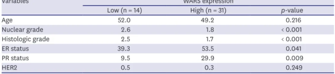
Correlations between clinicopathologic characteristics and WARS expression are shown in Figure 1B and Table 1. High WARS expression was significantly associated with ER (p = 0.041) and PR status (p = 0.009). On the other hand, low WARS expression was correlated with higher nuclear grade (p < 0.001) and histologic grade (p < 0.001), but not with the level of HER2 expression (Figure 1B and Table 1).
To evaluate whether WARS expression predicts survival, we explored a publicly available clinico-pathological dataset (GSE25066). In the group of patients who received
chemotherapy in the HR-positive breast cancer setting, 117 patients had tumors with a
‘chemotherapy-sensitive (Rx sensitive)’ parameter. Out of these, 43 patients 55.6% (n = 65)
0
DFS
C
40 80 100
30 0
60
20
90 60
WARS IHC HR-positive breast cancer
(SNUH, n = 45) HR-positive and chemosensitivity prediction
= Rx sensitive cases (GSE25066, n = 117)
0
DFS 40
80 100
30 0
60
20
90 60
HR-positive and taxane cases (GSE25066, n = 51)
WARS high (n = 65) WARS low (n = 52)
WARS high (n = 14) WARS low (n = 37) 0
IHC score
p < 0.001
p = 0.0885 (log-rank) p = 0.0667 (log-rank)
A
1 3 4
(n = 14CR ) 2
(n = 31nCR)
Time (mo) Time (mo)
0
Nuclear grade
WARs expression
p < 0.001 p < 0.001 p = 0.041 p = 0.009 p = 0.249
B
1 3 4
Low 2
High 0
Histologic grade
WARs expression 1
3 4
Low 2
High 0
ER status
WARs expression 20
60 100
Low 40
High 0
RR status
WARs expression 20
60 100
Low 40
High 0
HER2 status
WARs expression 0.5
1.5 2.5
Low
80 80 2.0
1.0
High
Figure 1. Association between WARS expression and chemotherapy response in HR-positive breast cancer. (A) Representative graphs of IHC score between nCR and CR groups in HR-positive breast cancer patients who received neoadjuvant chemotherapy (SNUH cohort, n = 45, left). Representative images of IHC staining using WARS antibody (×200). (B) Representative graphs show the clinicopathologic characteristics (nuclear grade, histologic grade, ER status, PR status, and HER2 status) in relation to WARS expression. The p-values were calculated using the 2-sided Student's t-test and Mann-Whitney U test (HER2 status). (C) Kaplan- Meier plot of DFS based on WARS expression in HR-positive breast cancer cohort (GSE25066) who received chemotherapy (n = 117, left) and taxane (n = 51, right).
WARS = tryptophanyl-transfer RNA synthetase; HR = hormone receptor; IHC = immunohistochemistry; CR = complete response; nCR = non-complete response;
SNUH = Seoul National University Hospital; DFS = disease-free survival.
showed a tendency to exhibit DFS rate (p = 0.088) (Figure 1C, left). Further, the correlation between WARS expression and taxane response was explored, which included 51 patients with centrally reviewed HR-positive patients. The results showed that WARS overexpression indicated a tendency of better DFS rate in HR-positive breast cancer patients who received taxane (WARS high, n = 14; WARS low, n = 37; p = 0.067) (Figure 1C, right). Collectively, our results suggest that WARS overexpression in HR-positive breast cancer is associated with favorable prognosis and improved chemotherapy response.
WARS overexpression sensitized docetaxel in HR-positive breast cancer cell lines
To understand the role of WARS in determining the sensitivity for chemotherapeutic agents, we first screened WARS expression levels in HR-positive breast cancer cell lines, including T47D, MCF7, and ZR-75-1. We observed that HR-positive breast cancer cells showed higher levels of WARS protein expression (upper bands of two bands in western blot data), compared to MCF10A immortalized normal breast cells (Figure 2A). The Cancer Cell Line Encyclopedia dataset also showed the same results, wherein overexpression of WARS was consistently observed in HR-positive cells compared to other subtypes of breast cancer cell lines (Figure 2B).
Since WARS overexpression was verified as a reliable biomarker for predicting good response to chemotherapy in our breast cancer patient cohort and the external public dataset (Figure 1A), we investigated whether suppression of WARS expression inhibited the cytotoxic efficacy of each chemotherapeutic agent, including docetaxel, adriamycin, and cyclophosphamide. Unexpectedly, we failed to detect significant cytotoxic effect of adriamycin or cyclophosphamide upon suppression of WARS expression (Supplementary Figure 1). On the other hand, we found that WARS downregulation dramatically increased cell viability against docetaxel in HR-positive breast cancer cell lines (Figure 2C and D). Upon analyzing the IC50 values for docetaxel treatment, WARS downregulation significantly increased the IC50 values from 2.2 to 4.0 nM and from 2.7 to 5.8 nM in T47D (p = 0.040) and ZR-75-1 cells (p = 0.034), respectively (Figure 2D). We also found that the IC50 values dramatically increased approximately 4-fold upon suppression of WARS expression in MCF7 (from 30.5 to 115.1 nM, p = 0.008), which is more resistant to docetaxel than T47D and ZR-75-1 cells.
Next, we established stable ZR-75-1 cells overexpressing WARS (Figure 2E, top). WARS overexpressing cells were sensitive to docetaxel (Figure 2E) and the IC50 value was also found to be significantly decreased from an average of 3.2 to 2.1 nM (Figure 2E, bottom, p = 0.047).
Collectively, these data indicated that WARS upregulation is associated with increased sensitivity to docetaxel in HR-positive breast cancer.
Table 1. Clinicopathologic characteristics in relation to WARS expression in HR-positive breast cancer patients
Variables WARS expression
Low (n = 14) High (n = 31) p-value
Age 52.0 49.2 0.216
Nuclear grade 2.6 1.8 < 0.001
Histologic grade 2.5 1.7 < 0.001
ER status 39.3 53.5 0.041
PR status 9.5 29.9 0.009
HER2 0.5 0.3 0.249
WARS = tryptophanyl-transfer RNA synthetase; HR = hormone receptor; ER = estrogen receptor; PR = progesterone receptor; HER2 = human epidermal growth factor receptor 2.
Suppression of WARS inhibits docetaxel-induced apoptosis in HR-positive breast cancer
To elucidate the mechanism underlying the enhanced HR-positive breast cancer cell death induced by WARS upregulation plus docetaxel, we simultaneously monitored phosphatidylserine exposure by Annexin-V/PI staining as a marker of apoptosis. Flow cytometric analysis revealed that WARS suppression caused a decrease in the percentage of apoptotic cell populations, including Annexin V+/PI− and Annexin V+/PI+ cells in the presence of docetaxel (Figure 3A). Viable cells (Annexin V/PI double negative) increased from 60% to 73% (p = 0.034) and apoptotic cell populations decreased from 38% to 22% (p = 0.001) in WARS-downregulated T47D cells compared to control cells upon docetaxel treatment (20 nM) for 48 hours. We obtained similar results in ZR-75-1 cells, in which the number of viable cells was significantly increased (p = 0.045), and apoptotic cells were reduced considerably A
T47D MCF7 MCF10A
ZR-75-1 WARS
β-actin
D T47D
0 IC50 value (nM)
p = 0.040 4
6
2 0
0 1 2 3
Cell viability (%)
Log [Docetaxel], nM 100
150
50
siControl siWARS
ZR-75-1 E
0 WARS mRNA (relative to GAPDH)
p = 0.042
1.0 2.0 1.5
0.5
pLOC WARS
siControl siWARS
CCLE DB
HR-positive cell lines ZR-75-1
WARS expression (RPKM) 0
B
20 40 50
T47D 30 10
MCF7 ZR-75-1
0 IC50 value (nM)
p = 0.034
4 8 6
2 0
0 1 2 3
Cell viability (%)
Log [Docetaxel], nM 100
150
50
siControl siWARS
siControl siWARS
0
0 1 2 3
Log [Docetaxel], nM 100
150
50
pLOCWARS
Cell viability (%)
0 WARS mRNA (relative to GAPDH)
p < 0.001
C
1.0 1.5
T47D 0.5
ZR-75-1 MCF7 p = 0.032 p = 0.023
siControl siWARS MCF7
0 IC50 value (nM)
p = 0.008 100
150
50 0
0 1 2 3
Cell viability (%)
Log [Docetaxel], nM 100
150
50
siControl siWARS
siControl siWARS
0 IC50 value (nM)
p = 0.047
2 4 3
1
pLOC WARS
Figure 2. Suppression of WARS enhanced resistance to docetaxel in HR-positive breast cancer cell lines. (A) Comparison of WARS protein levels in MCF10A immortalized normal cell line and HR-positive breast cancer cell lines (T47D, MCF7, and ZR-75-1) by western blotting. β-actin served as a loading control.
(B) Analysis of WARS mRNA expression levels (RPKM value) in HR-positive breast cancer cell lines in the Cancer Cell Line Encyclopedia dataset. (C) The representative graph confirms the inhibition of WARS mRNA expression by qPCR through siRNA experiments in T47D, ZR-75-1, and MCF7. (D) T47D, ZR-75-1, and MCF7 cells transfected with control siRNA or siWARS were treated with various concentrations of docetaxel. Cell viability was determined by CellTiter- Glo Luminescent Cell Viability assay (left). Representative graphs show the IC50 values (right). (E) The cytotoxic effect of docetaxel in ZR-75-1 cells stably overexpressing WARS. Overexpression of WARS mRNA as measured by qPCR (upper) in comparison to pLOC (vector control). Cell viability was assessed by CellTiter-Glo Luminescent Cell Viability assay. Representative graphs show the IC50 values (bottom). All data are representes as the mean ± standard error of mean (n = 3 biological replicates). The p-values were calculated by Student's t-test.
WARS = tryptophanyl-transfer RNA synthetase; HR = hormone receptor; qPCR = quantitative real-time polymerase chain reaction; si- = small interfering; IC50 = half maximal inhibitory concentration.
upon suppression of WARS expression compared to control-siRNA-treated cells (from 37%
to 25%, p = 0.031). The rate of apoptosis in MCF7 cells decreased from 23% to 18% upon treatment with docetaxel (100 nM) compared to control cells (Figure 3A, p = 0.041). There was no significant difference in the percentage of viable cells upon WARS depletion. Dosages of docetaxel that caused 30%–50% HR-positive breast cancer cell death were chosen for our experiments along with WARS depletion.
T47D
0 -
Apoptotic cell (%)
†
†
†
†
‡ *
30 40
10 20
10 20 - 10 20
siControl siWARS siControl
siWARS siControl siWARS siControl siWARS
A B
siControlsiWARSMCF7T47DZR-75-1
PI
0 10 20
0 50 100
Docetaxel (nM) Docetaxel (nM)
Docetaxel (nM)
siControlsiWARSsiControlsiWARS
Annexin V
ZR-75-1
0 -
Apoptotic cell (%)
*
*
*
*
†
†
30 40
10 20
10 20 - 10 20 Docetaxel
(nM)
MCF7
0 -
Apoptotic cell (%)
*
*
ns ns
* *
30
10 20
50 100 - 50 100 Docetaxel
(nM)
Docetaxel (nM) WARS c-PARP GAPDH
0 10 20 0 10 20
T47D siControl siWARS
Docetaxel (nM) WARS c-PARP GAPDH
0 10 20 0 10 20
ZR-75-1 siControl siWARS
Docetaxel (nM) WARS c-PARP GAPDH
0 50 100 0 50 100
MCF7 siControl siWARS
C T47D
0 -
Caspase 3/7 activity 1.5
0.5 1.0
+ - +
Docetaxel (nM)
p = 0.047 ns
ZR-75-1
0 -
Caspase 3/7 activity 1.5
0.5 1.0
+ - +
Docetaxel (nM)
p = 0.030 ns
MCF7
0 -
Caspase 3/7 activity 1.5
0.5 1.0
+ - +
Docetaxel (nM)
p = 0.038 ns
Figure 3. Suppression of WARS inhibits apoptosis induced by docetaxel in HR-positive breast cancer. (A) Flow cytometry images of WARS knockdown in HR- positive breast cancer cells treated with docetaxel (T47D and ZR-75-1, 10–20 nM; MCF7, 50–100 nM, respectively, left). Apoptosis was determined by Annexin V/
PI-staining. Representative graphs show the percentage (%) of apoptotic cells (Annexin V+/PI− and Annexin V+/PI+ distribution of cells, right). (B) Measurement of c-PARP and WARS expression by western blotting after docetaxel treatment in WARS knockdown cells. GAPDH served as a loading control. (C) Analysis of caspase 3/7 activity after docetaxel treatment (T47D and ZR-75-1, 20 nM; MCF7, 100 nM) in control siRNA- and WARS siRNA-treated cells by Caspase-Glo 3/7 reagent. All cells were pretreated with the pan caspase inhibitor z-VAD-FMK (10 μM) for 1 hour and then examined by Caspase-Glo assay. All data are represented as the mean ± standard error of mean (n = 3 biological replicates). The p-values were calculated by Student's t-test.
WARS = tryptophanyl-transfer RNA synthetase; HR = hormone receptor; PI = propidium iodide; PARP = poly (ADP-ribose) polymerase; GAPDH = glyceraldehyde 3-phosphate dehydrogenase; si- = small interfering; ns = not significant.
*p < 0.05, †*p < 0.01, ‡p < 0.001.
To further determine the rate of apoptosis, we analyzed cell lysates for cleaved PARP, a marker of apoptosis [14]. In agreement with the observed inhibition of cell death, docetaxel-induced PARP cleavage was decreased in WARS-depleted cells (Figure 3B). WARS knockdown was confirmed by western blotting (upper bands of two bands). It is well known that caspases play essential roles in apoptotic cell death. Therefore, we measured the activities of executioner caspases 3/7, which are known to exhibit increased activity in response to docetaxel [15].
In all tested HR-positive cells, the activity of caspases 3/7 significantly increased in WARS- overexpressing cells treated with docetaxel, whereas there was no difference in the WARS- downregulated cells (Figure 3C). Collectively, our results indicate that the novel combination of WARS upregulation and docetaxel enhances HR-positive breast cancer cell death via increased apoptotic signaling.
DISCUSSION
In this study, we identified a non-canonical function of WARS, one of the crucial proteins of the tRNA synthetase complex [6,16], in HR-positive breast cancer. We employed a stepwise approach to validate the predictive role of WARS in chemotherapy response and identified that WARS induced apoptosis, a key molecular mechanism, in docetaxel-based chemotherapy.
Although the role of WARS still remained to be evaluated in breast cancer, a recent proteomic approach examining 59 proteins suggested a potential predictive role of WARS in tumor recurrence and survival in triple-negative breast cancer [17]. In our study, for the first time, we verified the predictive role of WARS in chemotherapy in HR-positive breast cancer.
We confirmed that WARS overexpression predicts complete response to neoadjuvant chemotherapy in HR-positive breast cancer patients. This finding is concordant with a previous study which reported WARS as a novel predictive biomarker for unnecessary adjuvant chemotherapy for gastric cancer [8]. In addition, WARS was associated with low nuclear and histological grades, which are indicators of good prognosis. Although no significant correlation was found between WARS expression and other clinicopathologic parameters such as HER2 status, significant correlations were found with respect to ER and PR status.
Our immunohistochemistry findings demonstrating the strong association between WARS overexpression and favorable response to neoadjuvant chemotherapy prompted us to perform further molecular functional tests to investigate the mechanism by which WARS controls the cytotoxic effects of chemotherapy in HR-positive breast cancer. Our results showed that downregulation of WARS correlated strongly with resistance to docetaxel, and overexpression of WARS increased cell susceptibility to docetaxel in HR-positive breast cancer cells.
WARS has been proposed to be anti-angiogenic by regulating cell growth through phosphorylation of p53 by IFN-γ signaling [18], which eventually drives the cells to IFN- γ-induced cell death [9,18,19]. Therefore, in this study, we first hypothesized that WARS might sensitize HR-positive breast cancer cells to docetaxel through the apoptotic signaling pathway. Our caspase activity assay showed that docetaxel treatment induced significant caspase 3/7 activities, the key molecules of intrinsic cell death stimulation [15,20], in tumor cells overexpressing WARS, compared to the control group. In addition to the modulation of caspase activity by WARS, proteolytic cleavage of PARP which is the final executor of programmed cell death [14,21] was confirmed in HR-positive breast cancer cell lines with WARS overexpression. In the setting of docetaxel treatment and downregulation of WARS,
cleavage of PARP was significantly reduced in tumor cells. Moreover, cells showed a tendency of suppressed caspase 3/7 activity. Taken together, we conclude that WARS might modulate apoptotic pathways, mainly by directly targeting PARP rather than via the caspase pathway, especially in HR-positive breast cancer after docetaxel treatment.
Apart from the canonical role of tRNA synthetase in protein synthesis, it plays non-classic biological roles, including functions beyond translation, as a transcriptional activator of oncogenes [22] and a possible regulator of apoptosis [7,23,24]. When mammalian cells are exposed to apoptotic conditions, tRNA synthetase such as tyrosyl-tRNA synthetase, is secreted and split into two fragments thereby stimulating the production of TNF and tissue factors that promote apoptosis [25]. Therefore, we suggest that WARS might be able to enhance the cytotoxic effect of docetaxel by inducing the apoptotic pathway, especially via PARP activation, the final apoptotic executor, in HR-positive breast cancer.
While further in vitro and in vivo validation studies are required, we anticipate that WARS may be clinically applicable as a novel, predictive biomarker for response to chemotherapy in HR- positive breast cancer.
SUPPLEMENTARY MATERIAL
Supplementary Figure 1
The cytotoxic effect of adriamycin and cyclophosphamide in WARS knockdown T47D and MCF7 cells. Cell viability was analyzed using the CellTiter-Glo Luminescent Cell Viability Assay Kit, and IC50 value calculations were made using Hill's equation in the GraphPad Prism software 7.
Data are represented as the mean ± standard error of mean (n = 3 biological replicates).
Click here to view
REFERENCES
1. Sparano JA, Gray RJ, Makower DF, Pritchard KI, Albain KS, Hayes DF, et al. Adjuvant chemotherapy guided by a 21-gene expression assay in breast cancer. N Engl J Med 2018;379:111-21.
PUBMED | CROSSREF
2. Rouzier R, Perou CM, Symmans WF, Ibrahim N, Cristofanilli M, Anderson K, et al. Breast cancer molecular subtypes respond differently to preoperative chemotherapy. Clin Cancer Res 2005;11:5678-85.
PUBMED | CROSSREF
3. von Minckwitz G, Untch M, Blohmer JU, Costa SD, Eidtmann H, Fasching PA, et al. Definition and impact of pathologic complete response on prognosis after neoadjuvant chemotherapy in various intrinsic breast cancer subtypes. J Clin Oncol 2012;30:1796-804.
PUBMED | CROSSREF
4. Jin L, Zhu C, Qin X. Expression profile of tRNA-derived fragments in pancreatic cancer. Oncol Lett 2019;18:3104-14.
PUBMED | CROSSREF
5. Yu M, Lu B, Zhang J, Ding J, Liu P, Lu Y. tRNA-derived RNA fragments in cancer: current status and future perspectives. J Hematol Oncol 2020;13:121.
PUBMED | CROSSREF
6. Kwon NH, Fox PL, Kim S. Aminoacyl-tRNA synthetases as therapeutic targets. Nat Rev Drug Discov 2019;18:629-50.
PUBMED | CROSSREF
7. Smirnova EV, Lakunina VA, Tarassov I, Krasheninnikov IA, Kamenski PA. Noncanonical functions of aminoacyl-tRNA synthetases. Biochemistry (Mosc) 2012;77:15-25.
PUBMED | CROSSREF
8. Cheong JH, Yang HK, Kim H, Kim WH, Kim YW, Kook MC, et al. Predictive test for chemotherapy response in resectable gastric cancer: a multi-cohort, retrospective analysis. Lancet Oncol 2018;19:629-38.
PUBMED | CROSSREF
9. Adam I, Dewi DL, Mooiweer J, Sadik A, Mohapatra SR, Berdel B, et al. Upregulation of tryptophanyl- tRNA synthethase adapts human cancer cells to nutritional stress caused by tryptophan degradation.
OncoImmunology 2018;7:e1486353.
PUBMED | CROSSREF
10. Jin M. Unique roles of tryptophanyl-tRNA synthetase in immune control and its therapeutic implications.
Exp Mol Med 2019;51:1-10.
PUBMED | CROSSREF
11. Allison KH, Hammond MEH, Dowsett M, McKernin SE, Carey LA, Fitzgibbons PL, et al. Estrogen and progesterone receptor testing in breast cancer: American Society of Clinical Oncology/College of American Pathologists guideline update. Arch Pathol Lab Med 2020;144:545-63.
PUBMED | CROSSREF
12. Hatzis C, Pusztai L, Valero V, Booser DJ, Esserman L, Lluch A, et al. A genomic predictor of response and survival following taxane-anthracycline chemotherapy for invasive breast cancer. JAMA 2011;305:1873-81.
PUBMED | CROSSREF
13. Kim HY, Choi HJ, Lee JY, Kong G. Cancer target gene screening: a web application for breast cancer target gene screening using multi-omics data analysis. Brief Bioinform 2020;21:663-75.
PUBMED | CROSSREF
14. Cotter TG. Apoptosis and cancer: the genesis of a research field. Nat Rev Cancer 2009;9:501-7.
PUBMED | CROSSREF
15. Brentnall M, Rodriguez-Menocal L, De Guevara RL, Cepero E, Boise LH. Caspase-9, caspase-3 and caspase-7 have distinct roles during intrinsic apoptosis. BMC Cell Biol 2013;14:32.
PUBMED | CROSSREF
16. Rajendran V, Kalita P, Shukla H, Kumar A, Tripathi T. Aminoacyl-tRNA synthetases: Structure, function, and drug discovery. Int J Biol Macromol 2018;111:400-14.
PUBMED | CROSSREF
17. Campone M, Valo I, Jézéquel P, Moreau M, Boissard A, Campion L, et al. Prediction of recurrence and survival for triple-negative breast cancer (TNBC) by a protein signature in tissue samples. Mol Cell Proteomics 2015;14:2936-46.
PUBMED | CROSSREF
18. Sajish M, Zhou Q, Kishi S, Valdez DM Jr, Kapoor M, Guo M, et al. Trp-tRNA synthetase bridges DNA-PKcs to PARP-1 to link IFN-γ and p53 signaling. Nat Chem Biol 2012;8:547-54.
PUBMED | CROSSREF
19. Miyanokoshi M, Yokosawa T, Wakasugi K. Tryptophanyl-tRNA synthetase mediates high-affinity tryptophan uptake into human cells. J Biol Chem 2018;293:8428-38.
PUBMED | CROSSREF
20. Li J, Yuan J. Caspases in apoptosis and beyond. Oncogene 2008;27:6194-206.
PUBMED | CROSSREF
21. Cristofani R, Montagnani Marelli M, Cicardi ME, Fontana F, Marzagalli M, Limonta P, et al. Dual role of autophagy on docetaxel-sensitivity in prostate cancer cells. Cell Death Dis 2018;9:889.
PUBMED | CROSSREF
22. Kim MJ, Park BJ, Kang YS, Kim HJ, Park JH, Kang JW, et al. Downregulation of FUSE-binding protein and c-myc by tRNA synthetase cofactor p38 is required for lung cell differentiation. Nat Genet 2003;34:330-6.
PUBMED | CROSSREF
23. Ko YG, Kim EY, Kim T, Park H, Park HS, Choi EJ, et al. Glutamine-dependent antiapoptotic interaction of human glutaminyl-tRNA synthetase with apoptosis signal-regulating kinase 1. J Biol Chem 2001;276:6030-6.
PUBMED | CROSSREF
24. Wakasugi K, Schimmel P. Two distinct cytokines released from a human aminoacyl-tRNA synthetase.
Science 1999;284:147-51.
PUBMED | CROSSREF
25. Park SG, Jung KH, Lee JS, Jo YJ, Motegi H, Kim S, et al. Precursor of pro-apoptotic cytokine modulates aminoacylation activity of tRNA synthetase. J Biol Chem 1999;274:16673-6.
PUBMED | CROSSREF