https://doi.org/10.4174/astr.2019.96.4.169 Annals of Surgical Treatment and Research
Nomogram for accurate prediction of breast and axillary pathologic response after neoadjuvant chemotherapy in node positive patients with breast cancer
Hee Jun Choi1, Jai Min Ryu2, Isaac Kim2, Seok Jin Nam2, Seok Won Kim2, Jonghan Yu2, Jeong Eon Lee2, Se Kyung Lee2
1Department of Surgery, Samsung Changwon Hospital, Sungkyunkwan University School of Medicine, Changwon, Korea
2Division of Breast Surgery, Department of Surgery, Samsung Medical Center, Sungkyunkwan University School of Medicine, Seoul, Korea
INTRODUCTION
The use of neoadjuvant chemotherapy (NAC) is increasingly used for patients with operable breast cancer to allow for the use of more minimal surgery [1,2]. To avoid axillary lymph
node dissection (ALND) or mastectomy, it is preferable to have a sentinel lymph node (SLN) surgical procedure or breast conserving surgery (BCS), which results in fewer instances of problems and lower morbidity [1,35].
It should be noted that although about 30% patients had
Received August 14, 2018, Revised November 19, 2018, Accepted December 11, 2018
Corresponding Author: Se Kyung Lee
Division of Breast and Endocrine Surgery, Department of Surgery, Samsung Medical Center, Sungkyunkwan University School of Medicine, 81 Irwon-ro, Gangnam-gu, Seoul 06351, Korea
Tel: +82-2-3410-0051, Fax: +82-2-3410-6982 E-mail: [email protected]
ORCID code: https://orcid.org/0000-0003-1630-1783
Copyright ⓒ 2019, the Korean Surgical Society
cc Annals of Surgical Treatment and Research is an Open Access Journal. All articles are distributed under the terms of the Creative Commons Attribution Non- Commercial License (http://creativecommons.org/licenses/by-nc/4.0/) which permits unrestricted non-commercial use, distribution, and reproduction in any medium, provided the original work is properly cited.
Purpose: Many patients with cytology proven node-positive breast cancer receive a neoadjuvant chemotherapy (NAC) treatment. We developed a nomogram to predict the breast and axillary pathologic complete responses (pCR) in patients with a cytologically proven axillary node positive breast cancer with NAC.
Methods: We selected 995 patients who were diagnosed with an invasive breast cancer and axillary lymph nodes metastasis, and who were treated with NAC followed by a curative surgery at the Samsung Medical Center between January 2007 and December 2014. The baseline patient and tumor characteristics, chemotherapy regimen, and tumor and nodal responses were thoroughly analyzed and reviewed. A nomogram was developed using a binary logistic regression model with a cross validation.
Results: Axillary pCR was achieved in 47.3% and breast pCR was achieved in 24.3% of the patients after NAC. In this case, the both pCR was associated with an initial clinical tumor stage, negative progesterone receptor status, positive human epidermal growth factor receptor 2 status, and clinical radiologic nodal responses. A nomogram was developed based on the clinical and statistically significant predictors. It had good discrimination performance (area under the curve [AUC], 0.868; 95% confidence interval, 0.84–0.89) and calibration fit as noted in that case. The cross validation had an average AUC 0.853 (0.837–0.869).
Conclusion: Our nomogram might help to predict breast and axillary pCRs after NAC in patients with an initially node- positive breast cancer. Minimal surgery might be acceptable in patients for whom the nomogram indicates a high probability of achieving pCRs.
[Ann Surg Treat Res 2019;96(4):169-176]
Key Words: Complete response, Neoadjuvant treatment, Nomograms
no residual disease in the axilla after NAC, ALND has been recommended for most patients with a biopsy proven node
positive breast cancer regardless of their response to NAC treatment [6,7]. In this case, some evidence has suggested that the nodal stage after NAC reflects the prognosis more accurately than the initial axillary status [8]. Therefore, the ALND may not be needed for patients with a complete response (CR). These patients are able to avoid postoperative morbidities such as lymphedema, arm pain, and reduced arm movement [9,10].
If we can predict which patients will develop axillary pathologic complete response (pCR), we can prevent ALND and if we can predict the breast pCR, we can prevent mastectomy.
Additionally, both axillary and breast pCRs after a course of NAC are associated with improved oncologic survival [11].
Therefore we investigated the factors that predicted both pCRs and established a nomogram calculating the probability of both pCRs in patients who received NAC.
METHODS
This study is a registered medical record review based on a prospectively collected database. We selected 995 patients who were diagnosed with an invasive breast cancer and axillary lymph nodes metastasis by ultrasound of axilla, and who were treated with NAC followed by curative surgery at Samsung Medical Center between the timeframe of January 2006 and December 2015. Patients were eligible if they met the following criteria: (1) diagnosed with an enlarged axillary lymph node (LN) by the use of a breast ultrasonography and by fine needle aspiration cytology upon an initial examination, (2) completed all cycles of the planneddosage NAC, and (3) who had undergone a radical excision of a primary tumor and SLNB or ALND. Likewise, patients with bilateral breast cancer, previous ipsilateral axillary surgery, inflammatory breast cancer, or distant metastasis were excluded from the study.
Most patients (95.1%) received anthracycline and/or taxane
based regimens. These regimens included anthracycline plus cyclophosphamide, followed by anthracyclinebased, taxane
based, or trastuzumab regimens. The adjuvant radiotherapy was performed with tangential fields in all patients following by a BCS.
For the most part, the clinical response to treatment was evaluated by a breast magnetic resonance imaging (MRI) or a breast ultrasonography. In case of discordance between ultrasonography and MRI, we used the results of the MRI.
Clinical CR of the breast was defined as a disappearance of all of the tumor deposits on a MRI or breast ultrasonography.
We used RECIST (response evaluation criteria in solid tumor) criteria for clinical response evaluation. The quantification of response by using the categories of CR, partial response (PR), stable disease, and progressive disease (PD) served as a gross
estimate of the chemosensitivity. And clinical nodal response was divided the categories to breast MRI or breast sonography into disappeared, decreased or no changed/increased.
A sentinel node biopsy was performed with technetium
99m sulfur colloid diluted in normal saline solution and/or vital blue dye (0.8% indigo carmine). The site and timing of the agent administration were at the physician’s discretion:
the radiolabeled colloid was injected 2 to 6 hours before the scheduled surgery, and/or 5 mL of 0.8% indigo carmine was injected periareolarly, and the breast was massaged for 5 minutes. For the sulfurcolloid injection, a handheld gamma detection probe was used to scan the axilla transcutaneously, and was used to identify the most radioactive area. All radioactive and/or blue LNs and palpable LNs were excised and submitted as SLNs.
We measured, froze, and serially sectioned the excised SLNs transversely into 16 or 24 slices. After pathological evaluation of the sections, we fixed the remaining tissue in 10% formalin, embedded it in paraffin blocks, and finally prepared the hematoxylin and eosin (H&E)stained sections. We designated metastatic foci of 0.2 mm to 2 mm as micrometastases, and metastatic clusters smaller than 0.2 mm were considered isolated tumor cells, whether detected by the H&E or by the use of immunohistochemistry. All of the patients underwent a breast and axillary surgery within 6 weeks of completing the NAC. The type of breast surgery was selected according to the preferences of the surgeon and the patient.
We built a nomogram based on a binary logistic regression model with the significant and predefined predictors. The model was then used to predict the probabilities of the individual patients achieving both axillary and breast pCRs to NAC. The performance of the model was quantified with respect to significant factors related to discrimination and calibration. The intolerant abilities of the model were assessed by measuring the area under the receiveroperating characteristics curve. In this case, the calibration is the agreement between the frequencies of the observed outcomes, and the probabilities predicted by the model. The calibration plot was evaluated using the HosmerLemeshow goodnessof
fit test and visualized by accompanying plots. A nomogram was developed using a binary logistic regression model with a cross validation model. The cross validation has a 5fold cross
validation model with a random split analysis in a cohort of patients. The differences were assumed to be significant when the Pvalue was less than 0.05. This study was approved by the Institutional Review Board of Samsung Medical Center, Seoul, Korea (approval number: 201709051). Informed consent was waived because of the low risk posed by this investigation.
RESULTS
In this study, the median age at the time of surgery was 45.8 years in age. The demographic, clinicopathological, and
treatment characteristics of the patients included in this study are listed in Table 1. The axillary pCR was achieved in 47.3%
(n = 472) of the patients who underwent axillary surgery after NAC. The breast pCR was achieved in 24.3% (n = 242) of
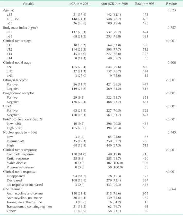
Table 1. Baseline patient and tumor characteristic of 995 patients treated with neoadjuvant chemotherapy
Variable pCR (n = 205) Non-pCR (n = 790) Total (n = 995) P-value
Age (yr) 0.623
≤35 31 (17.9) 142 (82.1) 173
>35, ≤55 148 (21.3) 548 (78.7) 696
>55 26 (20.6) 100 (79.4) 126
Body mass index (kg/m2) 0.757
≤25 137 (20.3) 537 (79.7) 674
>25 68 (21.2) 253 (78.8) 321
Clinical tumor stage <0.001
cT1 38 (36.2) 64 (63.8) 105
cT2 114 (22.3) 398 (77.7) 512
cT3 45 (14.0) 277 (86.0) 322
cT4 8 (14.3) 48 (85.7) 56
Clinical nodal stage 0.900
cN1 165 (20.4) 644 (79.6) 809
cN2 37 (21.3) 137 (78.7) 174
cN3 3 (25.0) 9 (75.0) 12
Estrogen receptor <0.001
Positive 56 (11.7) 421 (88.3) 477
Negative 149 (28.8) 369 (71.2) 518
Progesterone receptor <0.001
Positive 29 (8.3) 322 (91.7) 351
Negative 176 (27.3) 468 (72.7) 644
HER2 <0.001
Positive 95 (29.5) 227 (70.5) 322
Negative 110 (16.3) 563 (83.7) 673
Ki-67 proliferation index (%) <0.001
Low (≤20) 40 (9.2) 396 (90.8) 436
High (>20) 165 (29.6) 394 (70.4) 558
Nuclear grade (n = 866) 0.145
Low 3 (4.4) 65 (95.6) 68
Intermediate 35 (12.3) 250 (87.7) 285
High 64 (12.5) 449 (87.5) 513
Clinical tumor response <0.001
Complete response 170 (81.0) 40 (19.0) 210
Partial response 35 (8.3) 385 (91.7) 420
Stable disease 0 (0.0) 307 (100.0) 307
Progressive disease 0 (0.0) 58 (100.0) 58
Clinical node response <0.001
Disappeared 94 (54.7) 78 (45.3) 172
Decreased 108 (18.9) 279 (72.1) 387
No response or Increased 3 (0.7) 433 (99.3) 436
NAC regimen 0.064
Anthracycline and taxane 140 (21.4) 515 (78.6) 655
Anthracycline, no taxane 20 (14.4) 139 (85.6) 159
Taxane, no anthracycline 3 (15.8) 16 (84.2) 19
Transtuzumab containg regimen 31 (33.3) 62 (66.7) 93
Others 11 (15.9) 58 (84.1) 69
Values are presented as number (%).
pCR, pathologic complete response; HER2, human epidermal growth factor receptor 2; NAC, neoadjuvant chemotherapy.
the patients who underwent breast surgery. Generally speaking, both breast and axillary pCRs were achieved in 20.5% (n = 204) of the patients who underwent surgery. The patients with both pCRs tended to be estrogen receptor (ER) negative, progesterone receptor (PR) negative, human epidermal growth factor receptor 2 (HER2) positive, with high Ki67, good tumor response, good nodal response and early clinical tumor stage as compared with the patients with a nonpCR.
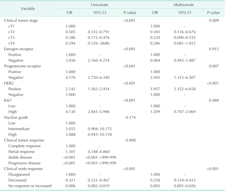
The both pCRs were associated with an initial clinical tumor stage, negative ER status, negative PR status, positive HER2 status, high Ki67 (Ki67 > 20) and clinical radiologic nodal responses in a univariate analysis. The multivariate logistic regression analysis showed that the both pCRs were associated with initial clinical tumor stage, negative PR status, positive HER2 status, and clinical radiologic nodal responses (Table 2).
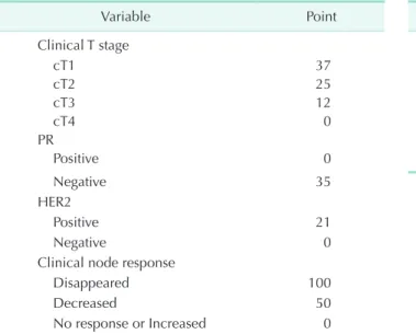
We made a nomogram based on the clinically and statistically
significant variables noted in this study. The significant variables that can be assessed in preoperative evaluations in the multivariate analysis were the noted years of a clinical tumor stage (cT1, cT2, cT3, or cT4), PR status (negative or positive), HER2 status (negative or positive), and the clinical radiologic nodal stage (disappeared, decreased or no changed/
increased) (Table 3). In this case, Fig. 1 illustrates the nomogram to calculate the probability of achieving both pCR. The total nomogram score is calculated by summing up the scores for each of the variables. In that instance, the total score can then be used to assign a probability of achieving both pCRs to individual patients using the scale.
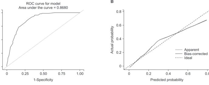
A nomogram was developed based on the clinical and statistically significant predictors. It had good discrimination performance (AUC, 0.868; 95% CI, 0.845–0.891) and calibration fit. The calibration plot showed a good and satisfactory
Table 2. Univariate and multivariate analysis for both pathologic complete response
Variable Univariate Multivariate
OR 95% CI P-value OR 95% CI P-value
Clinical tumor stage <0.001 0.009
cT1 1.000 - 1.000 -
cT2 0.505 0.322–0.791 0.303 0.136–0.676
cT3 0.286 0.172–0.476 0.220 0.090–0.535
cT4 0.294 0.126–.0686 0.286 0.081–1.012
Estrogen receptor <0.001 0.915
Positive 1.000 - 1.000 -
Negative 3.036 2.166–4.254 0.964 0.493–1.887
Progesterone receptor <0.001 0.007
Positive 1.000 - 1.000 -
Negative 4.176 2.750–6.340 2.945 1.333–6.507
HER2 <0.001 <0.001
Positive 2.142 1.563–2.934 3.957 2.352–6.658
Negative 1.000 - 1.000 -
Ki67 <0.001 0.488
Low 1.000 - 1.000 -
High 4.130 2.845–5.996 1.209 0.707–2.069
Nuclear grade 0.174
Low 1.000 -
Intermediate 3.033 0.904–10.175
High 3.088 0.943–10.118
Clinical tumor response 0.908
Complete response 1.000 -
Partial response 1.301 0.348–4.860
Stable disease <0.001 <0.001–>999.999 Progressive disease <0.001 <0.001–>999.999
Clinical node response <0.001 <0.001
Disappeared 1.000 - 1.000 -
Decreased 0.321 0.221–0.467 0.258 0.154–0.433
No response or increased 0.006 0.002–0.019 0.003 0.001–0.026
OR, odds ratio; CI, confidence interval; HER2, human epidermal growth factor receptor 2.
agreement between the predicted and observed probabilities according to an administered HosmerLemeshow test (Fig.
2). We had 5fold crossvalidation model with a random split analysis in a cohort of patients and Cross validation who had an average of AUC 0.853 (0.837–0.869) (Fig. 3).
DISCUSSION
Predicting pCR after NAC in patients with breast cancer and cytologically proven nodal metastasis is important for
understanding and improving the patient outcomes as well as for identifying patients in whom ALND and mastectomy might be omitted as a treatment option. We performed a registered medical record review based on a prospectively collected database and made a prediction model based on the clinicopathologic characteristics of a tumor to estimate the probability of achieving breast and axillary pCR. We used a 5fold crossvalidation model in a cohort of patients. We assigned the largest value of the Youden index as the cutoff and the nomogram score was 97 points. In this cutoff, sensitivity
Points
Clinical T stage
PR
HER2
Clinical node response
Total points
Predicted value
0 10 20 30 40 50 60 70 80 90 100
cT4 cT3
cT2 cT1
Positive
Negative
Positive Negative
0 20 40 60 80 100 120 140 150 180 200
No response/increased
Decreased
Disappeared
0.1 0.2 0.3 0.4 0.5 0.6 0.7 0.8
Fig. 1. Nomogram for the pre- diction of the probability of the pathologic complete response.
PR, progesterone receptor; HER2, human epidermal growth factor receptor 2.
Table 3. Model development of risk point
Variable Point Total points Predicted value
Clinical T stage 83 0.1
cT1 37 105 0.2
cT2 25 120 0.3
cT3 12 132 0.4
cT4 0 144 0.5
PR 155 0.6
Positive 0 167 0.7
Negative 35
HER2
Positive 21
Negative 0
Clinical node response
Disappeared 100
Decreased 50
No response or Increased 0
PR, progesterone receptor; HER2, human epidermal growth factor receptor 2.
was 91.2%, specificity was 67.2%, positive predictive value was 41.9%, negative predictive value was 96.7% and accuracy was 72.2%. The generated data demonstrated that the incidence of a low clinical T stage, negative PR, positive HER2 and clinical radiologic node response were all significant independent predictors of ypT0/ypN0 on an adjusted analysis.
No association in breast and axillary pCRs was found with a clinical tumor response in our study. This may indicate that it is the degree of the initial tumor size, which predicts a known biological response in the axilla and the breast. In our study, a stable disease and a PD seemed to be a meaningless designation for comparison, because there was no case of pCR, but it was difficult to find the meaning in the analysis. Moreover, the
cases of CR and PR were more than 50% in the incidence of a nonpCR. Interestingly to note, among 471 axilla pCR patients, 202 patients (42.9%) have breast and axilla pCRs. However, among the 242 breast pCR patients, 202 patients (84.2%) have breast and axilla pCRs. In line with this reasoning, only 40 patients (15.8%) indicative of breast pCR have nonaxilla pCR.
During our study period, it is noted that the trastuzumab was not added to the NAC as a standard treatment in patients with HER2 positive tumors. For the most part, the neoadjuvant trastuzumab therapy was approved in late 2013 in Korea, making it a relatively new treatment option for patients requiring this treatment. Therefore, only 10.0% of the patients in our study received trastuzumab with their NAC. In addition, A
Sensitivity
0 0 1.00
0.75
0.50
0.25
0.25 0.50 0.75
1-Specificity ROC curve for model Area under the curve = 0.8680
B
0
Actualprobability
0 0.6
0.4
0.2
0.2 0.4 0.6
Predicted probability
B = 1,000 repetitions, boot Mean absolute error = 0.032 n = 995 Apparent Bias-corrected Ideal
1.00 0.8
0.8
Fig. 2. The receiver-operating characteristics (ROC) curve and the calibration plot of the nomogram in the training set. (A) ROC curve with area under the curve = 0.868 (95% confidence interval, 0.845–0.891). (B) Calibration plot of the nomogram.
A
Sensitivity
0 0 1.0
0.8
0.6
0.4
0.4 0.6 0.8
1-Specificity
1.0 0.2
0.2
B
0
Actualprobability
0 0.6
0.4
0.2
0.2 0.4 0.6
Predicted probability
B = 1,000 repetitions, boot Mean absolute error = 0.046 n = 995 Apparent Bias-corrected Ideal
0.8 0.8
Fig. 3. The receiver-operating characteristics (ROC) curve of the nomogram in the cross-validation set. (A) ROC curve with area under the curve = 0.853 (95% confidence interval, 0.837–0.869). (B) Calibration plot of the nomogram.
the patients of the HER2 positive breast cancer had 29.5% (95 of 322) both pCR but these of HER2 negative breast cancer had 16.3% (110 of 673) both pCR. The American College of Surgeons Oncology Group Z1071 trial, which reflects current chemotherapy approaches including the use of trastuzumab for patients with HER2 positive tumors, showed differential nodal responses based on the tumor biology. The overall nodal pCR was 41.1%, but this varied from 21.1% in patients with Hormone receptor (HR) positive/HER2 negative tumors, to 49.4% in patients with a triplenegative breast cancer to 64.7% in patients with HER2 positive disease (P < 0.0001) [12].
Given these points, it was shown that recently a metaanalysis showed that improvement in eventfree survival for pCR vs non
pCR was substantial: (HR, 0.37). This association was greater for patients with a hormone receptornegative disease (HR, 0.29) than for those patients who had a hormone receptorpositive disease (HR, 0.52) [13]. Although the biologic tumor subtype was associated with pCR, we did not subsequently incorporate the biologic tumor subtype into the nomogram. Because the definition of biologic subtype varies somewhat among studies, we chose to include the receptor status (ER, PR, and HER2) of biologic subtype from the nomogram for simplicity purposes.
It is important to point out that our study corroborated these findings, with a noted positive HER2 status that was strongly predictive of a tumor and axillary response.
Many studies have evaluated nomograms for predicting the axillary response to NAC in node positive patients with breast cancer [1417]. A recent study showed that patients with high nuclear grade (odds ratio [OR], 13.4), HER2positive (OR, 4.7), ER negative (OR, 3.5), or PRnegative (OR, 4.3) tumors were more likely to achieve axillary pCR [14]. A study of 403 patients with biopsyconfirmed axillary metastases who received NAC, showed that patients who achieved a axillary pCR had received a better 5year overall survival (93 %) and disease
free survival (87%) than those who had acquired the residual axillary disease (72%, 60%) [7]. The axillary pCR is affected by offering a better oncological outcome of the patients. However a recent study demonstrated that the pCR defined as no residual invasive cancer in the breast and axillary nodes with presence or absence of in situ cancer (ypT0/is ypN0 or ypT0 ypN0), served to provide a better association with improved outcomes as compared to eradication of an invasive tumor from the breast
alone (ypT0/is) [18]. Therefore, the evaluation of both breast and axilla pCRs is more important than the evaluation of only the axilla pCR.
Although this study was performed in a single comprehensive cancer institution located in Korea and the number of patients was relatively small, this study did not investigate clinically suspected nodepositive patients, but instead investigated node
positive patients whose diagnoses were confirmed by cytology.
Therefore the results from the small number of participating patients are still meaningful, and allow us to confidently draw important results. Additionally this study was not a prospective randomized clinical trial, and for that reason, the distribution of patients might be uneven and can be presumed to have had some effect on the results of the regional control. Our study did not includ the regimens commonly used for neoadjuvant, therapy of docetaxel, carboplatin, trastuzumab, and pertuzumab in HER2 positive breast cancer, since it included patients who would have been in a followup of patients monitored up until 2014. Also, we had 238 patients with sentinel lymph node biopsy (SLNB) try and 85 patients with SLN negative. Analyses of several prospective trials show that the accuracy of SLNB can be improved using a dualtracer technique and the retrieval of 2 or more SLNs, with an ensured removal of the node initially confirmed to contain metastases [3]. Several studies currently are working on ways to ensure removal of the clipped nodes, while using techniques such as a targeted axillary dissection [4,19,20]. We did not perform a targeted axillary dissection in our study. If these better techniques were used, then a more accurate axillary evaluation would be possible in future studies.
In conclusion, the breast and axilla pCRs are associated significantly with a low clinical T stage, negative PR, positive HER2 and a clinical radiologic node response. Our nomogram might help predict breast and axilla pCRs after NAC in patients with initially nodepositive breast cancer. Patients with a high probability of achieving both breast and axillary pCRs might be candidates for less invasive surgery.
CONFLICTS OF INTEREST
No potential conflict of interest relevant to this article was reported.
REFERENCES
1. Fisher B, Brown A, Mamounas E, Wieand S, Robidoux A, Margolese RG, et al. Effect of preoperative chemotherapy on local
regional disease in women with operable breast cancer: findings from National Sur
gical Adjuvant Breast and Bowel Project
B18. J Clin Oncol 1997;15:248393.
2. Bear HD, Anderson S, Smith RE, Geyer CE Jr, Mamounas EP, Fisher B, et al. Se
quen tial preoperative or postoperative docetaxel added to preoperative doxoru
bicin plus cyclophosphamide for operable breast cancer: National Surgical Adjuvant Breast and Bowel Project Protocol B27. J Clin Oncol 2006;24:201927.
3. Boughey JC, Suman VJ, Mittendorf EA, Ahrendt GM, Wilke LG, Taback B, et al.
Sentinel lymph node surgery after neo
adjuvant chemotherapy in patients with nodepositive breast cancer: the ACOSOG Z1071 (Alliance) clinical trial. JAMA 2013;
310:145561.
4. Mittendorf EA, Caudle AS, Yang W, Krishnamurthy S, Shaitelman S, Chavez
MacGregor M, et al. Implementation of the american college of surgeons oncology group z1071 trial data in clinical practice:
is there a way forward for sentinel lym ph node dissection in clinically nodeposi tive breast cancer patients treated with neo
adjuvant chemotherapy? Ann Surg Oncol 2014;21:246873.
5. Kim KS, Kim Z, Shim EJ, Kim NH, Jung SY, Kim J, et al. The reality in the follow
up of breast cancer survivors: survey of Korean Breast Cancer Society. Ann Surg Treat Res 2015;88:1339.
6. Shen J, Gilcrease MZ, Babiera GV, Ross MI, MericBernstam F, Feig BW, et al.
Feasibility and accuracy of sentinel lym ph node biopsy after preoperative chemo
therapy in breast cancer patients with docu mented axillary metastases. Cancer 2007;109:125563.
7. Hennessy BT, Hortobagyi GN, Rouzier R, Kuerer H, Sneige N, Buzdar AU, et al.
Outcome after pathologic complete eradi
cation of cytologically proven breast can
cer axillary node metastases following pri mary chemotherapy. J Clin Oncol 2005;
23:930411.
8. Rouzier R, Extra JM, Klijanienko J, Falcou MC, Asselain B, VincentSalomon A, et
al. Incidence and prognostic significance of complete axillary downstaging after primary chemotherapy in breast cancer patients with T1 to T3 tumors and cyto
logically proven axillary metastatic lymph nodes. J Clin Oncol 2002;20:130410.
9. Mansel RE, Fallowfield L, Kissin M, Goyal A, Newcombe RG, Dixon JM, et al.
Randomized multicenter trial of sentinel node biopsy versus standard axillary treat ment in operable breast cancer: the ALMANAC Trial. J Natl Cancer Inst 2006;
98:599609.
10. Lucci A, McCall LM, Beitsch PD, Whit
worth PW, Reintgen DS, Blumen cranz PW, et al. Surgical compli cations associated with sentinel lymph node dissection (SLND) plus axillary lymph node dis
section compared with SLND alone in the American College of Surgeons On cology Group Trial Z0011. J Clin Oncol 2007;25:
365763.
11. Green MC, Buzdar AU, Smith T, Ibrahim NK, Valero V, Rosales MF, et al. Weekly paclitaxel improves pathologic complete remission in operable breast cancer when compared with paclitaxel once every 3 weeks. J Clin Oncol 2005;23:598392.
12. Boughey JC, McCall LM, Ballman KV, Mittendorf EA, Ahrendt GM, Wilke LG, et al. Tumor biology correlates with rates of breastconserving surgery and pathologic complete response after neoadjuvant chemotherapy for breast cancer: findings from the ACOSOG Z1071 (Alliance) Pro
spective Multicenter Clinical Trial. Ann Surg 2014;260:60814.
13. Broglio KR, Quintana M, Foster M, Olinger M, McGlothlin A, Berry SM, et al. Association of pathologic complete response to neoadjuvant therapy in HER2
positive breast cancer with longterm out comes: a metaanalysis. JAMA Oncol 2016;2:75160.
14. Vila J, Mittendorf EA, Farante G, Bassett RL, Veronesi P, Galimberti V, et al. Nomo
grams for predicting axillary response to neoadjuvant chemotherapy in clinically nodepositive patients with breast cancer.
Ann Surg Oncol 2016;23:35019.
15. Kantor O, Sipsy LM, Yao K, James TA. A predictive model for axillary node patho
logic complete response after neoadjuvant chemotherapy for breast cancer. Ann Surg Oncol 2018;25:130411.
16. Kim JY, Park HS, Kim S, Ryu J, Park S, Kim SI. Prognostic nomogram for prediction of axillary pathologic complete response after neoadjuvant chemotherapy in cytolo
gically proven nodepositive breast cancer.
Medicine (Baltimore) 2015;94:e1720.
17. Schipper RJ, Moossdorff M, Nelemans PJ, Nieuwenhuijzen GA, de Vries B, Strobbe LJ, et al. A model to predict pathologic com plete response of axillary lymph nodes to neoadjuvant chemo(immuno) therapy in patients with clinically node
positive breast cancer. Clin Breast Cancer 2014;14:31522.
18. Cortazar P, Geyer CE Jr. Pathological complete response in neoadjuvant treat
ment of breast cancer. Ann Surg Oncol 2015;22:14416.
19. Donker M, Straver ME, Wesseling J, Loo CE, Schot M, Drukker CA, et al. Marking axillary lymph nodes with radioactive iodine seeds for axillary staging after neoadjuvant systemic treatment in breast cancer patients: the MARI procedure. Ann Surg 2015;261:37882.
20. Caudle AS, Yang WT, Krishnamurthy S, Mittendorf EA, Black DM, Gilcrease MZ, et al. Improved axillary evaluation following neo adjuvant therapy for patients with nodepositive breast cancer using selec
tive evaluation of clipped nodes: imple
men tation of targeted axillary dissection.
J Clin Oncol 2016;34:10728.