INTRODUCTION
Berberine (BBR) is an isoquinoline alkaloid isolated from the Chinese herb Coptis chinensis (Huanglian) and is a promis- ing component of medicinal plants [1]. BBR represses tumor progression by suppressing abnormal cell proliferation, arrest- ing the cell cycle, and inducing apoptosis in breast cancer and osteosarcoma cells [2,3]. Furthermore, BBR suppresses the metastatic potential of breast cancer cells through downregul- ation of matrix metalloproteinase (MMP)-1 and MMP-9 [4,5]. BBR regulates various signaling pathways including mi-
togen-activated protein kinases (MAPKs), c-Jun N-terminal protein kinase (JNK), and nuclear factor κB (NF-κB) in a va- riety of cancer cells [6-8]. We also reported that BBR sup- presses 12-O-tetradecanoylphorbol-13-acetate-induced vas- cular endothelial growth factor expression through inhibition of the phosphatidylinositol-4,5-bisphosphate 3-kinase/protein kinase B (AKT) pathway in breast cancer cells [9]. Here, we ex- plored the regulatory mechanism of BBR on fibronectin (FN) expression in triple-negative breast cancer (TNBC) cells.
FN is one of the most abundant extracellular matrix glyco- proteins and is implicated in physiological and pathological processes such as cell differentiation, invasion, migration, and oncogenic transformation [10,11]. FN mRNA and protein expression is dramatically increased in the stroma of breast tumors compared with normal adult breast tissues [12]. The cell adhesive region of FN comprises at least two minimal amino acid sequences: an Arg-Gly-Asp (RGD) sequence and a Pro-His-Ser-Arg-Asn sequence [13,14]. The RGD site of FN binds to specific cell surface receptors called integrins, and the resulting complex plays a crucial role in the progression of breast cancer metastasis [13]. Administration of synthetic
Berberine Suppresses Fibronectin Expression through Inhibition of c-Jun Phosphorylation in Breast Cancer Cells
Yisun Jeong1, Daeun You1, Hyun-Gu Kang1, Jonghan Yu2,3, Seok Won Kim2,3, Seok Jin Nam2,3, Jeong Eon Lee1,2,3, Sangmin Kim2
1Department of Health Sciences and Technology, Samsung Advanced Institute for Health Sciences & Technology, Sungkyunkwan University, Seoul; 2Breast Cancer Center and 3Department of Surgery, Samsung Medical Center, Sungkyunkwan University School of Medicine, Seoul, Korea
ORIGINAL ARTICLE
Purpose: The exact mechanism regulating fibronectin (FN) ex- pression in breast cancer cells has not been fully elucidated. In this study, we investigated the pharmacological mechanism of berberine (BBR) with respect to FN expression in triple-negative breast cancer (TNBC) cells. Methods: The clinical significance of FN mRNA expression was analyzed using the Kaplan-Meier plotter database (http://kmplot.com/breast). FN mRNA and pro- tein expression levels were analyzed by real-time polymerase chain reaction and western blotting, respectively. Results: Using publicly available clinical data, we observed that high FN expres- sion was associated with poor prognosis in patients with breast cancer. FN mRNA and protein expression was increased in TNBC cells compared with non-TNBC cells. As expected, re-
combinant human FN significantly induced cell spreading and adhesion in MDA-MB231 TNBC cells. We also investigated the regulatory mechanism underlying FN expression. Basal levels of FN mRNA and protein expression were downregulated by a specific activator protein-1 (AP-1) inhibitor, SR11302. Interest- ingly, FN expression in TNBC cells was dose-dependently de- creased by BBR treatment. The level of c-Jun phosphorylation was also decreased by BBR treatment. Conclusion: Our findings demonstrate that FN expression is regulated via an AP-1–de- pendent mechanism, and that BBR suppresses FN expression in TNBC cells through inhibition of AP-1 activity.
Key Words: Berberine, Cell adhesion, Fibronectins, Transcription factor AP-1
Correspondence to: Sangmin Kim
Breast Cancer Center, Samsung Medical Center, Sungkyunkwan University School of Medicine, 81 Irwon-ro, Gangnam-gu, Seoul 06351, Korea Tel: +82-2-2148-7312, Fax: +82-2-3410-6982
E-mail: [email protected]
This research was supported by the Basic Science Research Program through the National Research Foundation of Korea (NRF) funded by the Ministry of Education (2016R1D1A1B01010508) and by a NRF grant funded by the Korean government Ministry of Science, ICT & Future Planning (MSIP) (2016R1A5A2945889).
Received: November 7, 2017 Accepted: December 12, 2017
Cancer
peptides containing the RGD sequence can inhibit FN cell adhesion function and metastatic potential in breast cancer models [15].
This study aimed to investigate the mechanism by which BBR inhibits FN expression in TNBC cells. We examined the clinical relevance of FN expression in breast cancer patients and demonstrated that BBR completely suppresses the basal levels of FN expression in TNBC cells through inhibition of activator protein (AP)-1 activity.
METHODS
Reagents
Dulbecco’s modified Eagle’s medium (DMEM), Roswell Park Memorial Institute (RPMI) 1640, and antibiotics were purchased from Life Technologies (Rockville, USA). Fetal bovine serum (FBS) was purchased from Hyclone (Logan, USA). BBR was purchased from Sigma (St. Louis, USA).
SR11302 was purchased from Tocris (Ellisville, USA). The secondary horseradish peroxidase (HRP)-conjugated anti- body and mouse monoclonal anti–β-actin antibody were purchased from Santa Cruz Biotechnology Inc. (Santa Cruz, USA). Rabbit monoclonal anti-FN antibodies to phospho (p)- c-Jun, total (t)-c-Jun, and c-Fos were purchased from AbCam (Cambridge, UK). The enhanced chemiluminescence (ECL) prime reagents were from Amersham (Buckinghamshire, UK).
Analysis of public database
Expression data were downloaded from a public database (Kaplan-Meier plotter database; http://kmplot.com/breast) [16]. The clinical relevance of FN mRNA expression in breast cancer patients was analyzed by Kaplan-Meier survival plots.
The hazard ratio with 95% confidence interval and log-rank p-values were calculated.
Cell culture and drug treatment
MDA-MB453, BT20, BT549, MDA-MB231, Hs578T, and MDA-MB157 human breast cancer cells were grown in a hu- midified atmosphere of 95% air and 5% CO2 at 37°C in DMEM supplemented with 10% FBS, 2 mM glutamine, 100 IU/mL penicillin, and 100 μg/mL streptomycin. BT474, T47D, ZR75-1, SKBR3, and HCC1143 human breast cancer cells were grown in RPMI1640 media under the same condi- tions.
For drug treatment, cells were maintained in culture medi- um without FBS for 24 hours and then treated with the indi- cated concentrations of BBR or SR11302 for 24 hours in fresh serum-free media.
Real-time polymerase chain reaction
Total RNA was extracted from cells using the TRIzol re- agent (Invitrogen, Carlsbad, USA), according to the manufac- turer’s instructions. Isolated RNA samples were then used for reverse transcriptase-polymerase chain reaction (RT-PCR).
Samples (1 µg of total RNA) were reverse-transcribed into cDNA in 20-µL reaction volumes using a first-strand cDNA synthesis kit for RT-PCR, according to the manufacturer’s in- structions (MBI Fermentas, Hanover, USA).
Gene expression was quantified by real-time PCR using a SensiMix SYBR Kit (Bioline Ltd., London, UK) with 100 ng of cDNA per reaction. The sequences of primer sets used for this analysis are as follows: human FN (forward, 5′-CCA CCC CCA TAA GGC ATA GG-3′; reverse, 5′-GTA GGG GTC AAA GCA CGA GTC ATC-3′) and glyceraldehyde-3-phos- phate dehydrogenase as an internal control (forward, 5′-ATT GTT GCC ATC AAT GAC CC-3′; reverse, 5′-AGT AGA GGC AGG GAT GAT GT-3′). An annealing temperature of 60°C was used for all primers. PCR was performed in a stan- dard 384-well plate format on an ABI 7900HT real-time PCR detection system. For data analysis, the raw threshold cycle (CT) value was first normalized to the housekeeping gene for each sample in order to obtain the ΔCT. The normalized ΔCT
was then calibrated to the control cell samples to obtain the ΔΔCT.
Western blotting
Cell culture media (supernatants) and cell lysates were used in immunoblot analysis for FN, c-Jun, c-Fos, and β-actin. The protein samples were boiled for 5 minutes in Laemmli sample buffer and electrophoresed in 8% (FN or β-actin) or 10% so- dium dodecyl sulfate polyacrylamide gel electrophoresis (SDS-PAGE). The separated proteins were transferred to poly- vinylidene fluoride membranes, and the membranes were blocked with 10% skim milk in tris-buffered saline (TBS) with 0.01% Tween-20 for 15 minutes. The blots were incubated overnight with antibodies against FN, c-Jun, c-Fos, and β-actin in 1% TBS/T buffer (0.01% Tween-20 in TBS) at 4°C.
The blots were then washed 3–4 times in TBS with 0.01%
Tween-20 and subsequently incubated with anti-rabbit perox- idase-conjugated antibody (1/2,000 dilution) in TBS/T buffer.
After 1 hour of incubation at room temperature, the blots were washed three times, and developed using ECL prime re- agents.
Cell adhesion and spreading assay
MDA-MB231 and Hs578T cells were grown to approxi- mately 80% confluence in DMEM containing 10% FBS, de- tached, and then suspended in serum-free medium. The cell
suspensions (5×10⁴ cells in 250 μL) were seeded into each well of 24-well plates precoated with 100 ng/mL recombinant human FN (rhFN). After incubation at 37°C for 3 hours, non- adherent cells were removed by washing with phosphate- buffered saline. Cell spreading was analyzed using a CK40 in- verted microscope (Olympus, Tokyo, Japan). Adherent cells were fixed in 4% formaldehyde and stained with 0.2% crystal violet in 10% ethanol. The stained cells were then dissolved in 2% SDS, and absorbance at 570 nm was measured using a spectrophotometer (Spectra Max 190; Molecular Devices, Sunnyvale, USA).
Statistical analysis
Statistical significance was determined using the Student t-test. Data are presented as mean±standard error of the mean. All quoted p-values are two-tailed, and differences were considered significant for p<0.05. Microsoft Excel was used for statistical analyses (Microsoft, Redmond, USA).
RESULTS
Aberrant FN expression is associated with poor prognosis in breast cancer patients
We analyzed whether FN expression is associated with sur- vival in breast cancer patients using the Kaplan-Meier plotter database (http://kmplot.com/breast) [16]. Abnormal FN ex- pression was associated with poor prognosis for relapse-free survival (p=0.006) (Figure 1A) and overall survival (p=
0.003) (Figure 1B) in breast cancer patients.
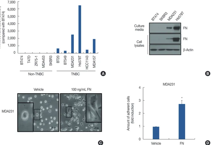
Next, we investigated FN mRNA expression levels in a vari- ety of breast cancer cells. The clinicopathological features of
breast cancer cell lines were determined from the American Type Culture Collection website (https://www.atcc.org). We also classified cells as non-TNBC (BT474, T47D, ZR75-1, MDA-MB453, and SKBR3) and TNBC (BT20, BT549, MDA- MB231, Hs578T, HCC1143, and MDA-MB157) to compare the levels of FN mRNA expression. As shown in Figure 2A, FN mRNA expression was significantly elevated in TNBC cells compared with non-TNBC cells. The FN mRNA expres- sion was 2587.8-fold (MDA-MB231 cells) and 6555.5-fold (Hs578T cells) greater than the control levels (BT474 cells) (Figure 2A). We also confirmed the level of FN protein ex- pression in non-TNBC cells (BT474 and SKBR3) and TNBC cells (MDA-MB231 and Hs578T) using culture media and whole cell lysates. As expected, FN protein levels were dra- matically increased in TNBC cells (Figure 2B). In a previous study, we reported that upregulation of FN by human epider- mal growth factor receptor 2 (HER2) overexpression aug- ments cell adhesion and invasion in breast cancer cells [15].
We therefore analyzed the morphological changes induced by treatment with 100 ng/mL rhFN. Extension of protrusions was increased in rhFN-treated cells compared with the con- trol cells (vehicle-treated) (Figure 2C). In addition, the adhe- sion rates of MDA-MB231 cells were significantly promoted by rhFN treatment (Figure 2D).
Basal FN expression is suppressed by SR11302 treatment Next, we investigated whether AP-1 activity is involved in FN expression. Cells in serum-free media were treated with a specific AP-1 inhibitor, SR11302, for 24 hours. As shown in Figure 3A, FN mRNA expression was dramatically decreased by SR11302 in both MDA-MB231 and Hs578T cells by 0.4±
Figure 1. Aberrant fibronectin (FN) expression is associated with poor prognosis in breast cancer patients. Clinical relevance of FN mRNA expression obtained from a public database (Kaplan-Meier plotter database; http://kmplot.com/breast). (A) Relapse-free survival. (B) Overall survival.
HR=hazard ratio.
1.0
0.8
0.6
0.4
0.2
0
1.0
0.8
0.6
0.4
0.2
0
0 50 100 150 200 250 0 50 100 150 200 250 300
Months
HR=1.2 (1.06–1.37) HR=1.29 (1.02–1.64)
Log-rank p=0.006 Log-rank p=0.003
Expression Expression
Low Low
High High
Months
Relapse-free survival Overall survival
A B
Figure 2. Level of fibronectin (FN) expression and role of FN in breast cancer cells. After serum starvation for 24 hours, the levels of FN mRNA and protein expression were analyzed by real-time polymerase chain reaction (A) and western blotting (B), respectively. (C) MDA-MB231 cells were seeded with or without 100 ng/mL recombinant human FN for 3 hours. Cell morphology was analyzed using a CK40 inverted microscope. (D) Rates of cell adhesion were analyzed by adhesion assay, as described in Methods. The results are representative of three independent experiments. Values shown are mean±standard error of the mean.
TNBC=triple-negative breast cancer. *p<0.01 vs. vehicle.
Figure 3. Basal fibronectin (FN) expression is suppressed by SR11302 treatment. (A) After serum starvation for 24 hours, cells were treated with 10 μM SR11302 for 24 hours. The level of FN mRNA expression was analyzed by real-time polymerase chain reaction. (B, C) After serum starvation for 24 hours, cells were treated with the indicated concentrations of SR11302 for 24 hours. The level of FN protein expression was analyzed by western blotting using conditioned culture media and lysates for both MDA-MB231 (B) and Hs578T (C) cells. The results are representative of three indepen- dent experiments. Values shown are mean±standard error of the mean.
Con=control; SR=SR11302. *p<0.01 vs. con; †p<0.05.
7,000 6,000 5,000 4,000 3,000 2,000 1,000 0
4
3
2
1
0
1.2 1.0 0.8 0.6 0.4 0.2 0
Non-TNBC
Vehicle
MDA231
†
*
MDA231 TNBC
FN
*
Hs578T
BT474 T47D ZR75-1 MDA453 SKBR3 BT20 BT549 MDA231 Hs578T HCC1143 MDA157
FN mRNA level (fold induction) (compared with BT474) Amount of adherent cells (fold induction)
FN mRNA level (fold induction)
A
D
A B C
B
C
Culture media
Culture
media Culture
media MDA231
FN
FN FN
Vehicle 100 ng/mL FN
FN
FN FN
β-Actin
β-Actin β-Actin
lysatesCell
lysatesCell Cell
lysates BT474
Con 5 10 (μM SR) Con 5 10 (μM SR)
SKBR3 MDA231Hs578T
MDA231 Hs578T
Con 10 μM SR
0.1-fold and 0.7±0.1-fold, respectively, compared with the control level (Figure 3A). In addition, FN protein expression was dose-dependently decreased by SR11302 in both MDA- MB231 (Figure 3B) and Hs578T cells (Figure 3C). These re- sults demonstrated that FN expression in TNBC cells is regul- ated through AP-1 activity.
Basal FN expression is suppressed by berberine treatment We then examined the effect of BBR on FN expression in TNBC cells. After serum starvation for 24 hours, cells were treated with the indicated concentrations of BBR for 24 hours in serum-free media. The chemical structure of BBR is repre- sented in Figure 4A. After 24 hours, we harvested conditioned culture media and prepared whole cell lysates for detection of FN mRNA and protein expression. As shown in Figure 4B, FN mRNA expression was decreased upon treatment with 50 μM BBR. In particular, FN mRNA expression was significant- ly decreased to 0.25±0.04-fold of the control level in MDA- MB231 cells. Furthermore, FN protein expression was dose- dependently decreased by BBR treatment in both MDA- MB231 (Figure 4C) and Hs578T cells (Figure 4D).
To verify the inhibitory mechanism of BBR on FN expres- sion, we examined the phosphorylation levels of various sig- naling molecules. We found that phosphorylation of c-Jun, a member of the AP-1 family, was significantly decreased by BBR treatment in Hs578T TNBC cells (Figure 4E); however, the level of c-Fos expression was not altered by BBR (Figure 4E). Thus, we demonstrated that BBR suppresses the FN mRNA and protein expression levels through inhibition of AP-1 activity.
DISCUSSION
FN plays a central role in processes associated with tumor cell proliferation and is highly expressed in malignant breast cancer cells, but not in nonmalignant breast epithelial cells [17,18]. The blood concentration of FN shows great promise as a marker for cancer and other diseases [19]. The level of plasma FN expression is directly associated with clinical events such as chemotherapy [19,20]. The plasma FN level is reportedly higher in patients with metastatic breast cancer than in those with no detectable disease [19]. In addition, ab- Figure 4. Basal fibronectin (FN) expression is suppressed by berberine (BBR) treatment. (A) The chemical structure of BBR. (B) After serum starvation for 24 hours, MDA-MB231 and Hs578T cells were treated with 50 μM BBR for 24 hours, and the level of FN mRNA expression was analyzed by real- time polymerase chain reaction. (C, D) After serum starvation for 24 hours, cells were treated with the indicated concentrations of BBR for 24 hours.
The level of FN protein expression was analyzed by western blotting using conditioned culture media and lysates of MDA-MB231 (C) and Hs578T (D) cells. (E) After serum starvation for 24 hours, cells were treated with 50 μM BBR for 4 hours. The levels of p-c-Jun, c-Fos, and β-actin expression in Hs578T cells were analyzed by western blotting. The results are representative of three independent experiments. Values shown are mean±standard error of the mean.
Con=control. *p<0.01 vs. con; †p<0.05.
1.2 1.0 0.8 0.6 0.4 0.2 0
MDA231
†
*
Hs578T
FN mRNA level (fold induction)
B A
D
C E
Culture media Culture
media FN FN p-c-Jun
FN
FN c-Fos
β-Actin
β-Actin β-Actin
lysatesCell lysatesCell
lysatesCell
Con 10 25 50 (μM BBR) Con 10 25 50 (μM BBR) Con 25 50 (μM BBR)
Hs578T
MDA231 Hs578T
Con 50 μM BBR
normal FN induction within tumor associated fibroblasts triggers tumor cell motility, cancer spread, and metastasis for- mation [21]. In the present study, we also observed that FN expression is directly associated with relapse-free survival and overall survival in breast cancer patients. Therefore, we pro- pose that suppression of FN expression is one of the most im- portant approaches for breast cancer treatment.
FN can trigger a variety of cellular signaling pathways and thus contribute to tumorigenesis [22]. The RGD site of FN binds to β1 integrin and augments the invasiveness of α5β1- integrin–expressing breast cancer cells [23]. FN-stimulated cell migration and invasion are achieved through activation of focal adhesion kinase in non-small cell lung carcinoma [24].
The basal level of FN expression is increased by HER2 overex- pression and subsequently triggers cell adhesion and invasion in breast cancer cells [15]. In addition, upregulation of FN gene expression by sex determining region Y-box 2 (SOX2) significantly increased the invasion of ovarian cancer cells [25]. Our results show that the basal levels of FN mRNA and protein expression are significantly increased in TNBC cells.
In addition, the rates of cell adhesion and spreading are also increased by FN treatment in MDA-MB231 TNBC cells. We thus demonstrate that FN expression is associated with char- acteristics of TNBC cells related to aggressiveness, such as cell invasion, adhesion, and migration.
Regions of the FN promoter contain binding sites for in- ducible transcription factors such as cAMP-responsive ele- ment-binding protein, AP-1, and AP-2 [26,27]. Angiotensin II (Ang II) activates FN gene transcription in vascular smooth muscle cells via the DNA binding activity of AP-1 [26,28]. In contrast, mutation of AP-1 disrupts nuclear binding and sup- presses Ang II-induced transcription in the native FN pro- moter [26]. In the present study, we explored the pharmaco- logical mechanism of BBR for suppressing FN expression in breast cancer cells. Consistent with the above results, we ob- served that SR11302, a specific AP-1 inhibitor, dose-depend- ently decreases the levels of FN mRNA and protein expression in TNBC cells, indicating that AP-1 activity plays an impor- tant role in FN expression in TNBC cells.
To date, BBR has been shown to have various pharmaco- logical functions as a traditional medicine or dietary supple- ment against fungal, bacterial, and viral infections and neo- plastic disease [1]. BBR induces cytotoxicity through upregu- lation of the proapoptotic genes Fas, FasL, p53, and Bax in co- lon cancer, hepatoma, and gastric carcinoma cells [29,30].
BBR suppresses several signaling pathways including MAPKs, JNK, and NF-κB in melanoma and breast cancer cells [6-8].
Here, we observed that BBR inhibits AP-1 activity via sup- pression of c-Jun phosphorylation in TNBC cells. We further
demonstrated that BBR downregulates the FN mRNA and protein expression in TNBC cells through inhibition of AP-1 activity.
In conclusion, we investigated the mechanism by which BBR regulates FN expression in TNBC cells. Our results showed that abnormal FN expression is associated with poor prognosis in breast cancer patients. In addition, FN mRNA and protein expression levels are higher in TNBC cells than in non-TNBC cells. FN plays an important role in TNBC breast cancer cell adhesion and spreading. Interestingly, BBR de- creases FN expression in TNBC cells through inhibition of c-Jun phosphorylation. Our results demonstrate that FN could be a novel therapeutic target in breast cancer, and that BBR might be a promising drug for treatment of TNBC through suppression of FN expression.
CONFLICT OF INTEREST
The authors declare that they have no competing interests.
REFERENCES
1. Ikram M. A review on the chemical and pharmacological aspects of genus Berberis. Planta Med 1975;28:353-8.
2. Liu Z, Liu Q, Xu B, Wu J, Guo C, Zhu F, et al. Berberine induces p53-de- pendent cell cycle arrest and apoptosis of human osteosarcoma cells by inflicting DNA damage. Mutat Res 2009;662:75-83.
3. Kim JB, Lee KM, Ko E, Han W, Lee JE, Shin I, et al. Berberine inhibits growth of the breast cancer cell lines MCF-7 and MDA-MB-231. Planta Med 2008;74:39-42.
4. Kim S, Han J, Lee SK, Choi MY, Kim J, Lee J, et al. Berberine suppresses the TPA-induced MMP-1 and MMP-9 expressions through the inhibi- tion of PKC-alpha in breast cancer cells. J Surg Res 2012;176:e21-9.
5. Li X, Zhao SJ, Shi HL, Qiu SP, Xie JQ, Wu H, et al. Berberine hydrochlo- ride IL-8 dependently inhibits invasion and IL-8-independently pro- motes cell apoptosis in MDA-MB-231 cells. Oncol Rep 2014;32:2777- 88.
6. Ho YT, Yang JS, Li TC, Lin JJ, Lin JG, Lai KC, et al. Berberine suppresses in vitro migration and invasion of human SCC-4 tongue squamous cancer cells through the inhibitions of FAK, IKK, NF-kappaB, u-PA and MMP-2 and -9. Cancer Lett 2009;279:155-62.
7. Jabbarzadeh Kaboli P, Rahmat A, Ismail P, Ling KH. Targets and mecha- nisms of berberine, a natural drug with potential to treat cancer with special focus on breast cancer. Eur J Pharmacol 2014;740:584-95.
8. Liang KW, Ting CT, Yin SC, Chen YT, Lin SJ, Liao JK, et al. Berberine suppresses MEK/ERK-dependent Egr-1 signaling pathway and inhibits vascular smooth muscle cell regrowth after in vitro mechanical injury.
Biochem Pharmacol 2006;71:806-17.
9. Kim S, Oh SJ, Lee J, Han J, Jeon M, Jung T, et al. Berberine suppresses TPA-induced fibronectin expression through the inhibition of VEGF secretion in breast cancer cells. Cell Physiol Biochem 2013;32:1541-50.
10. Hsiong SX, Huebsch N, Fischbach C, Kong HJ, Mooney DJ. Integrin-
adhesion ligand bond formation of preosteoblasts and stem cells in three-dimensional RGD presenting matrices. Biomacromolecules 2008;9:1843-51.
11. Mantovani A, Allavena P, Sica A, Balkwill F. Cancer-related inflamma- tion. Nature 2008;454:436-44.
12. Christensen L. The distribution of fibronectin, laminin and tetranectin in human breast cancer with special attention to the extracellular ma- trix. APMIS Suppl 1992;26:1-39.
13. Main AL, Harvey TS, Baron M, Boyd J, Campbell ID. The three-di- mensional structure of the tenth type III module of fibronectin: an in- sight into RGD-mediated interactions. Cell 1992;71:671-8.
14. Nagai T, Yamakawa N, Aota S, Yamada SS, Akiyama SK, Olden K, et al.
Monoclonal antibody characterization of two distant sites required for function of the central cell-binding domain of fibronectin in cell adhe- sion, cell migration, and matrix assembly. J Cell Biol 1991;114:1295- 305.
15. Jeon M, Lee J, Nam SJ, Shin I, Lee JE, Kim S. Induction of fibronectin by HER2 overexpression triggers adhesion and invasion of breast cancer cells. Exp Cell Res 2015;333:116-26.
16. Györffy B, Lanczky A, Eklund AC, Denkert C, Budczies J, Li Q, et al. An online survival analysis tool to rapidly assess the effect of 22,277 genes on breast cancer prognosis using microarray data of 1,809 patients.
Breast Cancer Res Treat 2010;123:725-31.
17. Helleman J, Jansen MP, Ruigrok-Ritstier K, van Staveren IL, Look MP, Meijer-van Gelder ME, et al. Association of an extracellular matrix gene cluster with breast cancer prognosis and endocrine therapy response.
Clin Cancer Res 2008;14:5555-64.
18. Nam JM, Onodera Y, Bissell MJ, Park CC. Breast cancer cells in three- dimensional culture display an enhanced radioresponse after coordi- nate targeting of integrin alpha5beta1 and fibronectin. Cancer Res 2010;70:5238-48.
19. Choate JJ, Mosher DF. Fibronectin concentration in plasma of patients with breast cancer, colon cancer, and acute leukemia. Cancer 1983;51:
1142-7.
20. Zerlauth G, Wolf G. Plasma fibronectin as a marker for cancer and oth- er diseases. Am J Med 1984;77:685-9.
21. Akiyama SK, Olden K, Yamada KM. Fibronectin and integrins in inva- sion and metastasis. Cancer Metastasis Rev 1995;14:173-89.
22. Bradshaw MJ, Smith ML. Multiscale relationships between fibronectin structure and functional properties. Acta Biomater 2014;10:1524-31.
23. Mierke CT, Frey B, Fellner M, Herrmann M, Fabry B. Integrin alpha- 5beta1 facilitates cancer cell invasion through enhanced contractile forces. J Cell Sci 2011;124:369-83.
24. Meng XN, Jin Y, Yu Y, Bai J, Liu GY, Zhu J, et al. Characterisation of fi- bronectin-mediated FAK signalling pathways in lung cancer cell migra- tion and invasion. Br J Cancer 2009;101:327-34.
25. Lou X, Han X, Jin C, Tian W, Yu W, Ding D, et al. SOX2 targets fibro- nectin 1 to promote cell migration and invasion in ovarian cancer: new molecular leads for therapeutic intervention. OMICS 2013;17:510-8.
26. Tamura K, Nyui N, Tamura N, Fujita T, Kihara M, Toya Y, et al. Mecha- nism of angiotensin II-mediated regulation of fibronectin gene in rat vascular smooth muscle cells. J Biol Chem 1998;273:26487-96.
27. Kumazaki T, Mitsui Y. Alterations in transcription factor-binding activi- ties to fibronectin promoter during aging of vascular endothelial cells.
Mech Ageing Dev 1996;88:111-24.
28. Beier UH, Holtmeier C, Weise JB, Görögh T. Fibronectin suppression in head and neck cancers, inflammatory tissues and the molecular mechanisms potentially involved. Int J Oncol 2007;30:621-9.
29. Mantena SK, Sharma SD, Katiyar SK. Berberine inhibits growth, induc- es G1 arrest and apoptosis in human epidermoid carcinoma A431 cells by regulating Cdki-Cdk-cyclin cascade, disruption of mitochondrial membrane potential and cleavage of caspase 3 and PARP. Carcinogenesis 2006;27:2018-27.
30. Hsu WH, Hsieh YS, Kuo HC, Teng CY, Huang HI, Wang CJ, et al. Ber- berine induces apoptosis in SW620 human colonic carcinoma cells through generation of reactive oxygen species and activation of JNK/
p38 MAPK and FasL. Arch Toxicol 2007;81:719-28.