© 2017 Korean Breast Cancer Society. All rights reserved. http://ejbc.kr | pISSN 1738-6756
INTRODUCTION
Neoadjuvant chemotherapy (NAC) is currently regarded as the standard and primary treatment for patients with locally advanced breast cancer [1,2]. Axillary lymph node (LN) status is an important prognostic factor in breast cancer patients, be- ing associated with the risk of locoregional recurrence and metastasis and guiding locoregional and systemic treatment decisions. NAC reduces breast tumor burden, increasing the ability to perform breast conservation and axillary conserva- tion surgery [3-6]. Another advantage of NAC is that long- term prognosis, including locoregional and survival out-
comes, is improved in patients who achieve pathologic com- plete response (pCR) in the breast and axilla [7,8], with nodal pCR being a more important prognostic factor than breast pCR [7,9]. NAC can convert 40% to 75% of patients present- ing with clinical axillary LN-positive disease to node-negative status [8,10]. In addition, axillary LN dissection (ALND) can be omitted for patients who achieve axillary pCR, avoiding postoperative complications such as lymphedema, arm pain, and reduced arm movement [11,12]. Identifying patients who do not require ALND requires a noninvasive method, ap- proximating the accuracy of ALND, to evaluate axillary LN response to NAC. To date, clinically node-positive patients have undergone ALND, regardless of nodal response, after NAC.
Other clinical trials have tested the suitability of sentinel LN biopsy (SLNB) after NAC for patients with clinically node- positive breast cancer. SLNB, however, has a relatively low true positive rate, ranging from 80% to 90%, while also having a relatively high false negative rate, up to 30% when only one
Improved Model for Predicting Axillary Response to Neoadjuvant Chemotherapy in Patients with Clinically Node-Positive Breast Cancer
Hyung Suk Kim, Man Sik Shin, Chang Jong Kim, Sun Hyung Yoo, Tae Kyung Yoo, Yong Hwa Eom, Byung Joo Chae, Byung Joo Song1
Division of Breast Surgery, Department of Surgery, Seoul St. Mary’s Hospital, College of Medicine, The Catholic University of Korea, Seoul; 1Division of Breast Surgery, Department of Surgery, Bucheon St. Mary’s Hospital, College of Medicine, The Catholic University of Korea, Bucheon, Korea ORIGINAL ARTICLE
Purpose: Pathological complete response (pCR) of axillary lymph node (LN) is frequently achieved in patients with clinically node- positive breast cancer after neoadjuvant chemotherapy (NAC).
Treatment of the axilla after NAC is not well established and the value of sentinel LN biopsy following NAC remains unclear. This study investigated the predictive value of axillary response fol- lowing NAC and evaluated the predictive value of a model based on axillary response. Methods: Data prospectively collected on 201 patients with clinically node-positive breast cancer who were treated with NAC and underwent axillary LN dissection (ALND) were retrieved. A model predictive of axillary pCR was developed based on clinicopathologic variables. The overall predic- tive ability between models was compared by receiver operating characteristic (ROC) curve analysis. Results: Of 201 patients who underwent ALND after NAC, 68 (33.8%) achieved axillary pCR.
Multivariate analysis using axillary LN pCR after NAC as the
dependent variable showed that higher histologic grade (p=
0.031; odds ratio [OR], 2.537; 95% confidence interval [CI], 1.087–5.925) and tumor response rate ≥47.1% (p=0.001; OR, 3.212; 95% CI, 1.584–6.515) were significantly associated with an increased probability of achieving axillary pCR. The area under the ROC curve for estimating axillary pCR was significantly higher in the model that included tumor response rate than in the model that excluded this rate (0.732 vs. 0.649, p=0.022).
Conclusion: Tumor response rate was the most significant inde- pendent predictor of axillary pCR in response to NAC. The mod- el that included tumor response rate was a significantly better predictor of axillary pCR than the model that excluded tumor re- sponse rate.
Key Words: Axilla, Breast neoplasms, Lymph nodes, Neoadjuvant therapy
Correspondence to: Byung Joo Song
Division of Breast Surgery, Department of Surgery, Bucheon St. Mary’s Hospital, College of Medicine, The Catholic University of Korea, 327 Sosa-ro, Wonmi-gu, Bucheon 14647, Korea
Tel: +82-32-340-2258, Fax: +82-32-340-2255 E-mail: [email protected]
Received: June 7, 2017 Accepted: October 2, 2017
Cancer
sentinel LN was removed [10,13]. Few previous studies have evaluated methods to improve the ability of axillary LN status to predict axillary pCR, and to improve the accuracy of SLNB in patients with clinically node-positive breast cancer after NAC. Therefore, additional tools may prove helpful in esti- mating axillary nodal response to NAC in patients with clini- cal node-positive breast cancer, and in identifying which pa- tients who do not require ALND. Models have been designed to predict the probabilities or risks of clinical outcomes, there- by assisting clinicians and patients in determining how to manage breast cancer [14,15]. These models have limitations, however, because they did not evaluate tumor response after NAC, but because they were not validated using data from an institution not involved in model development. This study evaluated factors predictive of axillary pCR and compared the model based on our data, which approximates the accuracy of axillary LN status, to identify patients with clinically node- positive breast cancer who achieved axillary pCR after NAC.
METHODS
Patient population
A total of 2,619 patients underwent surgery for malignant breast cancer at the Seoul St. Mary’s Hospital, The Catholic University of Korea from January 2010 to December 2015.
Data were prospectively collected from all patients and re- viewed retrospectively. Of the 2,619 patients, 260 had clinical
stage II or III primary breast cancer and underwent NAC fol- lowed by radical surgery (Figure 1). Core needle biopsy speci- mens of all primary tumors and axillary LNs were obtained before NAC. All patients had undergone breast magnetic res- onance imaging (MRI) before and during NAC, with the last MRI performed prior to undergoing surgery. Of these 260 pa- tients, 59 were excluded, including 43 without cytologically proven axillary LN metastasis, six who received another che- motherapy regimen, and 10 who discontinued NAC before completion. The remaining 201 patients were confirmed as having axillary LN metastasis and underwent radical opera- tion of the primary tumor with concurrent ALND. All the pa- tients received sequential chemotherapy or combination che- motherapy, consisting of anthracycline and taxane.
This study protocol was approved by the Institutional Re- view Board of Seoul St. Mary’s Hospital (KC 16RISI0859), which waived the requirement for informed consent because of the retrospective design of the study.
Definition of tumor response rate and clinical response Tumor and axillary LN response rates were evaluated on breast MRI by two experienced radiologists based on visual assessments and calculations. The tumor response rate was calculated as the percentage of tumors and axillary LNs show- ing reductions in size according to the Response Evaluation Criteria in Solid Tumors 1.1 criteria [16,17]. The longest tu- mor diameter and a short axis axillary LN diameter greater than 1.5 cm were defined as the target lesion. Breast MRI re- sults before NAC (baseline) and after NAC (before surgery) were compared. Individual lesion diameters are calculated as the sum of the diameters of all lesions. Clinical response was classified as complete response (CR), partial response, stable disease, or progressive disease (PD). CR was defined as the disappearance of all target lesions and partial response as a
≥30% reduction in the sum of the longest diameters of target lesions, relative to the sum of the diameters at baseline. PD was defined as a ≥20% increase in the sum of the longest di- ameters of target lesions, relative to the smallest sum in the study as reference; and stable disease was not defined as inter- mediary between partial response and PD [16,17].
Pathological diagnosis
Axillary LN status was evaluated by core needle biopsy be- fore NAC. Biopsy samples were assayed for expression of es- trogen receptor (ER), progesterone receptor (PR), human epi- dermal growth factor receptor 2 (HER2), and Ki-67, and their histologic grade was evaluated. Positive ER and PR status was defined as an Allred score ≥3 or nuclear staining ≥1%. HER2 status was determined by immunohistochemistry (IHC) or Figure 1. Study profile. Two hundred one patients with cytologically
positive axillary lymph node (LN) metastasis confirmed by core needle biopsy who received neoadjuvant chemotherapy (NAC) were enrolled in this study.
2,619 Patients who operated in our hospital (2010.01–2015.12)
260 Patients who underwent NAC in our hospital
(2010.01–2015.12) 59 Patients excluded 43 Cytologically negative
axillary LN metastasis 6 Another chemotherapy 10 Discontinue NAC 201 Patients cytologically
positive axillary LN metastasis
133 Non-axillary pathologic complete response 68 Axillary pathologic
complete response
fluorescence in situ hybridization (FISH), with positive HER2 status defined as an IHC score of 3+ or 2+ with HER2 gene amplification confirmed by FISH. The amplification ratio was defined as the HER2 gene locus copy number relative to chro- mosome 17 centromere copy number, with an amplification ratio ≥2.0 considered positive. Ki-67 was dichotomized by the percentage of cells expressing Ki-67 (<14% and ≥14%).
Breast cancers into the four different subtypes: luminal A (ER+ or PR+, HER2−, and Ki-67 <14%); luminal B ([ER+ or PR+, HER2−, and Ki-67 ≥14%] or [ER+ or PR+ and HER2+]); HER2 (ER− and PR− and HER2+); and triple-nega- tive breast cancer (ER− and PR− and HER2−). All IHC results were interpreted by a single pathologist. Responses of the pri- mary breast tumor and axillary LNs to NAC were recorded.
Axillary pCR was defined as the complete absence of previ- ously visible micrometastases and macrometastases (>0.2 mm) in axillary LNs following NAC.
Model construction and evaluation of performance
The predictive accuracy of models estimating residual nodal metastasis in patients with clinically node positive breast can- cer after NAC was determined by receiver operating charac- teristic (ROC) curve analysis. To develop a new model, the dataset was analyzed by univariate and multivariate logistic regression analysis. This new model was subsequently used to predict the likelihood of patients achieving axillary pCR to NAC. Construction of this new model included factors such as independent predictors (p<0.05) in the multivariate logis- tic regression model, as well as clinically significant predictors from other studies, as well as statistically relevant factors. The discriminatory performance of each model, defined as its abil- ity to distinguish among patients with different responses or events, was assessed by measuring the area under ROC curves (AUC). The statistical differences among different AUCs were also investigated.
Statistical analysis
Differences in continuous variables between groups of pa- tients who did and did not achieve axillary pCR were assessed by the t-test or Wilcoxon rank sum test, and differences in categorical variables were analyzed by the chi-square test or Fisher exact test. Categorical variables are presented as num- ber (%) or mean±standard deviation (SD), and continuous variables as median (interquartile range). Simple and multi- variate logistic regression models were calculated and used to analyze the relationship between covariates, as determined by odds ratio (OR) and 95% confidence interval (CI). The pre- dictive performance of each model was presented as sensitivi- ty, specificity, positive predictive value (PPV), and negative
predictive value (NPV), with differences between models cal- culated by comparing AUCs. All statistical analyses were per- formed using SAS version 9.4 (SAS Institute Inc., Cary, USA), with a p-value less than 0.05 considered statistically signifi- cant.
RESULTS
Patient characteristics
To investigate whether each factor was predictive of axillary LN pCR in response to NAC, the patients were assigned to groups that did and did not achieve LN pCR (Table 1). Of the 201 women investigated, 68 (33.8%) achieved axillary LN pCR, whereas 133 (66.2%) had residual axillary disease after NAC. Patients who achieved axillary pCR tended to be younger (<50 years). Tumors with higher histologic grade and higher Ki-67 expression were significantly more common in patients who did than did not achieve axillary pCR. In con- trast, negative ER and PR status, positive HER2 status, and tu- mors with early clinical and nodal stage did not differ signifi- cantly in the two groups.
Clinical response to neoadjuvant chemotherapy
Mean tumor diameters before and after NAC were 4.58±
2.24 cm and 1.92±1.89 cm, respectively, whereas mean axil- lary LN diameters before and after NAC were 1.85±0.85 cm and 0.77±0.50 cm, respectively (Tables 1 and 2). Tumor and axillary LN sizes throughout treatment were significantly smaller in patients who did than did not achieve axillary pCR.
The clinical CR rate was significantly higher in patients who did than did not achieve axillary pCR (16.2% vs. 3.8%, p=
0.004).
The mean overall tumor response rate was significantly higher in patients who did than did not achieve axillary pCR (57.9%±26.5% vs. 42.3%±22.2%, p<0.001). The median tu- mor response rate for all 201 patients was 47.1% (–10.1%–
100%). Using the median as the cutoff value, we found that tumor response rate was significantly higher in patients who did than did not achieve axillary pCR (70.6% [48/68] vs.
38.4% [51/133], p<0.001) (Table 2).
Predictors of axillary lymph node pathologic complete response
Table 3 shows univariate and multivariate analyses of fac- tors possible predictive of achieving axillary LN pCR. Univari- ate logistic regression analysis showed that patients with a high tumor response rate ( ≥47.1%) were more likely to achieve axillary pCR than patients with a lower tumor re- sponse rate (OR, 3.859; 95% CI, 2.059–7.230). Higher histo-
logic grade, higher Ki-67 score, clinical response, and axillary LN size after NAC were found to be significantly predictive of pCR. Multivariate analyses using axillary LN pCR after NAC as a dependent variable showed that higher histologic grade (p=0.031; OR, 2.537; 95% CI, 1.087–5.925) and higher (≥47.1%) tumor response rate (p=0.001; OR, 3.212; 95% CI, 1.584–6.515) were significantly associated with an increased probability of achieving axillary pCR. In contrast, older pa- tients were less likely than younger patients to achieve axillary pCR (p=0.018; OR, 0.433; 95% CI, 0.217–0.865). ER status, HER2 status, Ki-67 score and axillary LN size after NAC were not significantly associated with axillary LN pCR.
Assessment of the prediction model
Previous studies have shown that axillary pCR was associat- ed with younger age, high histologic grade, high levels of Ki- 67 expression, ER-negativity, and HER2-positivity [14,15]. We constructed a basic model based on these results and statisti- cally significant variables in our study, including age, ER-sta- tus, HER2-status, histologic grade, and Ki-67 expression, to determine whether this model could predict the probability of our patients achieving axillary pCR. We eliminated the nega- tive effect of our small-size population, which was shown to result in a wide CI. We then attempted to develop a new mod- Table 1. Comparison of patient clinicopathologic characteristics be-
tween the axillary LN pCR and non-axillary LN pCR before NAC Baseline characteristic
Axillary LN-pCR
p-value No (n=133)
No. (%) Yes (n=68) No. (%)
Age (yr)* 49.11±9.49 47.57±9.64 0.283
<50 64 (48.1) 42 (61.8) 0.067
≥50 69 (51.9) 26 (38.2)
Menopausal 0.666
Premenopausal 74 (55.6) 40 (58.8) Postmenopausal 59 (44.4) 28 (41.2)
Breast operation 0.071
Wide excision 47 (35.3) 33 (48.5)
Mastectomy 86 (64.7) 35 (51.5)
Clinical tumor stage 0.922
T1 10 (7.5) 6 (8.8)
T2 66 (49.6) 31 (45.6)
T3 52 (39.1) 28 (41.2)
T4 5 (3.8) 3 (4.4)
Clinical nodal stage 0.571
N1 85 (63.9) 45 (66.2)
N2 35 (26.3) 14 (20.6)
N3 13 (9.8) 9 (13.2)
Primary tumor size (cm)* 4.43±2.33 4.58±2.24 0.463 Axillary LN size (cm)* 1.82±0.95 1.85±0.85 0.651
Histologic type 0.628
IDC 125 (94.0) 66 (97.1)
ILC 6 (4.5) 2 (2.9)
Other 2 (1.5) 0
Histologic grade 0.002
Grade 1 or 2 99 (74.4) 36 (52.9)
Grade 3 34 (25.6) 32 (47.1)
ER 0.086
Negative 46 (34.6) 32 (47.1)
Positive 87 (65.4) 36 (52.9)
PR 0.151
Negative 72 (54.1) 44 (64.7)
Positive 61 (45.9) 24 (35.3)
HER2 0.917
Negative 89 (66.9) 46 (67.7)
Positive 44 (33.1) 22 (32.3)
Ki-67 0.031
Low 60 (45.1) 20 (29.4)
High 73 (54.9) 48 (70.6)
Subtype 0.374
Luminal A 48 (36.1) 15 (22.1)
Luminal B 38 (28.6) 23 (33.8)
HER2 24 (18.0) 14 (20.6)
TNBC 23 (17.3) 16 (23.5)
p-value of significant difference between Recurrence, by chi-square, Fisher exact, Student t-test or Wilcoxon rank sum test.
LN=lymph node; pCR=pathologic complete response; NAC=neoadjuvant chemotherapy; IDC=invasive ductal carcinoma; ILC=invasive lobular carci- noma; ER=estrogen receptor; PR=progesterone receptor; HER2=human epidermal growth factor receptor 2; TNBC=triple-negative breast cancer.
*Mean±SD.
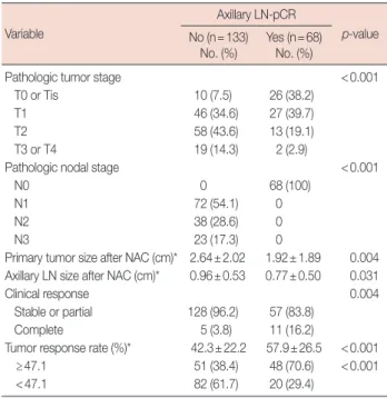
Table 2. Comparison of patient clinicopathologic response between the axillary LN pCR and non-axillary LN pCR after NAC
Variable
Axillary LN-pCR
p-value No (n=133)
No. (%) Yes (n=68) No. (%)
Pathologic tumor stage <0.001
T0 or Tis 10 (7.5) 26 (38.2)
T1 46 (34.6) 27 (39.7)
T2 58 (43.6) 13 (19.1)
T3 or T4 19 (14.3) 2 (2.9)
Pathologic nodal stage <0.001
N0 0 68 (100)
N1 72 (54.1) 0
N2 38 (28.6) 0
N3 23 (17.3) 0
Primary tumor size after NAC (cm)* 2.64±2.02 1.92±1.89 0.004 Axillary LN size after NAC (cm)* 0.96±0.53 0.77±0.50 0.031
Clinical response 0.004
Stable or partial 128 (96.2) 57 (83.8)
Complete 5 (3.8) 11 (16.2)
Tumor response rate (%)* 42.3±22.2 57.9±26.5 <0.001
≥47.1 51 (38.4) 48 (70.6) <0.001
<47.1 82 (61.7) 20 (29.4)
p-value of significant difference between Recurrence, by chi-square, Fisher exact and Wilcoxon rank sum test.
LN=lymph node; pCR=pathologic complete response; NAC=neoadjuvant chemotherapy.
*Mean±SD.
el, based on the independent predictors of axillary pCR shown in our multivariate logistic regression analysis. To determine whether a model that included tumor response rate would af- fect the axillary nodal response to NAC, we developed a mod- el dichotomizing tumor response rate as ≥47.1% or <47.1%.
We found that the model that included tumor response rate had a sensitivity of 42.7%, a specificity of 82.7%, a PPV of 55.8%, and an NPV of 73.8% in predicting axillary pCR (Table
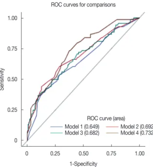
4). The ROC plots in Figure 2 showed that the model that in- cluded tumor response rate had an AUC of 0.732 (95% CI, 0.661–0.804), with better discriminatory ability than other models (p=0.022; 95% CI, 0.012–0.154) (Table 5, Figure 2).
We found that, compared with other predictive factors includ- ing clinical response and axillary LN size after NAC, the tu- mor response rate was the most important predictor and en- hanced the performance of our model.
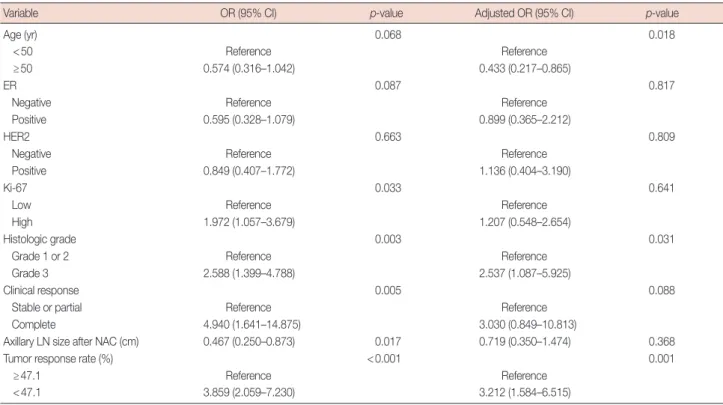
Table 3. Univariate and multivariate logistic regression analysis of variable factors for predicting axillary LN pCR
Variable OR (95% CI) p-value Adjusted OR (95% CI) p-value
Age (yr) 0.068 0.018
<50 Reference Reference
≥50 0.574 (0.316–1.042) 0.433 (0.217–0.865)
ER 0.087 0.817
Negative Reference Reference
Positive 0.595 (0.328–1.079) 0.899 (0.365–2.212)
HER2 0.663 0.809
Negative Reference Reference
Positive 0.849 (0.407–1.772) 1.136 (0.404–3.190)
Ki-67 0.033 0.641
Low Reference Reference
High 1.972 (1.057–3.679) 1.207 (0.548–2.654)
Histologic grade 0.003 0.031
Grade 1 or 2 Reference Reference
Grade 3 2.588 (1.399–4.788) 2.537 (1.087–5.925)
Clinical response 0.005 0.088
Stable or partial Reference Reference
Complete 4.940 (1.641–14.875) 3.030 (0.849–10.813)
Axillary LN size after NAC (cm) 0.467 (0.250–0.873) 0.017 0.719 (0.350–1.474) 0.368
Tumor response rate (%) <0.001 0.001
≥47.1 Reference Reference
<47.1 3.859 (2.059–7.230) 3.212 (1.584–6.515)
Statistics were carried out using logistic regression analysis.
LN=lymph node; pCR=pathologic complete response; OR=odds ratio; CI=confidence interval; ER=estrogen receptor; HER2=human epidermal growth factor receptor 2; NAC=neoadjuvant chemotherapy.
Table 4. Summary of the difference of prediction performance between the models Model Predicted result
Axillary LN-pCR No.
Sensitivity (95% CI) Specificity (95% CI) PPV (95% CI) NPV (95% CI) Observed result
Negative Positive
1 Negative 121 49 0.279 (0.173–0.386) 0.910 (0.861–0.959) 0.613 (0.441–0.784) 0.712 (0.644–0.780)
Positive 12 19
2 Negative 116 40 0.412 (0.295–0.529) 0.872 (0.815–0.929) 0.622 (0.481–0.764) 0.744 (0.675–0.812)
Positive 17 28
3 Negative 120 45 0.338 (0.226–0.451) 0.902 (0.852–0.953) 0.639 (0.482–0.796) 0.727 (0.659–0.795)
Positive 13 23
4 Negative 110 39 0.427 (0.309–0.544) 0.827 (0.763–0.891) 0.558 (0.423–0.693) 0.738 (0.668–0.809)
Positive 23 29
Model 1: age, estrogen receptor status, human epidermal growth factor receptor 2 status, histologic grade, Ki-67; Model 2: Model 1+clinical response; Model 3:
Model 1+axillary lymph node size after neoadjuvant chemotherapy (cm); Model 4: Model 1+tumor response rate.
LN=lymph node; pCR=pathologic complete response; CI=confidence interval; PPV=positive predictive value; NPV=negative predictive value.
DISCUSSION
Multivariate analysis of our patients showed that lower age (<50 years), higher histologic grade, and higher tumor re- sponse rate (≥47.1%) were significant independent predictors of an increased likelihood of achieving axillary pCR. Of these factors, tumor response rate was one of the most reliable and should be included in models predicting axillary response.
Other models for predicting axillary LN pCR have included factors unrelated to nodal status and did not include tumor response rate after NAC [14,15]. Our model, which included tumor response rate, was a better predictor of the probability of achieving axillary LN pCR. A comparison of models that did and did not include tumor response rate found that the model that included response rate, as evaluated by breast MRI, had a significantly improved predicted accuracy, with an AUC of 0.732 (95% CI, 0.661–0.804) and significantly bet- ter predictive power than other models (p=0.022; 95% CI, 0.012–0.154).
NAC has become a standard treatment in patients with clinically node positive breast cancer, resulting in an axillary response [18,19] and the conversion of 40% to 75% of patients from node-positive to node-negative status [8,10]. Patients who achieved axillary pCR had better 5-year overall (93% vs.
72%) and relapse-free (87% vs. 60%) survival rates than pa- tients with residual nodal disease [8]. However, current guide-
lines for the standard management of patients who achieve CR have not been adjusted accordingly, despite high nodal pCR rates [20]. Most patients with axillary LN metastases be- fore NAC undergo ALND, which has been associated with complication such as lymphedema, arm pain, and reduced arm movement due to shoulder dysfunction [11,12,21,22].
To better understand patient outcomes and to identify pa- tients who can omit ALND, it is necessary to improve the ac- curacy of axillary nodal status based on SLNB. SLNB has been used to predict pCR of axillary LNs after NAC in patients with breast cancer and cytologically confirmed nodal metastasis.
Accurate identification of the SLNB in patients likely to achieve nodal pCR, who may benefit from axilla-conserving surgery, is difficult. Cytologic node-positive breast cancer pa- tients who underwent SLNB after NAC and achieved nodal conversion were found to have a false negative rate as high as 20% if one SLN was removed, with the number of harvested SLNs determined by the false negative rate of SLNB after NAC [13]. The accurate determination of axillary nodal status may be improved by the detection of two or more SLNs, by using a dual-tracer for mapping, by using IHC for pathologic evalua- tion, and by ensuring the removal of the axillary LN initially identified as being a nodal metastasis by marking with a clip [23]. Therefore, in developing a model with improved perfor- mance, we added noninvasive predicting factors such as tu- mor response rate. Although clinical responses may be pre- dictive of axillary pCR in response to NAC, many patients do not achieve clinical CR, with most patients who receive NAC achieving partial response. Therefore, clinical response is pre- dictive of axillary pCR in few patients. Because partial re- sponse is defined as a ≥30% reduction in tumor size, clinical Figure 2. Receiver operating characteristics curve (ROC) of the each
models to predict axillary pathologic complete response. The area un- der the ROC curve is 0.732, 95% confidence interval (0.661–0.804) in model 4.
1.00
0.75
0.50
0.25
0
0 0.25 0.50 0.75 1.00
1-Specificity ROC curves for comparisons
Sensitivity
ROC curve (area)
Model 1 (0.649) Model 2 (0.692) Model 3 (0.682) Model 4 (0.732)
Table 5. Comparison difference of AUC each models
Model AUC Standard error 95% CI
Model 1 0.649 0.042 0.568–0.731
Model 2 0.692 0.041 0.612–0.771
Model 3 0.682 0.041 0.602–0.761
Model 4 0.732 0.037 0.661–0.804
Comparison each
models Difference AUC 95% CI p-value
Model 1 vs. Model 2 0.042 −0.008 to 0.092 0.097 Model 1 vs. Model 3 0.032 −0.021 to 0.086 0.243 Model 1 vs. Model 4 0.083 0.012 to 0.154 0.022 The difference of prediction performance between the models were presented the ROC curve (AUC) between the models.
Model 1: age, estrogen receptor status, human epidermal growth factor re- ceptor 2 status, histologic grade, Ki-67; Model 2: Model 1+clinical response;
Model 3: Model 1+axillary lymph node size after neoadjuvant chemotherapy (cm); Model 4: Model 1+tumor response rate.
AUC =area under receiver operating characteristic (ROC) curves; CI = confidence interval.
partial response results in various tumor response rates. Be- cause we found that tumor response rate was associated with axillary pCR and may be predictive in additional patients, we incorporated tumor response rate into our model. We also showed that tumor response could be easily determined by measuring tumor diameter and axillary LN diameter on breast MRI. Radiologic results have shown diagnostic value in evaluating axillary LN metastases after NAC, with combina- tions that included MRI showing greater sensitivity in detect- ing positive axillary LN metastases [24]. Breast MRI is includ- ed in the standard workup of patients undergoing NAC in our institution, with tumor response rate determined by measur- ing tumor and axillary LN diameter on breast MRI before and after NAC. Tumor response rate is an easily measured clinico- pathologic variable, allowing simple and rapid prediction of axillary pCR. This parameter can be used in making treat- ment decisions and in clinical trials [25]. Patients with a high- er tumor response rate are more likely to achieve axillary LN pCR. SLNBs negative for metastases indicate that ALND can be safely omitted, thereby avoiding the postoperative compli- cations of this procedure.
Our study had several advantages compared with previous studies predicting axillary pCR after NAC in patients with cy- tologically proven nodal metastasis [14,15,26]. Most impor- tantly, these previous studies did not include tumor and nodal response rates to NAC. Tumor response rate offers several ad- vantages compared with alternative methods for assessing ax- illary pCR after NAC. First, in contrast to SLNB, the predic- tion of axillary pCR based on tumor response rate is non-in- vasive, reducing associated morbidity. Second, because breast MRI is included in standard initial workup of patients with breast cancer before, tumor response rate can be readily cal- culated by comparing MRI results before and after NAC.
Moreover, this procedure is covered by the national health in- surance in Korea, eliminating the need for additional proce- dures, such as diagnostic tests and surgical procedures. Third, core needle biopsy was used to confirm all patients with axil- lary LN metastases before NAC, making our results more ac- curate than those of previous studies.
Our study also had several limitations. First, it was retro- spective in design, involving a limited number of patients at a single institution. The study results were not validated exter- nally, and median tumor response rate may have limited the generalizability of our findings. Second, our study included only patients who underwent NAC, followed by radical sur- gery including ALND. The false negative rate of SLNB is an important indicator of cytologically confirmed nodal metas- tasis. We did not compare the pathological status of the SLN to the remainder of LNs in the axilla following ALND. Third,
the patients with HER2-positive tumors did not include those who received NAC that included trastuzumab. Assessments of tumor response rates to NAC using combinations of radio- logic measurements are required, as are well-controlled, pro- spective studies in large numbers of patients.
In conclusion, this study evaluated the ability of various fac- tors to predict axillary LN pCR in breast cancer patients treat- ed with NAC and compared models based on these predic- tors. Tumor response rate was the most important predictor of axillary LN pCR in response to NAC. Use of models that include tumor response rates may avoid the need for unneces- sary axillary LN dissection.
CONFLICT OF INTEREST
The authors declare that they have no competing interests.
REFERENCES
1. Bear HD, Anderson S, Smith RE, Geyer CE Jr, Mamounas EP, Fisher B, et al. Sequential preoperative or postoperative docetaxel added to pre- operative doxorubicin plus cyclophosphamide for operable breast can- cer: National Surgical Adjuvant Breast and Bowel Project Protocol B-27. J Clin Oncol 2006;24:2019-27.
2. Cunningham JD, Weiss SE, Ahmed S, Bratton JM, Bleiweiss IJ, Tartter PI, et al. The efficacy of neoadjuvant chemotherapy compared to post- operative therapy in the treatment of locally advanced breast cancer.
Cancer Invest 1998;16:80-6.
3. Mauri D, Pavlidis N, Ioannidis JP. Neoadjuvant versus adjuvant system- ic treatment in breast cancer: a meta-analysis. J Natl Cancer Inst 2005;97:188-94.
4. Navarro-Cecilia J, Dueñas-Rodríguez B, Luque-López C, Ramírez- Expósito MJ, Martínez-Ferrol J, Ruíz-Mateas A, et al. Intraoperative sentinel node biopsy by one-step nucleic acid amplification (OSNA) avoids axillary lymphadenectomy in women with breast cancer treated with neoadjuvant chemotherapy. Eur J Surg Oncol 2013;39:873-9.
5. Rouzier R, Mathieu MC, Sideris L, Youmsi E, Rajan R, Garbay JR, et al.
Breast-conserving surgery after neoadjuvant anthracycline-based che- motherapy for large breast tumors. Cancer 2004;101:918-25.
6. Wolmark N, Wang J, Mamounas E, Bryant J, Fisher B. Preoperative chemotherapy in patients with operable breast cancer: nine-year results from National Surgical Adjuvant Breast and Bowel Project B-18. J Natl Cancer Inst Monogr 2001;30:96-102.
7. Gonzalez-Angulo AM, McGuire SE, Buchholz TA, Tucker SL, Kuerer HM, Rouzier R, et al. Factors predictive of distant metastases in patients with breast cancer who have a pathologic complete response after neo- adjuvant chemotherapy. J Clin Oncol 2005;23:7098-104.
8. Hennessy BT, Hortobagyi GN, Rouzier R, Kuerer H, Sneige N, Buzdar AU, et al. Outcome after pathologic complete eradication of cytologi- cally proven breast cancer axillary node metastases following primary chemotherapy. J Clin Oncol 2005;23:9304-11.
9. Rouzier R, Extra JM, Klijanienko J, Falcou MC, Asselain B, Vincent- Salomon A, et al. Incidence and prognostic significance of complete
axillary downstaging after primary chemotherapy in breast cancer pa- tients with T1 to T3 tumors and cytologically proven axillary metastatic lymph nodes. J Clin Oncol 2002;20:1304-10.
10. Boughey JC, Suman VJ, Mittendorf EA, Ahrendt GM, Wilke LG, Taback B, et al. Sentinel lymph node surgery after neoadjuvant chemo- therapy in patients with node-positive breast cancer: the ACOSOG Z1071 (Alliance) clinical trial. JAMA 2013;310:1455-61.
11. Lucci A, McCall LM, Beitsch PD, Whitworth PW, Reintgen DS, Blumencranz PW, et al. Surgical complications associated with sentinel lymph node dissection (SLND) plus axillary lymph node dissection compared with SLND alone in the American College of Surgeons Oncology Group Trial Z0011. J Clin Oncol 2007;25:3657-63.
12. Mansel RE, Fallowfield L, Kissin M, Goyal A, Newcombe RG, Dixon JM, et al. Randomized multicenter trial of sentinel node biopsy versus standard axillary treatment in operable breast cancer: the ALMANAC Trial. J Natl Cancer Inst 2006;98:599-609.
13. Kuehn T, Bauerfeind I, Fehm T, Fleige B, Hausschild M, Helms G, et al.
Sentinel-lymph-node biopsy in patients with breast cancer before and after neoadjuvant chemotherapy (SENTINA): a prospective, multicentre cohort study. Lancet Oncol 2013;14:609-18.
14. Jin X, Jiang YZ, Chen S, Shao ZM, Di GH. A nomogram for predicting the pathological response of axillary lymph node metastasis in breast cancer patients. Sci Rep 2016;6:32585.
15. Vila J, Mittendorf EA, Farante G, Bassett RL, Veronesi P, Galimberti V, et al. Nomograms for predicting axillary response to neoadjuvant che- motherapy in clinically node-positive patients with breast cancer. Ann Surg Oncol 2016;23:3501-9.
16. Eisenhauer EA, Therasse P, Bogaerts J, Schwartz LH, Sargent D, Ford R, et al. New response evaluation criteria in solid tumours: revised RECIST guideline (version 1.1). Eur J Cancer 2009;45:228-47.
17. Therasse P, Arbuck SG, Eisenhauer EA, Wanders J, Kaplan RS, Rubinstein L, et al. New guidelines to evaluate the response to treatment in solid tumors: European Organization for Research and Treatment of Cancer, National Cancer Institute of the United States, National Cancer Institute of Canada. J Natl Cancer Inst 2000;92:205-16.
18. Alvarado R, Yi M, Le-Petross H, Gilcrease M, Mittendorf EA, Bedrosian
I, et al. The role for sentinel lymph node dissection after neoadjuvant chemotherapy in patients who present with node-positive breast can- cer. Ann Surg Oncol 2012;19:3177-84.
19. Straver ME, Rutgers EJ, Russell NS, Oldenburg HS, Rodenhuis S, Wesseling J, et al. Towards rational axillary treatment in relation to neoadjuvant therapy in breast cancer. Eur J Cancer 2009;45:2284-92.
20. Vugts G, Maaskant-Braat AJ, de Roos WK, Voogd AC, Nieuwenhuijzen GA. Management of the axilla after neoadjuvant chemotherapy for clinically node positive breast cancer: a nationwide survey study in the Netherlands. Eur J Surg Oncol 2016;42:956-64.
21. Fleissig A, Fallowfield LJ, Langridge CI, Johnson L, Newcombe RG, Dixon JM, et al. Post-operative arm morbidity and quality of life: results of the ALMANAC randomised trial comparing sentinel node biopsy with standard axillary treatment in the management of patients with early breast cancer. Breast Cancer Res Treat 2006;95:279-93.
22. Kakuda JT, Stuntz M, Trivedi V, Klein SR, Vargas HI. Objective assess- ment of axillary morbidity in breast cancer treatment. Am Surg 1999;
65:995-8.
23. Caudle AS, Yang WT, Krishnamurthy S, Mittendorf EA, Black DM, Gilcrease MZ, et al. Improved axillary evaluation following neoadju- vant therapy for patients with node-positive breast cancer using selec- tive evaluation of clipped nodes: implementation of targeted axillary dissection. J Clin Oncol 2016;34:1072-8.
24. You S, Kang DK, Jung YS, An YS, Jeon GS, Kim TH. Evaluation of lymph node status after neoadjuvant chemotherapy in breast cancer patients: comparison of diagnostic performance of ultrasound, MRI and ¹⁸F-FDG PET/CT. Br J Radiol 2015;88:20150143.
25. Partridge SC, Gibbs JE, Lu Y, Esserman LJ, Tripathy D, Wolverton DS, et al. MRI measurements of breast tumor volume predict response to neoadjuvant chemotherapy and recurrence-free survival. AJR Am J Roentgenol 2005;184:1774-81.
26. Schipper RJ, Moossdorff M, Nelemans PJ, Nieuwenhuijzen GA, de Vries B, Strobbe LJ, et al. A model to predict pathologic complete re- sponse of axillary lymph nodes to neoadjuvant chemo (immuno) ther- apy in patients with clinically node-positive breast cancer. Clin Breast Cancer 2014;14:315-22.