Gorlin and Goltz in 1960 defined the condition as a syndrome comprising the principal triad of multiple basal cell nevi, jaw keratocysts, and skeletal anomalies.1-3 The condition is also known as Gorlin syndrome, nevoid basal cell carcinoma syndrome, and basal cell nevus syndrome.4-7 Gorlin-Goltz syndrome is an autosomal dominant inherited condition that exhibits high penetrance and variable expres- sivity, however this disorder can arise spontaneously.2,7-9 Almost 60% of the patients with Gorlin-Goltz syndrome have no known affected family members.2 The Gorlin- Goltz syndrome gene has been mapped to chromosome 9q22.3-q31.5,7 The prevalence of Gorlin-Goltz syndrome has been estimated about 1 per 60,000.10Males and females are equally affected.11The main clinical features of Gorlin- Goltz syndrome includes multiple keratocystic odontogenic tumor (75%), basal cell nevi (80% in whites and 38% in blacks), and skeletal anomalies (70%).2,12 We present a report of Gorlin-Goltz syndrome with familial pattern affecting father and daughter.
Case Report
A 39-year-old male patient reported to the dental insti- tute with the complaint of pus discharge from right lower posterior region of the jaw since one month ago. The patient noticed decayed teeth in the same region, which was not associated with pain. He had visited a private dental clinic, where decayed tooth had been extracted and medications had been given for about 5 days.
On general physical examination, the patient was well built, 182 cm tall with normal gait and satisfactory vital signs. The extraoral examination revealed hypertelorism, strabismus, and a cystic swelling on the left eyelid as well as his neck (Fig. 1). Intraoral examination revealed miss- ing teeth of the right mandibular first and second molars and pus discharge from the same region. No other skele- tal abnormalities were detected.
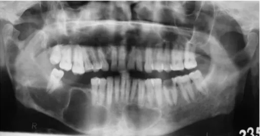
Panoramic radiograph revealed well defined multiple uni- locular radiolucencies with sclerotic borders in the mandi- bular body, ramus, and symphysis areas. The unilocular radiolucencies varied in diameter from minimum 3 cm to around 7 cm in diameter. The largest one was located in the body of the mandible on the right side extending supe- riorly from the edentulous area to the lower border of the mandible inferiorly. Three smaller radiolucencies measur-
Radiological features of familial Gorlin-Goltz syndrome
Shruthi Hegde, Shishir Ram Shetty
Department of Oral Medicine and Radiology, AB Shetty Memorial Institute of Dental Sciences, Nitte University, Mangalore, India ABSTRACT
Gorlin-Goltz syndrome is an autosomal dominant disorder principally characterized by cutaneous basal cell carcinomas, multiple keratocystic odontogenic tumors, and skeletal anomalies. This syndrome may be diagnosed early by dentist because keratocystic odontogenic tumors are usually one of the first manifestations of the syndrome. Early diagnosis and treatment are of utmost importance in reducing the severity of long term sequelae of this syndrome. This report presents a rare event of Gorlin-Goltz syndrome occurring in a 39-year-old male and his 8-year-old daughter. The clinical and investigative features of this familial disorder has been described in detail. (Imaging Sci Dent 2012;
42 : 55-60)
KEY WORDS : Basal Cell Nevus Syndrome; Odontogenic Cysts; Skeletal Anomalies
Received October 19, 2011; Revised December 2, 2011; Accepted January 21, 2012 Correspondence to : Dr. Shruthi Hegde
Department of Oral Medicine and Radiology, AB Shetty Memorial Institute of Dental Sciences, Nitte University , Deralakatte, Mangalore 575018, Karnataka, India Tel) 91-990-1321299, Fax) 91-824-2204776, E-mail) [email protected]
Copyright ⓒ 2012 by Korean Academy of Oral and Maxillofacial Radiology
This is an Open Access article distributed under the terms of the Creative Commons Attribution Non-Commercial License (http://creativecommons.org/licenses/by-nc/3.0) which permits unrestricted non-commercial use, distribution, and reproduction in any medium, provided the original work is properly cited.
Imaging Science in Dentistry∙pISSN 2233-7822 eISSN 2233-7830
ing around 3 cm in size were located in the posterior part of the right mandibular body and ramus as well in the left sym- physis region. A smaller radiolucency measuring around 1 cm in size was observed in the periapical region of the right maxillary third molar. There was no radiographic evidence of tooth displacement and root resorption (Fig. 2).
Cross sectional mandibular occlusal radiograph revealed a radiolucent area with minimum cortical plate expansion (Fig. 3A). The findings of panoramic radiograph raised the possibility of Gorlin-Goltz syndrome and further inves- tigations were carried out. Chest radiograph revealed the bifid fourth and eighth rib on the right side (Fig. 3B).
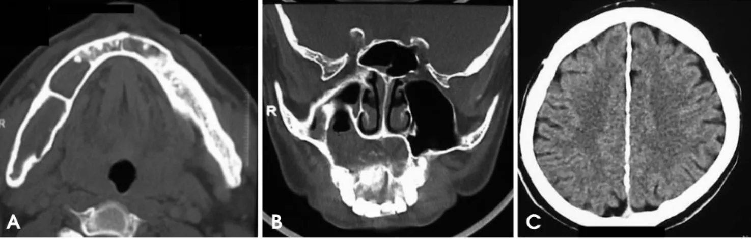
CT images showed hypodense areas in relation to the right mandibular body separated by a hyperdense septae.
Minor breach in the cortical integrity was observed in the lingual aspect of the ramus region. A single lytic lesion was observed in relation to symphysis region. The lesion involved the right half of maxilla including maxillary sinus.
Mucosal thickening in the right maxillary sinus was also observed (Figs. 4A and B). Calcification of falx cerebri and tentorium cerebella was also observed (Fig. 4C). Inci- sional biopsy of the lesion through intraoral approach re- vealed the following histopathological features: parakera- tinized, corrugated, 6-10 layers thick epithelium with pali- saded, polarized basal cell layer. The connective tissue showed daughter cysts suggestive of keratocystic odonto- genic tumor (Figs. 5A and B).
Owing to the familial tendency of this diagnosed condi- tion, the family members were subjected to thorough cli- nical and radiological examination. The patient’s 8-year- old daughter showed a clinical feature of hypertelorism (Fig. 6A). Her panoramic radiograph revealed well defined, unilocular radiolucency measuring approximately 2.5 cm in diameter with sclerotic border in her mandibular sym- physis region. The radiolucent area extended superiorly from the root apices of the mandibular anterior teeth to the lower border of the mandible inferiorly enclosing the left canine and premolar tooth buds (Fig. 6B).
Histopathological evaluation of the specimen obtained after intraoral biopsy procedure revealed histopathological features similar to that of her father (Fig. 7). No other family members were affected by this condition (Fig. 8).
A diagnosis of Gorlin-Goltz Syndrome was made on the basis of clinical, imaging, and histological findings. The father and daughter were advised surgical removal of the
A
B
C
Fig. 1.A. Photograph shows increased inner canthal distance. B.
Cystic swelling on the left eyelid is seen. C. Cystic swelling in the midline of the neck is seen.
Fig. 2.Panoramic radiograph shows multiple unilocular radiolucencies in mandibular body, ramus, and sym- physis region and a smaller uniloc- ular radiolucency in the right maxil- lary tuberosity area.
Fig. 3.A. The mandibular occlusal cross sectional radiograph shows a radiolucent area with minimum cor- tical plate expansion. B. The chest radiograph shows bifid fourth and eighth rib on the right side.
Fig. 4.A. Axial CT image shows hypodense areas in relation to the right mandibular body separated by hyperdense septae. B. Coronal CT image shows the lesion involving the right half of maxilla including maxillary sinus. C. Axial CT image shows calcification of falx cerebri.
A B C
Fig. 5.Photomicrographs show the parakeratinized, corrugated, 6-10 layers thick epithelium with palisaded, polarized basal cell layer.
Also, daughter cysts in connective tissue suggestive of an keratocystic odontogenic tumor is observed (H&E stain, A. ×40, B. ×100) .
A B
A B
keratocystic odontogenic tumor. Unfortunately due to certain logistical and financial reasons, the patients could not keep up with appointments.
Discussion
Gorlin-Goltz syndrome was first reported by Jarish in 1894.13The syndrome has been designated by a variety of different terms including Gorlin syndrome, nevoid basal cell carcinoma syndrome, basal cell nevus syndrome, syn- drome of jaw cysts, and jaw cyst - basal cell nevus - bifid rib syndrome.4-7,12Gorlin suggested the term nevoid basal
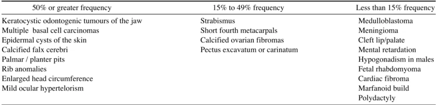
cell carcinoma syndrome, however all affected adults did not have basal cell carcinomas. The condition is known as Gorlin syndrome because of Gorlin’s contributions to the understanding of the condition.7,12 Clinical features of Gorlin-Goltz syndrome arises at the first, second or third decade.2In the present case, the features were identified at fourth decade and in daughter at the first decade. Gorlin- Goltz syndrome has an autosomal dominant mode of inher- itance, but can arise spontaneously, or can have a variable phenotypic penetration.14 Similar pattern of inheritance was observed in our cases. Gorlin and Goltz in 1960 gave a complete description of the syndrome.1This syndrome is associated with a wide spectrum of developmental ano- malies and neoplasms.9The most common and significant features are summarized in Table 1.
Keratocystic odontogenic tumor can be the first feature of the syndrome. The tumor is typically found as inciden- tal radiographic findings. The tumor may manifest clini- cally if it become infected or cause symptoms such as swelling. The tumors were responsible for detection of
Fig. 6.A. The daughter’s photograph reveals hypertelorism. B. Her panoramic radiograph shows well defined radiolucency in the mandi- bular symphysis region with displacement of the erupting canine.
A B
Fig. 7.Photomicrograph shows the parakeratinized, corrugated, 6- 8 layers thick epithelium with palisaded, polarized basal cell layer (H&E stain, ×100).
Fig. 8. Pedigree chart of the affected family shows the mode of inheritance of Gorlin-Goltz syndrome.
the syndrome in our cases. Keratocystic odontogenic tumors in Gorlin-Goltz syndrome usually comprise unilo- cular or multilocular radiolucencies of the mandibular body, angle, or ramus.2 In children and adolescents, the cysts may cause displacement of developing teeth and delayed dental development.9 The present case also showed the similar radiological features such as the displacement of the tooth bud in the daughter’s mandibular symphysis region. The most frequent skin lesions of Gorlin-Goltz syndrome are cutaneous basal cell carcinomas, benign dermal cysts, and palmar and plantar keratosis or pits.2,15 Apart from the cutaneous dermal cyst, there was no skin lesion in the father. Almost 70% of patients with this syn- drome have some degree of cranio-facial anomalies. These can comprise frontal and parietal bossing and broad nasal root which may be associated with occular hypertelorism.2 Hypertelorism was also observed in these cases. Thoracic cage anomalies such as bifid and fused ribs may be present.
Syndactyly or polydactyly of toes may occur.2Although no digital abnormalities were observed in both of our cases, bifid ribs were observed in the father. Ectopic calcification of falx cerebri, tentorium cerebella, and bridged sella may also be detected radiologically.2The diagnosis of Gorlin- Goltz syndrome requires the presence of two major or one major and two minor criteria.2,3,7,16 In the present cases, the following major and minor criteria were present: histo- logically proven keratocystic odontogenic tumors of the jaws, calcification of the falx cerebri, bifid ribs, and hyper- telorism. The patients affected by Gorlin-Goltz syndrome must be evaluated by several relevant specialists to preci- sely confirm the diagnosis, detect the likely genetic basis, provide appropriate genetic counseling, and manage the various clinical manifestations. Early diagnosis and treat- ment may reduce the severity of the long term sequelae of Gorlin-Goltz syndrome including malignancy and oromax- illofacial deformation and destruction.
References
1. Gorlin RJ, Goltz RW. Multiple nevoid basal-cell epithelioma, jaw cysts and bifid rib. A syndrome. N Engl J Med 1960; 262 : 908-12.
2. Manfredi M, Vescovi P, Bonanini M, Porter S. Nevoid basal cell carcinoma syndrome: a review of the literature. Int J Oral Maxillofac Surg 2004; 33 : 117-24.
3. Casaroto AR, Loures DC, Moreschi E, Veltrini VC, Trento CL, Gottardo VD, et al . Early diagnosis of Gorlin-Goltz syn- drome: case report. Head Face Med 2011; 7 : 2.
4. Markt JC. Implant prosthodontic rehabilitation of a patient with nevoid basal cell carcinoma syndrome: a clinical report.
J Prosthet Dent 2003; 89 : 436-42.
5. Honavar SG, Shields JA, Shields CL, Eagle RC, Demirci H, Mahmood EZ. Basal cell carcinoma of the eyelid associated with Gorlin-Goltz syndrome. Ophthalmology 2001; 108 : 1115-23.
6. Doede T, Seidel J, Riede FT, Vogt L, Mohr FW, Schier F.
Occult, life-threatening, cardial tumor in syndactylism in Gorlin Goltz syndrome. J Pediatr Surg 2004; 39 : e17-9.
7. Deepa MS, Paul R, Balan A. Gorlin Goltz syndrome: a review. J Indian Acad Oral Med Radiol 2003; 15 : 203-9.
8. Lo Muzio L, Nocini P, Bucci P, Pannone G, Consolo U, Pro- caccini M. Early diagnosis of nevoid basal cell carcinoma syndrome. J Am Dent Assoc 1999: 130 : 669-74.
9. Melo ES, Kawamura JY, Alves CA, Nunes FD, Jorge WA, Cavalcanti MG. Imaging modality correlations of an odonto- genic keratocyst in the nevoid basal cell carcinoma syndrome:
a family case report. Oral Surg Oral Med Oral Pathol Oral Radiol Endod 2004; 98 : 232-6.
10. Lee BD, Kim JH, Choi DH, Koh KS, Lee SR. Recurrent odon- togenic keratocysts in basal cell nevus syndrome: report of a case. Korean J Oral Maxillofac Radiol 2004; 34: 203-7.
11. Kimonis VE, Goldstein AM, Pastakia B, Yang ML, Kase R, DiGiovanna JJ, et al. Clinical manifestations in 105 persons with nevoid basal cell carcinoma syndrome. Am J Med Genet 1997; 69 : 299-308.
12. Gorlin RJ, Cohen MM, Levin LS. Syndromes of the head and neck. 3rd ed. New York: Oxford University Press; 1990. p.
372-8.
13. Jarish W. Zur lehre von den autgeschwulsten. Archiv Jur Dermatologic Syphilogic 1894; 28 : 163-222.
Table 1.Most common clinical features of the Gorlin-Goltz syndrome1,11
50% or greater frequency 15% to 49% frequency Less than 15% frequency
Keratocystic odontogenic tumours of the jaw Strabismus Medulloblastoma
Multiple basal cell carcinomas Short fourth metacarpals Meningioma
Epidermal cysts of the skin Calcified ovarian fibromas Cleft lip/palate
Calcified falx cerebri Pectus excavatum or carinatum Mental retardation
Palmar / planter pits Hypogonadism in males
Rib anomalies Fetal rhabdomyoma
Enlarged head circumference Cardiac fibroma
Mild ocular hypertelorism Marfanoid build
Polydactyly
14. Patil K, Mahima VG, Gupta B. Gorlin syndrome: a case report. J Indian Soc Pedod Prev Dent 2005; 23 : 198-203.
15. Rai S, Gauba K. Jaw cyst-Basal cell nevus-Bifid rib syndrome:
a case report. J Indian Soc Pedod Prev Dent 2007; 25: 137-9.
16. Jones EA, Sajid MI, Shenton A, Evans DG. Basal cell carcino- mas in gorlin syndrome: a review of 202 patients. J Skin Can- cer 2011; 2011 : 217378.