J. of Korean Bone & Joint Tumor Soc.
Volume 15, Number 2, December, 2009
※통신저자: 김김 진진 아아
경기도 부천시 원미구 소사동 2번지 가톨릭대학교 의과대학 부천성모병원 병리과
Tel: 032) 340-7091, Fax: 032) 340-2219, E-mail: [email protected]
소아의 견갑골에 생긴 투명세포연골육종 - 1예 보고 -
가톨릭대학교 의과대학 병리학교실, 정형외과학교실1 이경지∙이안희∙김진아∙김형민1∙이교영
투명세포연골육종은 모든 연골육종의 2%를 차지하는 매우 드문 저등급성 종양이다. 주로 긴뼈의 뼈끝에서 발생하며 대퇴골과 상완골의 근위부에서 가장 흔하고, 견갑골을 포함한 납작 뼈에는 드물다. 25-50세 사이의 연령에서 호발하며, 20세 이전의 발생은 흔치 않다. 조직학적 으로 종양 세포는 소엽상 무리를 지어 관찰되며, 투명하고 풍부한 세포질을 특징적으로 가진 다. 저자들은 8세 여아의 견갑골에서 발생된 투명세포연골육종을 보고하고자 한다.
색인 단어: 견갑골, 투명세포연골육종
Clear cell chondrosarcoma is a rare variant of low-grade chondrosarcoma. It is charac- terized by round to polygonal chondrocytes with abundant clear cytoplasm arranged in a lobulated growth pattern. Unlike conven- tional chondrosarcoma, clear cell chondrosar- coma has a strong tendency to arise in the epiphyseal ends of long tubular bones, espe- cially the proximal femur and the proximal humerus2). Therefore, clear cell chondrosar- comas of the scapula, flat bone, are rare.
Clear cell chondrosarcoma frequently affects individuals in the third to fifth decades of life and it is distinctly uncommon in young patients6). Here, we report such a rare tumor involving the scapula in an 8-year-old
girl.
CASE REPORT
An 8-year-old girl presented with protru- sion of the right clavicle for 6 months dura- tion. On examination, she had a subtle asymmetry in the shoulders. Laboratory data were remarkable for an elevated serum alkaline phospatase of 385U/L. Magnetic resonance image of the left shoulder revealed an expansive mass based on the left scapular spine projected into the glenoid and coronoid processes. The tumor had an isoin- tense signal in T1-weighted images, and an intense heterogenous signal on T2-weighted
images (Fig. 1).
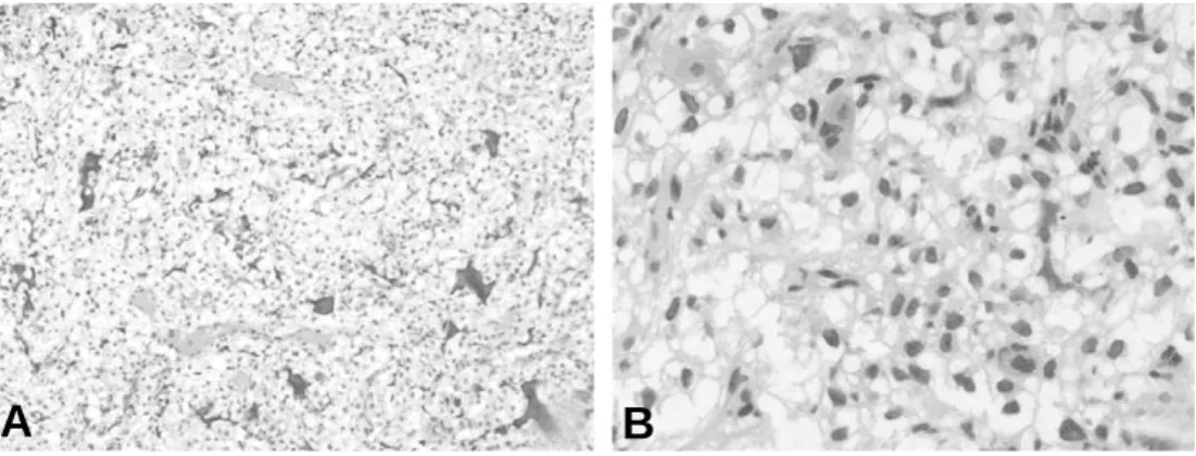
Curettage and bone graft with allo-chip bone was performed. Histologically, the tumor showed compact sheets of tumor cells with round or ovoid shaped, abundant clear cytoplasm (Fig. 2A). The nucleus was cen- trally located and the cytoplasmic border was distinct. Mitotic figures were rare.
Osteoid production was conspicuous. In some areas, osteoid seams or bony trabeculae sur- rounded by osteoblasts were noted, which
were considered to be benign and reactive (Fig. 2B). PAS was strongly positive within the clear cells. Vimentin was positive but S- 100 protein was negative. Histopathologic examination of the curettage specimen con- firmed the diagnosis of clear cell chondrosar- coma. Following surgery, the alkaline phos- phatase returned to normal. The patient is disease free after 6 months’follow-up.
Fig. 1. Magnetic resonance images of the left shoulder shows an isointense signal mass in T1-weight- ed coronal MR image (A) and in T1- weighted axial MR image (B). T2-weighted coronal MR image shows a heterogenous pattern (C). Plain X-ray also shows a relatively well defined lesion with mineralization (D).
A B
C D
DISCUSSION
Benign cartilaginous tumors including osteochondroma, enchondroma, and chon- dromyxoid fibroma are rather common in childhood. However, chondrosarcoma, a malignant cartilage-forming tumor, is rarely seen in the first and second decades of life10). Clear cell chondrosarcoma is extremely rare3). Unlike conventional chondrosarcoma, clear cell chondrosarcoma commonly involves the epiphysis of long bone2). The proximal femur is the most frequent site of involve- ment, followed by the proximal humerus, whereas involvement of the scapula is rare1). Only approximately 55 cases of conventional chondrosarcoma in the scapula have been reported4,9) and only one case of clear cell chondrosarcoma has been reported to date1). Clear cell chondrosarcoma usually occurs in adults in the third to fifth decades of life.
Patients with clear cell chondrosarcoma under 15 years of age are extremely rare7).
The clinical symptoms are not specific and most of the patients complain of local pain of variable duration. Because of low-grade malignant and slow growing nature of the
tumor, the clinical courses were generally long-standing5). Ogose et al.8) reviewed and analyzed the patients with clear cell chon- drosarcoma, figured out that clear cell chon- drosarcoma produce alkaline phosphatase, which can be used as a tumor marker. Our patient had an elevated alkaline phosphatase level, after removal of the tumor, the level decreased.
Histologic appearance is characteristic and diagnostic. Clear cell chondrosarcoma is characterized by the presence of large cells with extensively vacuolated clear cytoplasm and a lobulated architecture2). Nuclei are round and centrally located in the clear cytoplasm. The tumor cells are surrounded by a loose, scarce to moderate amount of cartilaginous matrix. Mitotic figures are rare. In some cases, conventional chon- drosarcoma can be seen. Multinucleated giant cells are commonly found in clear cell chondrosarcoma. Clear cell chondrosarcoma contains variable amounts of hyaline carti- lage, osteoid production, and focal cystic changes such as aneurismal bone cyst for- mation, making it difficult to get diagnosis.
Solitary bone cyst, aneurysmal bone cyst,
Fig. 2. Histologic appearance of clear cell chondrosarcoma. Compact sheets of tumor cells with round or ovoid shaped, clear cytoplasm and central small nuclei. Irregular osteoid are observed (A).
High-power view of tumor cells showed abundant clear cytoplasm with centrally located nuclei and distinct cytoplasmic borders. A few multinucleated giant cells are observed (B).
A B
chondroblastoma, osteoblastoma, metastatic clear cell carcinoma, and osteosarcoma should be included in the differential diag- nosis.
Because clear cell chondrosarcoma is rare in the childhood, and the scapula is an uncommon location, our patient in this report seems to be the youngest patient reported, to our knowledge. Although the occurrence of clear cell chondrosarcoma aris- ing from the scapula is extremely rare in children, awareness of this entity in the young age group and its occurrence in the scapula is important. Our case highlights the facts that clear cell chondrosarcoma could occur at an odd site and in children.
REFERENCES
01) Collins MS, Koyama T, Swee RG, and Inwards CY: Clear cell chondrosarcoma: radiographic, com- puted tomographic, and magnetic resonance find- ings in 34 patients with pathologic correlation.
Skeletal Radiol, 32(12): 687-694, 2003.
02) Dahlin DC and Ivins JC: Benign chondroblas- toma. A study of 125 cases. Cancer, 30(2): 401-413, 1972.
03) Donati D, Yin JQ, Colangeli M, et al.: Clear cell chondrosarcoma of bone: long time follow-up of 18 cases. Arch Orthop Trauma Surg, 128(2): 137-142, 2008.
04) Griffin AM, Shaheen M, Bell RS, Wunder JS, and Ferguson PC: Oncologic and functional out- come of scapular chondrosarcoma. Ann Surg Oncol, 15(8): 2250-2256, 2008.
05) Laporte C, Anract P, Tomeno B, and Forest M:
Clear cell chondrosarcoma. Study of 13 clinical cases and review of the literature. Rev Chir Orthop Reparatrice Appar Mot, 82(8): 691-699, 1996.
06) Mahesha V, Goyal R, Vaiphei K, Nada R, and Gupta AK: Primary chondrosarcoma of ethmoid bone in a 6-year-old child. Ann Diagn Pathol, 10(3): 154-156, 2006.
07) Ohno T, Park P, Oguro K, et al.: Ultrastructural study of a clear cell chondrosarcoma. Ultrastruct Pathol, 10(4): 321-330, 1986.
08) Ogose A, Hotta T, Kawashima H, et al.:
Elevation of serum alkaline phosphatase in clear cell chondrosarcoma of bone. Anticancer Res, 21(1B): 649-655, 2001.
09) Pant R, Yasko AW, Lewis VO, Raymond K, and Lin PP: Chondrosarcoma of the scapula: long-term oncologic outcome. Cancer, 104(1): 149-158, 2005.
10) Young CL, Sim FH, Unni KK, and McLeod RA:
Chondrosarcoma of bone in children. Cancer, 66(7):
1641-1648, 1990.
Clear Cell Chondrosarcoma of the Scapula in a Child -A Case Report-
Kyungji Lee, M.D., An-Hi Lee, M.D., Jeana Kim, M.D., Hyoung-Min Kim, M.D.
1, Kyo-Young Lee, M.D.
Department of Hospital Pathology, Department of orthopedic surgery
1, College of Medicine, The Catholic University of Korea, Seoul, Korea
Clear cell chondrosarcoma is a rare, low-grade variant of chondrosarcoma that comprises approximately 2% of all chondrosarcomas. This tumor usually involves the epiphysis and epimetaphysis of long bones, especially the proximal part of the femur or humerus, whereas involvement of the scapula is rare. It occurs at any age, but the peak is third to fifth decade, and is rarely seen in the first and second decades of life. Histologically, tumor cells with abundant clear cytoplasm and benign giant cells are usually found. We report on a case of clear cell chon- drosarcoma of the scapula in an 8-year-old girl.
Key Words: Scapula, Clear cell chondrosarcoma
Address reprint requests to Jeana Kim, M.D.
Deaprtment of Hospital Pathology, Bucheon St. Mary’s Hospital, College of Medicine, The Catholic University of Korea, Wonmigu Sosa 2 dong 2 Bucheon city, Gyeonggi-province
TEL: 82-32-340-7091, FAX: 82-32-340-2219, E-mail: [email protected]
Abstract