Intrinsic Vertebral Markers for Spinal Level Localization in Anterior Cervical Spine
Surgery: A Preliminary Report
Deepak Kumar Jha
1, Anil Thakur
2, Mukul Jain
3, Arvind Arya
3, Chandrabhushan Tripathi
4, Rima Kumari
5, Suman Kushwaha
61Department of Neurosurgery, Institute of Human Behavior and Allied Sciences, Delhi, India
2Department of Otolaryngology, PMCH, Dhanbad, India
3Department of Neuro-anaesthesia, Institute of Human Behavior and Allied Sciences, Delhi, India
4Department of Bio-statistics, Institute of Human Behavior and Allied Sciences, Delhi, India
5Department of Neuro-radiology, Institute of Human Behavior and Allied Sciences, Delhi, India
6Department of Neurology, Institute of Human Behavior and Allied Sciences, Delhi, India
Study Design: Prospective clinical study.
Purpose: To observe the usefulness of anterior cervical osteophytes as intrinsic markers for spinal level localization (SLL) during sub-axial cervical spinal surgery via the anterior approach.
Overview of Literature: Various landmarks, such as the mandibular angle, hyoid bone, thyroid cartilage, first cricoid ring, and C6 carotid tubercle, are used for gross cervical SLL; however, none are used during cervical spinal surgery via the anterior approach. We present our preliminary assessment of SLL over anterior vertebral surfaces (i.e., intrinsic markers) in 48 consecutive cases of anterior cervical spinal surgeries for the disc-osteophyte complex (DOC) in degenerative diseases and granulation or tumor tissue associated with infectious or neoplastic diseases, respectively, at an ill-equipped center.
Methods: This prospective study on patients undergoing anterior cervical surgery for various sub-axial cervical spinal pathologies aimed to evaluate the feasibility and accuracy of SLL via intraoperative palpation of disease-related morphological changes on anterior vertebral surfaces visible on preoperative midline sagittal T1/2-weighted magnetic resonance images.
Results: During a 3-year period, 48 patients (38 males,10 females; average age, 43.58 years) who underwent surgery via the anterior approach for various sub-axial cervical spinal pathologies, including degenerative disease (n= 42), tubercular infection (Pott’s disease;
n=3), traumatic prolapsed disc (n=2), and a metastatic lesion from thyroid carcinoma (n=1), comprised the study group. Intrinsic marker palpation yielded accurate SLL in 79% of patients (n=38). Among those with degenerative diseases (n=42), intrinsic marker palpation yielded accurate SLL in 76% of patients (n=32).
Conclusions: Intrinsic marker palpation is an attractive potential adjunct for SLL during cervical spinal surgeries via the anterior approach in well-selected patients at ill-equipped centers (e.g., those found in developing countries). This technique may prove helpful when radiographic visualization is occasionally inadequate.
Keywords: Cervical Spondylosis; Spine; Degenerative disease; Osteophyte; Herniated disc
Copyright Ⓒ 2016 by Korean Society of Spine Surgery
This is an Open Access article distributed under the terms of the Creative Commons Attribution Non-Commercial License (http://creativecommons.org/licenses/by-nc/3.0/) which permits unrestricted non-commercial use, distribution, and reproduction in any medium, provided the original work is properly cited.
Asian Spine Journal • pISSN 1976-1902 eISSN 1976-7846 • www.asianspinejournal.org
Received Feb 20, 2016; Revised Apr 8, 2016; Accepted May 6, 2016 Corresponding author: Deepak Kumar Jha
Department of Neurosurgery, Institute of Human Behavior and Allied Sciences, Delhi 110095, India Tel: +91-9868527900, Fax: +91-1122114066, E-mail: [email protected]
ASJ
A SJ
Introduction
For at least three decades, the anterior cervical approach has been used in the surgical treatment of various patholo- gies, particularly for degenerative diseases of the cervical spine [1]. The anterior approach is advantageous because it allows better access to the spine [2]. This approach grants surgical access to almost the entire cervical spine and often helps maintain the normal spinal curvature [2]. Although external landmarks can be used to estimate a general ori- entation along the cervical spine, conventional or digital X-ray or fluoroscopy is used for intraoperative spinal level localization (SLL) [3]. The three-dimensional techniques of radiography and intraoperative computed tomography (CT) are relatively new in the field of SLL [4,5]. These newer technologies have addressed the occasional chal- lenges of SLL encountered in patients with short necks and lower cervical spinal diseases in whom earlier methods failed due to the shadowing effects of the shoulders in lat- eral projections [6].The use of intraoperative radiography is the most important factor in avoiding surgical treatment of the wrong spinal level [7]. However, many hospitals and institutes, particularly those located in peripheral regions of developing countries, do not have access to the aforementioned radiographic localization equipment [8].
Although various cervical landmarks have been reported, including the mandibular angle, hyoid bone, thyroid car- tilage, and first cricoid ring, the C6 carotid tubercle is the only spinal/vertebral landmark that has been studied with respect to SLL [9]. In other words, none of the studied anterior vertebral/intervertebral surface landmarks, except the C6 carotid tubercle, have been used for SLL [10]. We present our preliminary report of a prospective study of an assessment of anterior vertebral surfaces with the disc- osteophyte complex (DOC), observed in cases of degen- erative diseases, and granulation or tumor tissue, observed in cases of infective or neoplastic diseases, respectively, in 48 consecutive patients treated via anterior cervical spinal surgeries at an ill-equipped center.
Materials and Methods
At our center, we used a mobile X-ray unit (high fre- quency, 150 mA; Allengers Medical Systems Ltd., Chan- digarh, India) for SLL, given the unavailability of digital C-arm or fluoroscopy equipment. We observed time in- tervals of approximately 25 minutes between radiographic
exposure and arrival of X-ray films in the operating room (OR). This interval led to the unnecessary prolongation of anesthesia and overall surgical duration. Therefore, we prospectively designed this study to evaluate the feasibil- ity and accuracy of SLL via intraoperative palpation of disease-related morphological changes on anterior verte- bral surfaces visible on preoperative midline sagittal T1/2- weighted magnetic resonance (MR) images. Our facility does not have an MR imaging facility, and MR imaging is performed at private sector facilities outside the premise of our center. All patients provided informed consent prior to their inclusion.
The inclusion criteria were as follows:
(1) Various pathologies for which sub-axial cervical spi- nal surgery through an anterior approach was needed.
(2) Consent to undergo surgeries at the available facilities.
Preoperatively, midline sagittal T1/2-weighted MR images of the cervical spine were evaluated in collabora- tion with a neuroradiologist; special attention was given to the morphologies of anterior vertebral body surfaces, intervertebral spaces (e.g., disc bulges), and cervical os- teophytes at the disease level and adjacent levels. Distinct findings on the midlines of anterior surfaces, such as DOC, granulation, or tumor tissue, were considered as in- trinsic markers. A standard anterior cervical approach was adopted; right transverse (≤2-level surgeries) or oblique (>2-level surgeries) incisions were made according to the target spinal level, without radiography [11]. Right-side incisions were made on the upper, middle and lower cer- vical creases for the C3–4, C5–6, and C6–7 levels, respec- tively. During surgery, following dissection up to the pre- vertebral fascia, the midline was identified between the bilateral longus colli muscles. Exposed vertebral bodies and intervertebral spaces in the midline region were pal- pated by the main surgeon (D.K.J.) and an assistant (resi- dent). The findings were matched with those observed on midline sagittal T1/2-weighted MR images. Depending on palpation findings, the surgical level was identified and a lateral radiograph was taken via a mobile X-ray unit after placing a 16-gauge needle in the disc space. Further sur- gery was performed only after radiographic confirmation of the disease level (i.e., receipt of X-ray film in the OR).
Palpation findings and SLL accuracy via anterior vertebral surface palpation were noted in each case.
Repeat X-ray or CT scans (for lower cervical levels where X-rays were inconclusive) were performed on postopera- tive day 1 to further confirm the surgery level and implant
Table 1. Patient characteristics Serial
no. Age
(yr) Sex Diagnosis Surgical procedure performed Intrinsic
markers
Duration of sympt oms (mo)
Nurick Score
1 35 M C6–7 PIVD C6–7 Smith Robinson’s operation with iliac crest autograft N 3 3
2 42 M C5–6 PIVD C5–6 Smith Robinson’s operation with iliac crest autograft N 12 2
3 60 M C3–4 PIVD C3–4 Smith Robinson’s operation with iliac crest autograft S 24 4
4 36 M C5–6 PIVD C5–6 Smith Robinson’s operation with iliac crest autograft B 24 3
5 25 M C5–6 PIVD C5–6 Smith Robinson’s operation with iliac crest autograft S 1.5 3
6 40 M C3–4 PIVD C3–4 Smith Robinson’s operation BL 1 3
7 50 M C4–5 Potts
spine with cord compression
C4–5 Corpectomy with C3–6 fixation with titanium cage
and plates with HAC S 2 4
8 45 M C5–6 PIVD C5–6 Smith Robinson’s operation with PEEK with HAC graft BL 3 3
9 32 M C5–7 PIVD C5–6, 6–7 Smith Robinson’s operation with PEEK & Zero-P
grafting BL 60 2
10 45 M C5–6 PIVD C5–6 Smith Robinson’s operation with iliac crest autograft S 1.5 4
11 50 F C5–6 PIVD C5–6 Smith Robinson’s operation with iliac crest autograft S 6 2
12 35 M C4–5 PIVD C4–5 Smith Robinson’s operation with iliac crest autograft BL 3 3
13 45 F C5–6 PIVD C5–6 Smith Robinson’s operation with iliac crest autograft BL 12 3
14 41 F C4–5, 5–6 PIVD C4–5, 5–6 Smith Robinson’s operation with iliac crest autograft BL 2 2
15 62 M C3–4 PIVD C3–4 Smith Robinson’s operation with iliac crest autograft S 4 3
16 38 M C5–6 PIVD C5–6 Smith Robinson’s operation with iliac crest autograft S 2 2
17 53 M C5–6 Traumatic
PIVD C5–6 Smith Robinson’s operation with iliac crest autograft S 3 4
18 33 F C5–6 PIVD C5–6 Smith Robinson’s operation with iliac crest autograft BL 0.5 2
19 60 M C3–4, 4–5 PIVD C3–4, 4–5 Smith Robinson’s operation with iliac crest autograft BL 8 4 20 55 M C5 Potts spine C5 Corpectomy with C4–6 fusion and fixation with titanium
cage and plates with HAC S 1 4
21 61 F C5–6 PIVD C5–6 Smith Robinson’s operation with iliac crest autograft B 10 3
22 40 F C5–6 PIVD C5–6 Smith Robinson’s operation with PEEK graft BL 3 3
23 61 M C4–5–6 PIVD C4–5, 5–6 Smith Robinson’s operation with iliac crest
autograft S 1 3
24 56 F C7 Thyroid
secondary C7 Vertebrectomy with C6–D1 fusion and fixation with
titanium cage and plates with HAC S 2 3
25 63 M C5–6 PIVD C5–6 Anterior discectomy with fusion and fixation with PEEK and HAC granules
BL 1 4
26 25 M C5–6 PIVD C5–6 Smith Robinson’s operation with iliac crest autograft B 3 4
27 59 M C5–6 PIVD C5–6 Corpectomy with fusion and fixation of C4 and C7 by
titanium cage and plates with HAC S 2 4
28 40 M C4–5 PIVD C4–5 Anterior discectomy with fusion with HAC with collagen block
S 2 4
29 52 M C5–6, C6–7
PIVD C5–6, 6–7 Anterior discectomy with fusion by iliac crest
autograft BL 4 3
30 51 F C5–6 Right para-
central PIVD C5–6 Anterior discectomy with iliac crest autograft S 4 2
(Continued to the next page)
position used for fusion and fixation.
Patients were categorized into five groups based on preoperative MR images: group 1, intrinsic marker at the disease level; group 2, intrinsic marker at one level above;
group 3, intrinsic marker at one level below; group 4, in- trinsic marker at disease or adjacent level, but not distinc- tive; group 5, no intrinsic marker at disease or adjacent level, patients with no intrinsic markers (group 5) were also included in the observation of incidence.
For further analysis, the study subjects were classified in to two groups as follows: group A, intrinsic marker at the disease level (group 1); and group B, intrinsic marker not
at the disease level (groups 2, 3, 4, and 5). As all studied variables (age, Nurick score, and duration) were continu- ous, Student’s t-test for independent samples and the Mann-Whitney U test were used where appropriate.
Results
Forty-eight consecutive patients who met the inclusion criteria of various sub-axial cervical spinal pathologies during the last 3 years and required surgery via the an- terior approach comprised the study group (Table 1).
The patients included 38 men and 10 women with ages Serial
no. Age
(yr) Sex Diagnosis Surgical procedure performed Intrinsic
markers
Duration of sympt oms (mo)
Nurick Score
31 30 M C4–5 Traumatic
listhesis with C5–6 PIVD
C5 Corpectomy with C4–6 fusion and fixation by titanium cage and plates with HAC
S 0.5 4
32 43 M C5–6 PIVD C5–6 Smith Robinson’s operation with iliac crest autograft BL 2 2
33 34 F C4–5 PIVD C4–5 Smith Robinson’s operation with PEEK with HAC
grafting U 2 2
34 60 M C5–6 PIVD C5–6 Anterior cervical discectomy with PEEK with HAC
grafting S 30 3
35 27 M C5–6 PIVD with
Myelopathy C5–6 Anterior cervical discectomy with PEEK with HAC
grafting S 2 3
36 40 M C5–6 PIVD C5–6 Smith Robinson’s operation with iliac crest autograft BL 3 3
37 30 M C5–6 PIVD C5–6 Anterior cervical discectomy with PEEK with HAC grafting
S 4 4
38 52 M C5–6, 6–7 PIVD C5–6, 6–7 Anterior discectomy with PEEK with HAC grafting S 6 4
39 45 M C4–5 PIVD C4–5 Anterior discectomy with HAC with collagen graft S 2 2
40 27 M C3–4 PIVD C3–4 Smith Robinson’s operation with iliac crest autograft BL 6 3
41 48 M C5–6 PIVD C5–6 Anterior discectomy with fusion and fixation by
titanium cage and plates with HAC B 6 3
42 46 M C4–5, 5–6 PIVD C4–5, 5–6 Anterior discetomy with fusion and fixation with
titanium cages and HAC S 1 2
43 54 M C4–5, 5–6 PIVD C4–5, 5–6 Anterior discetomy with fusion and fixation with HAC
S 12 4
44 30 M C5–6 PIVD C5–6 Anterior discectomy with fusion and fixation HAC U 4 2
45 40 M C5–6 PIVD C5–6 Anterior discectomy with PEEK with HAC grafting BL 2 3
46 44 M C5–6 PIVD C5–6 Anterior discectomy with PEEK with HAC grafting S 0.5 3
47 30 F C6–7 Potts spine C6–7 Corpectomy with C5–D1 fusion and fixation with
itanium cage and plates with HAC S 1 4
48 22 M C3–4 PIVD with
myelopathy C3–4 Smith Robinson’s operation with iliac crest autograft BL 2 4 M, male; C, cervical; PIVD, prolapsed inter-vertebral disc; N, no intrinsic marker; S, same level; B, one level below; F, female; BL, minimally palpable but not definite; PEEK, polyether ether ketone; HAC, hydroxyl-apatite crystals; U, one level above.
Table 1. Continued
ranging from 22 to 63 years (average, 43.58 years). The most common pathology was degenerative disease (n=42), followed by Pott’s disease (n=3), traumatic prolapsed disc (n=2), and a metastatic lesion from thyroid carcinoma (n=1). Various surgical procedures were performed via the anterior approach, including decompression, spinal alignment, and fusion with or without fixation, as per the indications (Table 1).
Our results are summarized in Table 2. Overall, distinct intrinsic markers were palpated at the disease level, at one level above, and at one level below in 62.5% (n=30), 4.17%
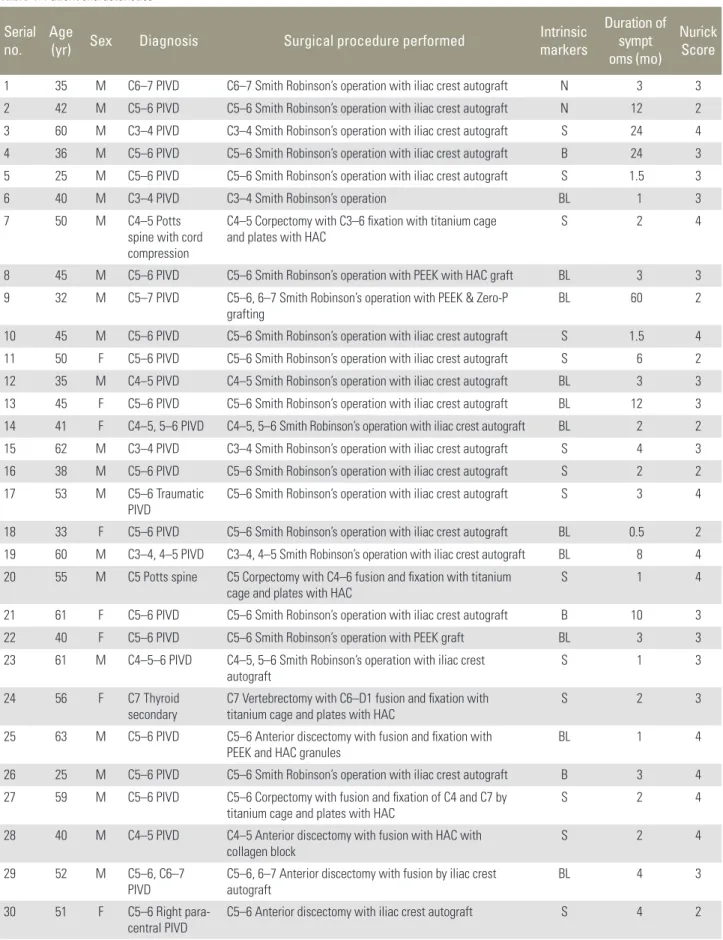
(n=2), and 12.5% (n=6) of patients, respectively. Intrinsic markers were present, but not distinctively palpable, in 18.75% (n=9) of patients and were absent in the remain- ing one patient (2.08%). Of the 42 patients with degenera- tive diseases, midline anterior vertebral surface intrinsic markers (Fig. 1) were palpated at the disease level, one level above, and one level below in 57.14% (n=24), 4.76%
(n=2), and 14.29% (n=6) of patients, respectively. Among the nine patients (21.43%) with degenerative diseases, DOC caused minimal bulges that could not be differenti- ated by palpation from adjacent levels (Fig. 2); as noted above, one patient (2.38%) had no intrinsic marker. A disrupted anterior longitudinal ligament, together with extruded disc material, tubercular granulation tissue, and highly vascular pink soft tissue, was observed over the anterior vertebral surfaces, along with vertebral destruc- tion, in patients suffering from traumatic disc disease (n=2), Pott’s disease (n=3), and a metastatic lesion from thyroid carcinoma (n=1), respectively (Fig. 3); SLL was straightforward in all these cases (n=6). There were no differences in the interpretation of palpation findings be- tween the surgeons with respect to SLL, except among pa- tients in group 4 (n=9). Overall, SLL via intrinsic marker palpation was accurate in 79.2% of patients (n=38) from groups 1, 2, and 3 but was inconclusive because of mini-
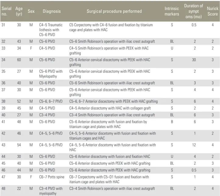
Fig. 2. (A) C5–6 prolapsed intervertebral disc causing anterior cord compression with hyperintensity in the cord, suggestive of compres- sive myelopathy/cord edema. (B) Outline of an anterior vertebral sur- face, showing only a minimal anterior bulge at the C5–6 interspace.
A B
Fig. 1. (A) C6–7 prolapsed intervertebral disc with anterior cord com- pression. (B) Outline of an anterior vertebral surface, showing an an- terior bulge (arrow) of the C5–6 inter-space due to a disc-osteophyte complex.
A B
Table 2. Results of SLL based of various groups of patients
Serial no. Group No. of cases (n=48)
1 Group 1: intrinsic marker at the level of disease 30
2 Group 2: intrinsic marker at 1 level above 2
3 Group 3: intrinsic marker at 1 level below 6
4 Group 4: intrinsic marker at or adjacent level, but not distinctive 9
5 Group 5: no intrinsic marker at or adjacent level 1
SLL, spinal level localization.
mal or absent intrinsic markers in the remaining 20.8% of patients (n=10) from groups 4 and 5. Among the patient subset with degenerative diseases (n=42), SLL via intrin- sic marker palpation was accurate in 76.2% of patients (n=32) but inconclusive in 23.8% of patients (n=10). After excluding patients with degenerative diseases, SLL via intrinsic marker palpation yielded conclusive results in the remaining six patients, whose presenting conditions included traumatic (n=2), infective (n=3), or neoplastic (n=1) pathologies.
Table 3 demonstrates that the average patient age (±standard deviation) was 43.58±11.54 years. The ap-
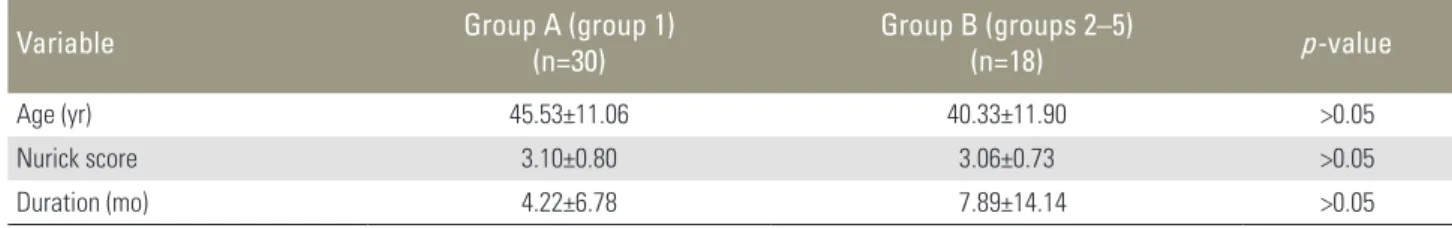
proximate mean severity score (Nurick score) was 3±0.77, whereas the average disease duration was 6±10.13 months Table 4 demonstrates that the average age in group A (45.53±11.06 years) was higher than that in group B (40.33±11.90 years), although this difference was not statistically significant (p>0.05). The mean Nurick scores of both groups (3.10±0.80 and 3.06±0.73) were similar (p>0.05), whereas the average disease duration in group A (4.22±6.78 months) was considerably shorter than that in group B (7.89±14.14 months), with high variability in both groups; however, this difference was not statistically significant (p>0.05).
Discussion
Degenerative cervical spine diseases are characterized by changes in spinal alignment, progressive changes in biological properties of the disc and annulus, changes in the anterior and posterior longitudinal ligaments, and the formation of osteophytes all around the cervical spine ver- tebral bodies, leading to neural and vascular compression [12]. The resulting bony spurs may form along the ventral or dorsal margin of the cervical spine. Posterior osteo- phytes have been studied in detail, mainly because of their proximity to the neural foramina and cord and frequent tendency to cause neuropathy and/or myelopathy; how- ever, anterior osteophytes are rarely mentioned in the lit- erature [6,13]. Brain and Colleagues [14,15] suggested that clinical outcomes of cervical spondylotic myelopathy re- sulted from disc protrusion, osteophyte formation, and as- sociated soft tissue abnormalities. In most instances, these formations extend only a few millimeters from each body [16]. An increase in joint motion causes osteophyte growth acceleration, which is most pronounced at the C5–C6 and C6–C7 levels [15-19]. These two levels are also the most often affected by spondylosis, with the C6–C7 level more commonly affected than the C5–C6 level [20]. Normal spinal cord movement is affected by ventral osteophytes, Fig. 3. (A) C7 vertebral body collapse with an associated prevertebral
and intraspinal isointense soft tissue mass that causes cord compres- sion with involvement of the posterior spinal elements. (B) Outline of an anterior vertebral surface showing a bulge (arrow) caused by the soft tissue mass in the pre-vertebral space.
A B
Table 3. Age, Nurick score, and symptom duration of patients
Variable Groups 1–5 (n=48)
Age (yr) 43.58±11.54
Nurick score 3.08±0.77
Duration (mo) 6.07±10.13
Values are presented as mean±stanadard deviation.
Table 4. Analysis of age, Nurick score, and symptom duration of patients
Variable Group A (group 1)
(n=30) Group B (groups 2–5)
(n=18) p-value
Age (yr) 45.53±11.06 40.33±11.90 >0.05
Nurick score 3.10±0.80 3.06±0.73 >0.05
Duration (mo) 4.22±6.78 7.89±14.14 >0.05
Values are presented as mean±stanadard deviation.
causing the occurrence of ventral deformities [21,22].
Bony vertebrae respond to variations in strain energy den- sity and stress, which may induce a remodeling process and lead to osteophyte formation. Further increases in the strain energy density and stress have been observed in the anterior region of the vertebral cortex compared to the posterior region and may correlate with the presence of anterior osteophytes in the cervical vertebral column, particularly at the diseased/symptomatic level [23,24].
Therefore, we assumed that these changes in the anterior region of the cortex facilitate intraoperative identification of the target spinal level via palpation in patients with degenerative diseases. The presence of distinct intrinsic markers conclusive for SLL in 79.2% of our study group is noteworthy and demonstrates the potential of SLL.
In anterior cervical spinal surgeries, the pre-vertebral fascia, a thin and transparent layer in subjects not affected by disease, is exposed after the initial dissection. In the midline between the bilateral longus colli muscles, the surgeon can see and palpate structures under the pre-ver- tebral fascia, including the anterior longitudinal ligament, anterior surfaces of the vertebral bodies, and annular liga- ments, along with disc bulges. The anterior vertebral col- umn is involved in various infective or neoplastic diseases of the cervical spine, leading to changes over the anterior vertebral surfaces [23]. During surgery, these lesions are visible under the prevertebral fascia and can be palpated for SLL. These obvious markers of neoplastic or infec- tive diseases involving the anterior column are routinely examined in detail on MR images for the purposes of di- agnosis and surgical planning. Although CT scan images can better delineate the bony outlines of vertebrae, the pressure required to palpate bony outlines may not be safe for patients with diseases causing spinal instability and soft tissue lesions over anterior vertebral surfaces [25]. In addition, intrinsic markers visible on MR images repre- sent a combination of bony and soft tissue structures that form the visible, palpable anterior vertebral surface un- der the pre-vertebral fascia [25]. In a total of six patients with traumatic, infectious (Pott’s disease), and neoplastic (metastatic) lesions, intrinsic markers comprising soft tissue lesions were very obvious under the pre-vertebral fascia. Although some reports have described the involve- ment of such lesions with the prevertebral fascia, their use for intraoperative SLL has not previously been described [23,25].
Since its introduction, radiographic localization of the
spinal level has become a routine practice during anterior cervical spinal surgery. Posterior cervical spinal surger- ies also conventionally use the C2 and C7 spinal levels for SLL. Although most developing countries have some outstanding centers of excellence, these state-of-the-art facilities are few and far between, and suburban and rural areas continue to have very limited direct access even to primary general neurosurgical services [8]. Recent reports have shown that significant numbers of community health centers in rural areas of developing countries do not have X-ray facilities [8]. Given the disparities in available pub- lic sector healthcare infrastructure and human resources in most developing countries, many socioeconomically poor patients suffering from various cervical spinal pa- thologies remain untreated because of the high expenses in private sector hospitals or long waiting times at over- burdened public sector hospitals [8]. SLL via intrinsic marker palpation will enable many spinal/neurosurgeons at ill-equipped centers to perform anterior cervical spi- nal surgeries in selected patients with distinct intrinsic markers. Although this practice may sound impractical in the current era, it is likely to help the poorest patients in developing countries who could lose their livelihood be- cause of endemic tuberculosis or degenerative diseases of the cervical spine.
The use of surface landmarks, such as the mandibular angle, hyoid bone, thyroid cartilage, first cricoid ring, and C6 carotid tubercle, as general reference points for SLL has been reported in the literature; however, these landmarks do not correspond to the exact cervical spine levels [9,10,26]. Although these landmarks can facilitate skin incision placement, definitive SLL cannot be based on these landmarks alone. Except for the C6 tubercle, positions of all these surface landmarks are likely to change with respect to shifts in the vertebral body posi- tions caused by head and neck movements [10]. Despite various reports on these landmarks, observations vary significantly depending on the patients’ age, sex, and so- matometric measurements [9,10,26]. However, none of these factors affect intrinsic markers, which are located directly on the vertebral body. We consider the findings of palpation to be subjective and note that experience in the utilization of this technique with simultaneous radio- graphic localization in a few cases, particularly during the training period, will help young spinal surgeons and neu- rosurgeons acknowledge the utility of this technique and select appropriate cases. Although an older age and short
symptom duration appeared to be associated with the presence of same-level palpable intrinsic markers/DOCs in our study, these associations were not significant, and a larger number of patients may be needed to draw definite conclusions.
After the lumbar spine, the cervical spine is the second most common site of wrong-level spinal surgery; however, the exact incidence is unknown because of medicolegal implications [20]. The Joint Commission (JCAHO) Proto- col, SMaX Program [7], and IRACE are various protocols aimed at preventing wrong-level surgeries, and all include intraoperative radiography for SLL as a uniform constitu- ent. However, intraoperative radiography does not always guarantee identification of the correct surgical level [27].
The limitations of intraoperative radiography include inadequate radiological visualization, which is common in the lower cervical spine because of the anatomical rela- tionship among the musculoskeletal structures of the pec- toral girdle [28,29]. Transient caudal shoulder displace- ment has been helpful for intraoperative radiographic visualization of the lower cervical spine [30]. Intrinsic markers can be very useful in situations where conven- tional radiographic methods fail to provide unequivo- cal results. Although available radiographic localization equipment must be used for SLL, we should strive to identify inexpensive alternatives that would be immensely useful in developing countries who cannot afford state of art modern equipments [8]. We do not suggest that such alternatives should replace or substitute radiological local- ization and note that such techniques have no role in well- equipped centers. Similarly, we should remember that patients who lack intrinsic markers according to imaging studies (>20% in our series) should only undergo surgery when radiological level localization equipment is avail- able. Unfortunately, the poor state of healthcare facilities in developing countries is a reality [8], and until the situa- tion improves to a desired level, clinical methods will play a role in SLL.
Conclusions
We conclude that distinct bony or soft tissue intrinsic markers caused by degenerative or other disease processes are common over the midline anterior vertebral surfaces of the cervical spine. These markers may be utilized as adjunct identifiers for SLL in selected patients undergo- ing cervical spinal surgeries via the anterior approach at
ill-equipped centers in developing countries where ra- diographic localization equipment is not available. This technique may also be useful during lower cervical spinal surgeries, where radiographic visualization is occasionally inadequate.
Conflict of Interest
No potential conflict of interest relevant to this article was reported.
References
1. Oh CH, Yoon SH. Past, present, and future of cervi- cal arthroplasty. Keio J Med 2013;62:47-52.
2. Kim WK. Middle cervical approach. In: Korean Spi- nal Neurosurgery Society, editor. The textbook of spine. Seoul: Koonja Publisher; 2008. p.377-90.
3. Lawrence BD, Jacobs WB, Norvell DC, Hermsmeyer JT, Chapman JR, Brodke DS. Anterior versus poste- rior approach for treatment of cervical spondylotic myelopathy: a systematic review. Spine (Phila Pa 1976) 2013;38(22 Suppl 1):S173-82.
4. Bydon M, Xu R, Amin AG, et al. Safety and efficacy of pedicle screw placement using intraoperative com- puted tomography: consecutive series of 1148 pedicle screws. J Neurosurg Spine 2014;21:320-8.
5. Klingler JH, Sircar R, Scheiwe C, et al. Comparative study of C-arms for intraoperative 3-dimensional imaging and navigation in minimally invasive spine surgery. Part I . Applicability and image quality.
Clin Spine Surg 2016 Jun 28 [Epub]. https://doi.
org/10.1097/BSD.0000000000000186
6. Srinivasan US, Lawrence R. Posterior osteophyte evolution and its impact in cervical spondylosis: a literature review. J Spine 2013;S2:007.
7. North American Spine Society. Prevention of wrong- site surgery: sign, mark and X-ray (SMaX). La- Grange, IL: North American Spine Society; 2001.
8. Srinivisan R. Healthcare in India: vision 2020, issues and prospects. New Delhi: Planning Commission;
2005 [cited 2015 Apr 16]. Available from: http://www.
planningcommission.nic.in/reports/genrep/.../26_
bg2020.doc.
9. Mirjalili SA, McFadden SL, Buckenham T, Stringer MD. Vertebral levels of key landmarks in the neck.
Clin Anat 2012;25:851-7.
10. Lee JH, Lee HS, Lee DY, Lee DO. The efficacy of ca- rotid tubercle as an anatomical landmark for identifi- cation of cervical spinal level in the anterior cervical surgery: comparison with preoperative C-arm fluo- roscopy. Clin Orthop Surg 2013;5:129-33.
11. Smith GW, Robinson RA. The treatment of certain cervical-spine disorders by anterior removal of the intervertebral disc and interbody fusion. J Bone Joint Surg Am 1958;40:607-24.
12. Ferrara LA. The biomechanics of cervical spondylo- sis. Adv Orthop 2012;2012:493605.
13. Orhan KS, Acar S, Ulusan M, Aydoseli A, Guldiken Y.
Persistent cough associated with osteophyte forma- tion and vagus nerve impingement following cervical spinal surgery: case report. J Neurosurg Spine 2013;
19:167-9.
14. Brain WR, Knight GC, Bull JW. Discussion of rup- ture of the intervertebral disc in the cervical region.
Proc R Soc Med 1948;41:509-16.
15. Brain WR, Northfield D, Wilkinson M. The neuro- logical manifestations of cervical spondylosis. Brain 1952;75:187-225.
16. Truumees E. Pain and neurologic dysfunction. In:
The Cervical Spine Research Society Editorial Com- mittee, Clark CR, editor. The cervical spine. 4th ed.
Philadelphia: Lippincott William & Wilkins; 2005.
p.957-78.
17. Arnasson O, Carlsson CA, Pellettieri L. Surgical and conservative treatment of cervical spondylotic ra- diculopathy and myelopathy. Acta Neurochir (Wien) 1987;84:48-53.
18. Kim RC. Spinal cord pathology. In: Nelson JS, Parisi JE, Schochet SS, editors. Principles and practice of neuropathology. St. Louis: CV Mosby; 1993. p.398- 435.
19. Mullin J, Shedid D, Benzel E. Overview of cervical spondylosis pathophysiology and biomechanics.
World Spinal Column J 2011;2:89-97.
20. Panjabi M, White A 3rd. Biomechanics of nonacute cervical spinal cord trauma. Spine (Phila Pa 1976) 1998;13:838-42.
21. Epstein JA, Epstein BS, Lavine LS, Carras R, Rosen- thal AD. Cervical myeloradiculopathy caused by arthrotic hypertrophy of the posterior facets and laminae. J Neurosurg 1978;49:387-92.
22. Patten JP. Neurological differential diagnosis. Lon- don: Springer-Verlag; 1977. p.139-71.
23. Kumaresan S, Yoganandan N, Pintar FA, Maiman DJ, Goel VK. Contribution of disc degeneration to osteo- phyte formation in the cervical spine: a biomechani- cal investigation. J Orthop Res 2001;19:977-84.
24. Nathan H. Osteophyte of the vertebral column: an anatomical study of their development according to age, race, and sex with considerations as to their etiology and significance. J Bone Joint Surg Am 1962;44:243-68.
25. Davis WL, Harnsberger HR. CT and MRI of the nor- mal and diseased perivertebral space. Neuroradiol- ogy 1995;37:388-94.
26. Oh CH, Ji GY, Yoon SH, et al. Surface landmarks do not correspond to exact levels of the cervical spine:
references according to the sex, age and height. Ko- rean J Spine 2014;11:178-82.
27. Hsiang J. Wrong-level surgery: a unique problem in spine surgery. Surg Neurol Int 2011;2:47.
28. Goodkin R, Laska LL. Wrong disc space level sur- gery: medicolegal implications. Surg Neurol 2004;61:
323-41.
29. Wong DA, Herring SA. Protecting patients and pre- venting medical errors: a progress report on NASS’
patient safety initiative. SpineLine 2003;4:24-6.
30. Witiw CD, Citow JS, Ginsberg HJ, et al. A novel device to simplify intraoperative radiographic visu- alization of the cervical spine by producing transient caudal shoulder displacement: a 2-center case series of 80 patients. J Neurosurg Spine 2013;19:697-700.