INTRODUCTION
Bilateral spermatic cord torsion in the newborn is extremely rare, especially asynchronous torsion, and a true emergency because of the risk of anorchia (1, 2). The first case of bilat- eral torsion of the spermatic cord was reported in 1967 by Frederick and associates on a newborn who was explored 10 hours after birth (3). To our knowledge, to date, only 5 cases of asynchronous bilateral neonatal torsion have been docu- mented in the English literature (4-7). We present such a case in a 4-day-old boy with subsequent operative discovery of prior in utero torsion of the contralateral spermatic cord.
CASE REPORT
A 2,930 g full-term male newborn with right scrotal swelling since birth was transferred to our institude for fur- ther evaluation and treatment on the 4th day after birth. He was afebrile and other vital signs were normal. The left testis with a knot of spermatic cord was small-sized, hard, nontender and nontranslucent. Color Doppler ultrasound of testes re- vealed no pulsations.
Bilateral transscrotal approach was immediately performed.
The right testis had a 90°extravaginal torsion and revealed dark gray, hemorrhagic and necrotic. The left testis had a 360°extravaginal torsion and was gray and atrophic (Fig. 1).
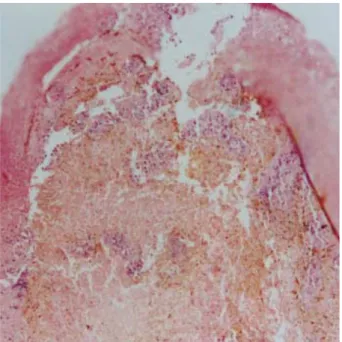
For the future Leydig cell function and salvage of some viable tissue of the testes, both testes were left in place after detor- sion. Histologic examination of biopsies of both testes reveal-
ed infarction (Fig. 2). His postoperative recovery was unevent- ful.
At 2 months old, both testes were palpable, although they were smaller than normal. The parent were advised of the high probability of sterility and the possible need for exoge- nous androgen replacement to attain secondary sex character- istics at puberty.
DISCUSSION
Perinatal torsion of the spermatic cord or testis (PTT) is defined as spermatic cord torsion occurring prenatally and within the postnatal first 30 days (8, 9). PTT may be unilat- eral or bilateral. Bilateral PTT is a extremely rare condition and a true urologic emergency because incomplete physical examination or failure to diagnose this condition promptly may result in functional anorchia (2). Therefore, PTT must be recognized by the physicians who examine the newborn immediately after delivery.
The etiology of PTT is not clear. Speculation concerning etiology has included high birth weight, difficult labor or breech presentation, and an over-reactive cremasteric reflex (4).
PTT is extravaginal as opposed to intravaginal torsion in older children and adults (5). In contrast to the postnatal tes- ticular torsion, PTT is asymptomatic, insidious, and the only abnormality is an enlarged, firm scrotal mass (8, 9).
The diagnosis of the spermatic cord torsion has been assess- ed by physical examination, transillumination test, and ultra-
Sang Don Lee, Chang Sek Cha
Department of Urology, Pusan National University Hospital, Busan, Korea
Address for correspondence Sang Don Lee, M.D.
Department of Urology, School of Medicine, Pusan National University, 1-10 Ami-dong, Seo-gu, Busan 602-739, Korea
Tel : +82.51-240-7348, Fax : +82.51-255-7133 E-mail : [email protected]
712 J Korean Med Sci 2002; 17: 712-4
ISSN 1011-8934
Copyright � The Korean Academy of Medical Sciences
Asynchronous Bilateral Torsion of the Spermatic Cord in the Newborn
: A Case Report
Asynchronous bilateral torsion of the spermatic cord in the newborn is extremely rare. We report such a case in a 4-day-old boy with subsequent operative discov- ery of prior in utero torsion of the contralateral spermatic cord. The diagnosis was made by physical examination, transillumination test, color Doppler ultrasound, and confirmed by emergent surgical exploration. To our knowledge, the present case is the 6th case of asynchronous bilateral torsion of the spermatic cord in the English literature, and the first case in Korea.
Key Words : Spermatic Cord; Torsion; Infant, Newborn
Received : 23 August 2001 Accepted : 16 November 2001
Asynchronous Bilateral Torsion of the Spermatic Cord in the Newborn 713
sonography/Doppler ultrasound.
The differential diagnosis of a testicular mass that does not transmit light in the neonatal period includes testicular tu- mor, hematocele, torsion of the testicular or epididymal ap- pendages, incarcerated hernia, scrotal abcess, ectopic spleen or adrenal, and other conditions such as orchitis and epididymi- tis (5, 9, 10).
With increasing clinical recognition of PTT, the manage- ment of these cases has aroused some controversies including the surgical approach, timing of operation, need for contralat- eral exploration and orchiopexy, and treatment of necrotic testes, especially in bilateral torsions. The preferred surgical approach is controversial. However, an inguinal incision is generally recommended although a scrotal incision may be justified in cases of an emergency operation for postnatal tor- sion or in cases of suspected bilateral neonatal torsion.
In unilateral or bilateral PTT, an emergency detorsion and orchiopexy should be carried out in an attempt to salvage the affected testis or testes. When both testes are affected, explo- ration to confirm the diagnosis and remove the non-viable testes may be performed electively even several weeks after birth. However, in unilateral torsion, surgery should be un- dertaken during the first few days of life as contralateral tor- sion can occur within the first 48 hr of life (4). We believe that prompt exploration and contralateral orchiopexy gener- ally is easy to do, carries a negligible risk, and is the only de- finitive way to establish the diagnosis and rule out other potential pathological conditions. Although asynchronous bilateral PTT is extremely rare, this report indicates that the spermatic cord is at risk for torsion during a length of time from in utero to sometime in the postnatal period.
The specific treatment of the spermatic cord torsion with infarction is very complex. The argument for removal is that it reduced the possibility of infection arising in the necrotic tissue and that the replacement of dead tissue contravenes established surgical rules. Our case showed smaller testes without blood flow on a color Doppler ultrasound even though
detorsion and orchiopexy had been performed. In considera- tion of this outcome, the removal of obviously necrotic testes appears to be reasonable. However, some reports have sup- ported that detorsion and orchiopexy, even if the testes are necrotic, is advocated to secure Leydig cell function for andro- gen secretion unless the child presents signs of systemic tox- icity (9, 10). Also, there is little evidence of any harm ensu- ing from retaining damaged testes. We believe that these fac- tors, as well as the psychological and cosmetic benefits of even a smaller-than-normal testis within the scrotum, must be con- sidered at the time of exploration.
REFERENCES
1. Olguner M, Akgur FM, Aktug T, Derebek E. Bilateral asynchronous perinatal testicular torsion: a case report. J Pediatr Surg 2000; 35:
1348-9.
2. Tripp BM, Homsy YL. Prenatal diagnosis of bilateral neonatal tor- sion: a case report. J Urol 1995; 3: 1990-1.
3. Frederick PL, Dushku N, Eraklis AJ. Simultaneous bilateral torsion of the testes in a newborn infant. Arch Surg 1967; 94: 299-300.
4. LaQuaglia MP, Bauer SB, Eraklis AE, Feins N, Mandell J. Bilater- al neonatal torsion. J Urol 1987; 138: 1051-4.
5. Kay R, Strong DW, Tank ES. Bilateral spermatic cord torsion in the neonate. J Urol 1980; 123: 293.
6. Feins N. To pex or not to pex. J Pediatr Surg 1983; 18: 697-9.
7. Brandt MT, Sheldon CA, Wacksman J, Mattews P. Prenatal testic- ular torsion: principles of management. J Urol 1992; 147: 670-2.
8. Das S, Singer A. Controversies of perinatal torsion of the spermatic Fig. 2.The biopsy specimen of right testis shows hemorrhagic congestion and ischemic coagulative necrosis in the entire field (H&E,×40).
Fig. 1.Bilateral spermatic cord torsion. (A) the right testis is dark gray. (B) the left testis is small and atrophic due to the spermat- ic cord torsion in utero.
A B
1 2 3
714 S.D. Lee, C.S. Cha
cord: a review, survey and recommendations. J Urol 1990; 143:
231-3.
9. Atallah MW, Ippolito JJ, Rubin BW. Intrauterine bilateral torsion
of the spermatic cord. J Urol 1976; 116: 128-9.
10. Sheridan WG, Davies DGL. Extravaginal testicular torsion. Brit J Clin Prac 1988; 42: 128-30.