Introduction
The presence of diastolic dysfunction has been previously documented in children in the acute phase of Kawasaki disease (KD).1-3) Diastolic dysfunction at the long-term follow-up in children with a history of KD (KDHx group) and coronary ar- tery lesions (CALs) have also been reported.4) However, previ- ously, left ventricular (LV) mass index or pulsed wave or tissue Doppler parameters such as the ratio of mitral inflow Doppler velocity during early diastole (E) and late diastole (A) (E/A) or mitral annular tissue Doppler velocity during early diastole (E´) was utilized to assess LV diastolic function.4) Pulsed-wave or tissue Doppler parameters are technically affected by angle de- pendency,5) and also affected by movement of adjacent myo-
ORIGINAL ARTICLE J Cardiovasc Ultrasound 2018;26(1):26-32
• Received: November 20, 2017 • Revised: December 27, 2017 • Accepted: February 26, 2018
• Address for Correspondence: Soo Jung Kang, Department of Pediatrics, CHA Bundang Medical Center, CHA University School of Medicine, 59 Yatap-ro, Bundang-gu, Seongnam 13496, Korea Tel: +82-31-780-5248, Fax: +82-31-780-5239, E-mail: [email protected]
• This is an Open Access article distributed under the terms of the Creative Commons Attribution Non-Commercial License (http://creativecommons.org/licenses/by-nc/4.0) which permits unrestricted non-commercial use, distribution, and reproduction in any medium, provided the original work is properly cited.
cardial segments,6) thereby detection of subclinical diastolic dys- function at long-term follow-up of children in the KDHx group by these parameters may be challenging.
With LV diastolic dysfunction, the resultant rise in left atrial (LA) pressure would lead to LA remodeling, reflected in im- paired LA function.7) During systole, the LA functions as a res- ervoir of pulmonary venous return.8) LA reservoir function could be reflected in phasic LA volume changes such as LA total emptying fraction, which is (maximum LA volume - mini- mum LA volume) / maximum LA volume.8) Also, LA reser- voir function could be assessed using myocardial deformation imaging as peak LA longitudinal strain (PLALS) at systole.9) In addition, LA stiffness index, which is defined as the amount of
Long Term Outcomes of Left Atrial Reservoir Function in Children with a History of Kawasaki Disease
Soo Jung Kang, MD, PhD1, Jihyun Ha, MD1, Seo Jung Hwang, MT2, and Hyo Jin Kim, MT2
Departments of 1Pediatrics, 2Diagnostic Laboratory Medicine, CHA Bundang Medical Center, CHA University School of Medicine, Seongnam, Korea
Background: Decreased left atrial (LA) reservoir function is reported to be associated with elevated left ventricular (LV) end diastolic pressure and LV diastolic dysfunction. Echocardiographic parameters that reflect LA reservoir function include LA total emptying fraction [(maximum LA volume - minimum LA volume) / maximum LA volume], peak LA longitudinal strain (PLALS) at systole, and LA stiffness index (E/E´/PLALS). We aimed to investigate the long-term outcomes of LV diastolic func- tion in children with a history of Kawasaki disease (KD) (KDHx group) by assessing LA reservoir function.
Methods: Retrospectively, echocardiograms performed at a mean follow-up period of 5 years after the acute phase of KD in 24 children in the KDHx group were compared to those from 20 normal control subjects. LA total emptying fraction, PLALS, LA stiffness index, LV peak longitudinal systolic strain (ε), and strain rate (SR) were evaluated with conventional echocardiographic parameters.
Results: The mean age at long term follow-up echocardiography in children in the KDHx group was 6.8 years. Five children (20.8%) had coronary artery lesions (CALs) in the acute stage of KD. No children showed CALs at a mean follow-up period of 5 years after the acute phase of KD. There were no significant differences in the conventional echocardiographic parameters and in LA total emptying fraction, PLALS, LA stiffness index, LV peak longitudinal systolic ε, and SR, between the children in the KDHx and control group.
Conclusion: LV diastolic function assessed by LA reservoir function parameters at long-term follow-up in children in the KDHx group appears to be favorable.
KEY WORDS: Kawasaki disease · Atrial function · Long term · Prognosis.
pressure required to change a unit of LA volume during the passive filling of LA,10) could be noninvasively obtained as E/
E´/PLALS.11) An increased LA stiffness index reflects impaired LA reservoir function.12)13) In adults, LA total emptying frac- tion, PLALS, and LA stiffness index all have been utilized to assess the severity of LV diastolic dysfunction.7)11)14)15)
To date, LA reservoir function has not been evaluated at long- term follow-up in children in the KDHx group. Therefore, we assessed LA reservoir function at long-term follow-up of chil- dren in the KDHx group using LA total emptying fraction, PLALS, and LA stiffness index, along with conventional echo- cardiographic parameters, and compared it with those of a control group.
Methods
Study population
From 2007–2011, 487 children were admitted to CHA Bun- dang Medical Center for treatment of KD. Of these children, only the children with available long-term (≥ mean of 5 years after the acute phase of KD) follow-up echocardiograms were included in this study. The proposal for this study was ap- proved by the Institutional Review Board of CHA University Bundang Medical Center (IRB: 2017-11-023), and since our study was retrospective, waiver of informed consent was ob- tained. At the time of admission, the diagnosis of KD was made according to the published American Heart Association (AHA) guidelines.16) For controls, data of children who were referred to the pediatric cardiology outpatient clinic for evalu- ation of chest pain and underwent echocardiography were ret- rospectively studied. Children who had structural heart anom- alies or cardiac causes of chest pain were excluded from our study group.
Clinical data
Data on duration of fever before intravenous immune glob- ulin (IVIG) treatment were collected. Initial laboratory data obtained from a blood sample taken before IVIG treatment (white blood cell count, percentage of segmented neutrophils, serum albumin and alanine aminotransferase levels, C-reactive protein levels, and NT-pro B type natriuretic peptide levels) were also evaluated. All children in the KDHx group were admin- istered IVIG (2 g/kg) at the acute phase of KD. Aspirin (80 mg/
kg/day) was started with IVIG and was continued for 3–4 days after the child became afebrile, and the dose was reduced to 5 mg/kg/day and given for 6–8 weeks.16) IVIG nonresponders were defined as children experiencing persistent or recrudescent fever ≥ 36 h after completing the initial IVIG infusion.16) Exclu- sion criteria from the KDHx group were children who did not have late follow-up echocardiograms after the acute phase of KD.
Echocardiographic data
The long-term (≥ 5 years after the acute phase of KD) fol-
low-up echocardiograms of children in the KDHx group were compared with those of the control group. All echocardiograms of the children in our study were obtained using a commercial echocardiography system (Acuson SC 2000, Siemens Medical, Mountain View, CA, USA). Both conventional echocardiogra- phy and myocardial deformational analyses were performed.
Analysis of all conventional and deformation data were per- formed by a single pediatric cardiologist, who at the time of analysis was blinded to the group allocation of study subjects (KDHx versus control group).
Conventional echocardiography
Conventional echocardiographic parameters were obtained according to published recommendations.17) LV ejection frac- tion, E/A, E´, E/E´, and LV mass indexed to body surface area were obtained. The internal diameters of the proximal right coronary artery, proximal left anterior descending coronary ar- tery, and left main coronary artery were measured. The defini- tion of CALs were when calculated z-scores of the coronary ar- teries were ≥ 2.5 in one or more of the measured coronary sites.18) The z-scores of the coronary arteries were calculated according to the method of McCrindle et al.18) using the measured coronary dimensions and the published nonlinear regression equations.18)
Myocardial deformation analysis
Two-dimensional echocardiographic images that were ob- tained at a mean of 70 frames per second and stored for pro- spective analysis were used for deformation imaging analysis.
The software used for deformation analysis was velocity vector imaging (version 3.0, Siemens Medical). Three consecutive car- diac cycles were analyzed. PLALS, LV peak longitudinal systol- ic strain (ε), and LV peak systolic strain rate (SR) were all ob- tained offline using the apical four-chamber view. To obtain PLALS, the endocardial border of the LA was initially traced manually at the end of systole just before mitral valve open- ing.9)19) Subsequently, automatic tracking of the endocardial border of the LA using the velocity vector imaging software generated PLALS (Fig. 1A). The average of PLALS from the roof, interatrial septum, and the lateral wall of LA were used for analysis (Fig. 1B). LA stiffness index was obtained nonin- vasively by the formula E/E´/PLALS.11) The maximum LA vol- ume, which was also generated automatically with PLALS by the velocity vector imaging software,20)21) was obtained and in- dexed to the body surface area. Using the maximum and mini- mum LA volumes automatically generated by the velocity vector imaging software, the LA total emptying fraction was calculated as (maximum LA volume - minimum LA volume) / maximum LA volume.8) To obtain LV peak longitudinal systolic ε and SR, the endocardial border of the LV was traced manual- ly and tracked automatically by the software at the onset of the QRS wave on the electrocardiogram. The average of ε and SR curves from six segments (three from the interventricular septum and three from the LV free wall) were used for analysis.
Statistics
SPSS version 24 (IBM SPSS Statistics 24, Armonk, NY, USA) was used for data analysis. All data are expressed as mean ± standard deviation or median (range). Student’s t-test or the Mann-Whitney U test, was used to compare continuous data between children in the KDHx and control groups. Chi-square analysis was used to compare categorical variables between the two groups. A p-value < 0.05 was considered significant. Cor- relation of LA total emptying fraction, PLALS, and LA stiff- ness index with clinical data was assessed by Pearson’s or Spearman’s correlation. To obtain intraobserver variability, one observer repeated an offline analysis of LA total emptying frac- tion, PLALS, LA stiffness index, LV longitudinal peak systolic ε, and SR in echocardiograms of 15 randomly selected chil- dren after 4 weeks. The intraobserver variability was evaluated by the mean percentage error.22)
Results
A total of 44 children were included in our study (24 in the KDHx group and 20 in the control group). The mean age and time from onset of KD to follow-up of the children in the KDHx group was 6.8 years and 5 years, respectively. Clinical and echocardiographic data of the children in the KDHx and control group are shown in Tables 1 and 2, respectively. Age at follow-up, percentage of boys, body surface area, heart rate, systolic and diastolic blood pressures were all similar between the children in the KDHx and control group. Six children (25%) showed incomplete KD. No children in our study were IVIG nonresponders. In the KDHx group, 5 children (20.8%) had CALs in the acute stage of KD. On echocardiograms per- formed at a mean follow-up period of 5 years after the acute phase of KD, no children showed CALs. As shown in Table 2, there were no significant differences in LV ejection fraction, LV mass index, mitral E/A, mitral E/E´, and maximum LA volume index between the children in the KDHx group at long-term
A B
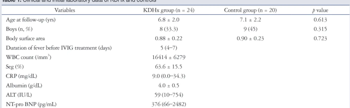
Fig. 1. Peak left atrial longitudinal strain (PLALS) at systole. A: Example of PLALS at systole by velocity vector imaging at long-term follow-up of a child with a history of Kawasaki disease. B: The average of PLALS from 3 atrial segments (interatrial septum, roof, and lateral wall) is shown.
Table 1. Clinical and initial laboratory data of KDHx and controls
Variables KDHx group (n = 24) Control group (n = 20) p value
Age at follow-up (yrs) 6.8 ± 2.0 7.1 ± 2.2 0.613
Boys (n, %) 8 (33.3) 9 (45) 0.315
Body surface area 0.88 ± 0.22 0.90 ± 0.23 0.723
Duration of fever before IVIG treatment (days) 5 (4–7)
WBC count (/mm3) 16414 ± 6279
Seg (%) 63.6 ± 15.5
CRP (mg/dL) 9.0 (0.0–34.3)
Albumin (g/dL) 4.0 ± 0.5
ALT (IU/L) 59 (10–754)
NT-pro BNP (pg/mL) 376 (66–2482)
Data are expressed as mean ± standard deviation, or median (range). KDHx: children with a history of Kawasaki disease, IVIG: intravenous immune globulin, WBC: white blood cell, Seg: percentage of segmented neutrophils, CRP: C-reactive protein, ALT: alanine aminotransferase, NT-pro BNP: N-terminal pro-B- type natriuretic peptide
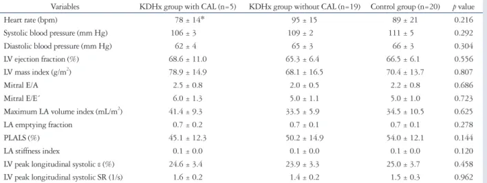
follow-up and the control group. Likewise, there were no sig- nificant differences in LA emptying fraction, PLALS, LA stiff- ness index, LV peak longitudinal systolic ε, and SR between the children in the KDHx group at long-term follow-up and control group. In our study, LV systolic dysfunction was deter- mined as LV peak longitudinal systolic ε < 18%, according to published reference values.23) No children in the KDHx group showed significant mitral regurgitation or LV systolic dysfunc- tion. When the children in the KDHx group at long term fol- low-up was divided into subgroups according to initial CAL status at acute phase of KD, no significant differences were found in LA total emptying fraction, PLALS, LA stiffness index, LV peak longitudinal systolic ε, and SR among the children in the
KDHx group with and without initial CAL at long term fol- low-up and control group (Table 3). No significant correlations were noted between LA emptying fraction, PLALS, LA stiff- ness index, and clinical and laboratory parameters. The mean percentage error for intraobserver variabilities in LA total emp- tying fraction, PLALS, LA stiffness index, LV longitudinal peak systolic ε, and SR were 16, 17, 18, 14, and 15%, respectively.
Discussion
We found no significant differences in LA reservoir function assessed by LA emptying fraction, PLALS, LA stiffness index, between the children in the KDHx and control groups. In ad- dition, LV peak longitudinal systolic ε and SR were similar be-
Table 2. Echocardiographic data at long term follow-up of KDHx and controls
Variables KDHx group (n = 24) Control group (n = 20) p value
Heart rate (bpm) 91 ± 16 89 ± 21 0.216
Systolic blood pressure (mm Hg) 108 ± 5 111 ± 5 0.292
Diastolic blood pressure (mm Hg) 64 ± 5 66 ± 3 0.304
LV ejection fraction (%) 66.0 ± 7.4 66.5 ± 6.1 0.556
LV mass index (g/m2) 70.4 ± 16.5 70.4 ± 13.7 0.807
Mitral E/A 2.1 ± 0.6 2.2 ± 0.8 0.686
Mitral E/E´ 5.2 ± 1.2 5.0 ± 1.0 0.723
Maximum LA volume index (mL/m2) 35.1 ± 7.3 34.5 ± 10.5 0.625
LA emptying fraction 0.7 ± 0.1 0.7 ± 0.1 0.278
PLALS (%) 49.1 ± 14.3 54.0 ± 12.1 0.144
LA stiffness index 0.1 ± 0.0 0.1 ± 0.0 0.120
LV peak longitudinal systolic ε (%) 24.0 ± 3.3 25.0 ± 3.7 0.458
LV peak longitudinal systolic SR (1/s) 1.5 ± 0.2 1.5 ± 0.3 0.962
Data are expressed as mean ± standard deviation. Strain and strain rate values are presented as absolute values. KDHx: children with a history of Kawasaki dis- ease, mitral E/A: ratio of mitral inflow Doppler velocity during early diastole (E) and late diastole (A), mitral E/E´: ratio of E and mitral annular tissue Doppler velocity during early diastole (E´), LA: left atrial, LV: left ventricular, PLALS: peak LA longitudinal strain, ε: strain, SR: strain rate
Table 3. Echocardiographic data at long term follow-up of KDHx with and without CALs at the acute phase and controls
Variables KDHx group with CAL (n=5) KDHx group without CAL (n=19) Control group (n=20) p value
Heart rate (bpm) 78 ± 14* 95 ± 15 89 ± 21 0.216
Systolic blood pressure (mm Hg) 106 ± 3 109 ± 2 111 ± 5 0.292
Diastolic blood pressure (mm Hg) 62 ± 4 65 ± 3 66 ± 3 0.304
LV ejection fraction (%) 68.6 ± 11.0 65.3 ± 6.4 66.5 ± 6.1 0.556
LV mass index (g/m2) 78.9 ± 14.9 68.1 ± 16.5 70.4 ± 13.7 0.807
Mitral E/A 2.5 ± 0.8 2.0 ± 0.5 2.2 ± 0.8 0.686
Mitral E/E´ 6.0 ± 1.3 5.0 ± 1.1 5.0 ± 1.0 0.723
Maximum LA volume index (mL/m2) 41.4 ± 9.3 33.5 ± 5.9 34.5 ± 10.5 0.625
LA emptying fraction 0.7 ± 0.2 0.7 ± 0.1 0.7 ± 0.1 0.278
PLALS (%) 45.1 ± 12.3 50.2 ± 14.9 54.0 ± 12.1 0.144
LA stiffness index 0.1 ± 0.0 0.1 ± 0.0 0.1 ± 0.0 0.120
LV peak longitudinal systolic ε (%) 24.6 ± 3.4 23.9 ± 3.3 25.0 ± 3.7 0.458
LV peak longitudinal systolic SR (1/s) 1.6 ± 0.2 1.4 ± 0.2 1.5 ± 0.3 0.962
Data are expressed as mean ± standard deviation. Strain and strain rate values are presented as absolute values. *p < 0.05 compared with the control group.
KDHx: children with a history of Kawasaki disease, CAL: coronary artery lesion, mitral E/A: ratio of mitral inflow Doppler velocity during early diastole (E) and late diastole (A), mitral E/E’: ratio of E and mitral annular tissue Doppler velocity during early diastole (E´), LA: left atrial, LV: left ventricular, PLALS: peak LA longitudinal strain, ε: strain, SR: strain rate
tween the children in the KDHx and control groups. Since LA reservoir function reflects LV diastolic dysfunction,7)11)14)15) as- sessing LA reservoir function could determine the optimal fol- low-up intervals in children in the KDHx group, especially those without a history of CALs.
LA reservoir function could be assessed by LA total emptying fraction, PLALS, and LA stiffness index.8)12) Phasic changes of LA volume such as LA total emptying fraction have been shown to be associated with the severity of diastolic dysfunction.7) LA pressure increase due to elevated LV end-diastolic pressure may result in LA stretch and fibrosis, limiting the ability of LA filling and emptying.24)25) Acute LV diastolic dysfunction has been reported previously in children in the acute phase of KD.1-3) However, our results of similar LA emptying fraction between the children in the KDHx and control groups suggest that the LV diastolic dysfunction observed in the acute stage of KD may be resolved by the long term follow-up of KD.
PLALS has been shown to be more strongly correlated to LV diastolic function than tissue Doppler parameters26) in adults, due to strain (ε) and SR imaging being relatively independent of heart motion and nearby segment contraction.5)8)27) Howev- er, PLALS is known to be affected by significant acute mitral regurgitation,3) as acute mitral regurgitation could directly di- late the LA, independently of LV filling pressures.28) In addi- tion, PLALS would decrease in the presence of LV systolic dys- function, when impairment of mitral annular movement from the base to the apex in systole13) would decrease PLALS. In this study, no children in the KDHx group showed significant mi- tral regurgitation or LV systolic dysfunction. Therefore, in our study, we could assume that PLALS would reflect LV diastolic function, independent of the effects of significant mitral regur- gitation or LV systolic dysfunction, and that LV diastolic func- tion appear to be preserved.
Our study is the first to evaluate LA stiffness index derived by noninvasive methods11) at long term follow-up of children in the KDHx group and controls. In adults, LA stiffness index has been shown to be able to distinguish diastolic heart failure from diastolic dysfunction.11) Also, in adult atrial fibrillation patients undergoing LA ablation, LA stiffness index has been shown to estimate the compliance of the LA myocardium because it is the ratio of change in volume to change in pressure during LA passive filling.10) Our results of similar LA stiffness index in the children in the KDHx and control groups at long term follow- up suggest that LA compliance may be normalized by long term follow-up of KD.
Previously, LV diastolic dysfunction detected during the long- term follow-up of children in the KDHx group had been stud- ied with conflicting results. Both structural abnormalities4)29) and functional impairment30)31) of coronary arteries have been associated with diastolic dysfunction in children in the KDHx group. Arnold et al.29) identified diastolic abnormalities in seg- ments supplied by stenotic coronary arteries in 17 children in the KDHx group who had CALs. Selamet Tierney et al.4)
showed the association of coronary abnormalities with diastol- ic dysfunction using evidence of prolonged deceleration time and decreased E´ velocity on follow-up of children in the KDHx group and coronary abnormalities. Additionally, decreased coro- nary flow reserve in children in the KDHx group without overt coronary structural abnormalities have been reported.30)31) Based on these observations, we could speculate a subclinical decrease in LV myocardial function and resulting atrial remod- eling leading to altered LA reservoir function in children in the KDHx group due to the functional impairment of coro- nary arteries. However, the results in this study did not show significant differences in LA reservoir function, LV peak longi- tudinal systolic ε, and SR in the children in the KDHx and control groups. Possible explanations of this similar outcomes between the two groups may be the small number of the study population, or the limited number of children with CALs in the KDHx group at the acute phase of KD (5/24, 16.7%).
Another reason for the similar LA reservoir function and LV peak longitudinal systolic ε and SR at long term follow-up in the children in the KDHx and control groups may be that LV diastolic dysfunction may be more evidently detected in the acute phase of KD (less than 1 month of onset of KD)4) than in the later stages of the disease. In the acute stage of KD, myo- carditis may cause myocardial interstitial edema,2) resulting in LV diastolic dysfunction. However, in the convalescent phase, normalization of echocardiographic parameters such as mitral E/A and E/E´, have been reported.1) Histologic abnormalities in the follow-up period of KD, such as inflammatory cell infil- tration, interstitial fibrosis, and disarray have been shown in specimens of KD patients with giant aneurysms.32) However, to date, it is unclear whether myocarditis in the acute stage of KD causes myocardial dysfunction at long-term follow-up of KD, independent of CALs.33) Resolution of acute LV systolic dysfunc- tion in children in the KDHx group 1–3 years after KD onset have been reported.34) Our results suggest that LV diastolic func- tion also may be normalized in the long-term follow-up of chil- dren in the KDHx group without CALs, as implied previously.33) Limitations
Limitations of this study are the retrospective design, the small number of subjects in the study population which might have affected our statistical results, and the unavailability of invasive hemodynamic data as a reference value for assessment of LV di- astolic function. However, since children in the KDHx group at long-term follow-up were clinically asymptomatic, invasive cardiac catherization was not necessary, as previously stated.16) In addition, all deformation analyses were performed using the apical four-chamber view only, and the limited number of ana- lyzed segments might have affected our statistical results.
Conclusion
LV diastolic function assessed by LA reservoir function pa- rameters at long-term follow-up in children in the KDHx group
appears to be favorable.
References
1. Takeuchi D, Saji T, Takatsuki S, Fujiwara M. Abnormal tissue doppler images are associated with elevated plasma brain natriuretic peptide and in- creased oxidative stress in acute Kawasaki disease. Circ J 2007;71:357-62.
2. Kurotobi S, Kawakami N, Shimizu K, Aoki H, Nasuno S, Takahashi K, Kogaki S, Ozono K. Brain natriuretic peptide as a hormonal marker of ventricular diastolic dysfunction in children with Kawasaki disease. Pedi- atr Cardiol 2005;26:425-30.
3. Kang SJ, Kwon YW, Hwang SJ, Kim HJ, Jin BK, Yon DK. Clinical utility of left atrial strain in children in the acute phase of Kawasaki disease.
J Am Soc Echocardiogr 2018;31:323-32.
4. Selamet Tierney ES, Newburger JW, Graham D, Baker A, Fulton DR, Colan SD. Diastolic function in children with Kawasaki disease. Int J Cardiol 2011;148:309-12.
5. Dandel M, Hetzer R. Echocardiographic strain and strain rate imaging- -clinical applications. Int J Cardiol 2009;132:11-24.
6. D’hooge J, Heimdal A, Jamal F, Kukulski T, Bijnens B, Rademakers F, Hatle L, Suetens P, Sutherland GR. Regional strain and strain rate measurements by cardiac ultrasound: principles, implementation and limita- tions. Eur J Echocardiogr 2000;1:154-70.
7. Otani K, Takeuchi M, Kaku K, Haruki N, Yoshitani H, Tamura M, Abe H, Okazaki M, Ota T, Lang RM, Otsuji Y. Impact of diastolic dys- function grade on left atrial mechanics assessed by two-dimensional speckle tracking echocardiography. J Am Soc Echocardiogr 2010;23:961-7.
8. Blume GG, Mcleod CJ, Barnes ME, Seward JB, Pellikka PA, Bas- tiansen PM, Tsang TS. Left atrial function: physiology, assessment, and clinical implications. Eur J Echocardiogr 2011;12:421-30.
9. Wakami K, Ohte N, Asada K, Fukuta H, Goto T, Mukai S, Narita H, Kimura G. Correlation between left ventricular end-diastolic pressure and peak left atrial wall strain during left ventricular systole. J Am Soc Echocar- diogr 2009;22:847-51.
10. Khurram IM, Maqbool F, Berger RD, Marine JE, Spragg DD, Ashikaga H, Zipunnikov V, Kass DA, Calkins H, Nazarian S, Zim- merman SL. Association between left atrial stiffness index and atrial fibril- lation recurrence in patients undergoing left atrial ablation. Circ Arrhythm Electrophysiol 2016;9:e003163.
11. Kurt M, Wang J, Torre-Amione G, Nagueh SF. Left atrial function in diastolic heart failure. Circ Cardiovasc Imaging 2009;2:10-5.
12. Machino-Ohtsuka T, Seo Y, Tada H, Ishizu T, Machino T, Yamasaki H, Igarashi M, Xu D, Sekiguchi Y, Aonuma K. Left atrial stiffness re- lates to left ventricular diastolic dysfunction and recurrence after pulmonary vein isolation for atrial fibrillation. J Cardiovasc Electrophysiol 2011;22:
999-1006.
13. Barbier P, Solomon SB, Schiller NB, Glantz SA. Left atrial relaxation and left ventricular systolic function determine left atrial reservoir function.
Circulation 1999;100:427-36.
14. Posina K, McLaughlin J, Rhee P, Li L, Cheng J, Schapiro W, Gulotta RJ, Berke AD, Petrossian GA, Reichek N, Cao JJ. Relationship of pha- sic left atrial volume and emptying function to left ventricular filling pressure:
a cardiovascular magnetic resonance study. J Cardiovasc Magn Reson 2013;
15:99.
15. Kurt M, Tanboga IH, Aksakal E, Kaya A, Isik T, Ekinci M, Bilen E.
Relation of left ventricular end-diastolic pressure and N-terminal pro-brain natriuretic peptide level with left atrial deformation parameters. Eur Heart J Cardiovasc Imaging 2012;13:524-30.
16. Newburger JW, Takahashi M, Gerber MA, Gewitz MH, Tani LY, Burns JC, Shulman ST, Bolger AF, Ferrieri P, Baltimore RS, Wilson WR, Baddour LM, Levison ME, Pallasch TJ, Falace DA, Taubert KA; Committee on Rheumatic Fever, Endocarditis, and Kawasaki
Disease, Council on Cardiovascular Disease in the Young, American Heart Association. Diagnosis, treatment, and long-term management of Kawasaki disease: a statement for health professionals from the Committee on Rheumatic Fever, Endocarditis, and Kawasaki disease, council on Cardiovas- cular Disease in the Young, American Heart Association. Pediatrics 2004;
114:1708-33.
17. Lai WW, Geva T, Shirali GS, Frommelt PC, Humes RA, Brook MM, Pignatelli RH, Rychik J; Task Force of the Pediatric Council of the American Society of Echocardiography; Pediatric Council of the American Society of Echocardiography. Guidelines and standards for performance of a pediatric echocardiogram: a report from the Task Force of the Pediatric Council of the American Society of Echocardiography. J Am Soc Echocardiogr 2006;19:1413-30.
18. McCrindle BW, Li JS, Minich LL, Colan SD, Atz AM, Takahashi M, Vetter VL, Gersony WM, Mitchell PD, Newburger JW; Pediatric Heart Network Investigators. Coronary artery involvement in children with Kawasaki disease: risk factors from analysis of serial normalized mea- surements. Circulation 2007;116:174-9.
19. Kim DG, Lee KJ, Lee S, Jeong SY, Lee YS, Choi YJ, Yoon HS, Kim JH, Jeong KT, Park SC, Park M. Feasibility of two-dimensional global longitudinal strain and strain rate imaging for the assessment of left atrial function: a study in subjects with a low probability of cardiovascular disease and normal exercise capacity. Echocardiography 2009;26:1179-87.
20. Jarnert C, Melcher A, Caidahl K, Persson H, Rydén L, Eriksson MJ.
Left atrial velocity vector imaging for the detection and quantification of left ventricular diastolic function in type 2 diabetes. Eur J Heart Fail 2008;10:
1080-7.
21. Mor-Avi V, Lang RM, Badano LP, Belohlavek M, Cardim NM, De- rumeaux G, Galderisi M, Marwick T, Nagueh SF, Sengupta PP, Sica- ri R, Smiseth OA, Smulevitz B, Takeuchi M, Thomas JD, Vannan M, Voigt JU, Zamorano JL. Current and evolving echocardiographic tech- niques for the quantitative evaluation of cardiac mechanics: ASE/EAE con- sensus statement on methodology and indications endorsed by the Japanese Society of Echocardiography. J Am Soc Echocardiogr 2011;24:277-313.
22. Kutty S, Padiyath A, Li L, Peng Q, Rangamani S, Schuster A, Dan- ford DA. Functional maturation of left and right atrial systolic and dia- stolic performance in infants, children, and adolescents. J Am Soc Echocardiogr 2013;26:398-409.e2.
23. Levy PT, Machefsky A, Sanchez AA, Patel MD, Rogal S, Fowler S, Yaeger L, Hardi A, Holland MR, Hamvas A, Singh GK. Reference ranges of left ventricular strain measures by two-dimensional speckle-track- ing echocardiography in children: a systematic review and meta-analysis. J Am Soc Echocardiogr 2016;29:209-25.e6.
24. Bytyçi I, Bajraktari G, Ibrahimi P, Berisha G, Rexhepaj N, Henein MY. Left atrial emptying fraction predicts limited exercise performance in heart failure patients. Int J Cardiol Heart Vessel 2014;4:203-7.
25. Moe GW, Grima EA, Angus C, Wong NL, Hu DC, Howard RJ, Armstrong PW. Response of atrial natriuretic factor to acute and chronic increases of atrial pressures in experimental heart failure in dogs. Role of changes in heart rate, atrial dimension, and cardiac tissue concentration. Cir- culation 1991;83:1780-7.
26. Cameli M, Sparla S, Losito M, Righini FM, Menci D, Lisi M, D’Ascenzi F, Focardi M, Favilli R, Pierli C, Fineschi M, Mondillo S. Correlation of left atrial strain and Doppler measurements with invasive measurement of left ventricular end-diastolic pressure in patients stratified for different val- ues of ejection fraction. Echocardiography 2016;33:398-405.
27. Perk G, Tunick PA, Kronzon I. Non-Doppler two-dimensional strain imaging by echocardiography--from technical considerations to clinical appli- cations. J Am Soc Echocardiogr 2007;20:234-43.
28. Hsiao SH, Huang WC, Lin KL, Chiou KR, Kuo FY, Lin SK, Cheng CC. Left atrial distensibility and left ventricular filling pressure in acute ver- sus chronic severe mitral regurgitation. Am J Cardiol 2010;105:709-15.
29. Arnold R, Goebel B, Ulmer HE, Gorenflo M, Poerner TC. An exercise tissue Doppler and strain rate imaging study of diastolic myocardial dysfunc- tion after Kawasaki syndrome in childhood. Cardiol Young 2007;17:478- 86.
30. Muzik O, Paridon SM, Singh TP, Morrow WR, Dayanikli F, Di Carli MF. Quantification of myocardial blood flow and flow reserve in children with a history of Kawasaki disease and normal coronary arteries using posi- tron emission tomography. J Am Coll Cardiol 1996;28:757-62.
31. Hauser M, Bengel F, Kuehn A, Nekolla S, Kaemmerer H, Schwaiger M, Hess J. Myocardial blood flow and coronary flow reserve in children with
“normal” epicardial coronary arteries after the onset of Kawasaki disease as- sessed by positron emission tomography. Pediatr Cardiol 2004;25:108-12.
32. Yonesaka S, Takahashi T, Eto S, Sato T, Otani K, Ueda T, Sato A, Kitagawa Y, Konno Y, Kinjo M. Biopsy-proven myocardial sequels in Ka- wasaki disease with giant coronary aneurysms. Cardiol Young 2010;20:
602-9.
33. McCrindle BW, Rowley AH, Newburger JW, Burns JC, Bolger AF, Gewitz M, Baker AL, Jackson MA, Takahashi M, Shah PB, Kobayas- hi T, Wu MH, Saji TT, Pahl E; American Heart Association Rheu- matic Fever, Endocarditis, and Kawasaki Disease Committee of the Council on Cardiovascular Disease in the Young; Council on Cardio- vascular and Stroke Nursing; Council on Cardiovascular Surgery and Anesthesia; and Council on Epidemiology and Prevention. Diagnosis, treatment, and long-term management of Kawasaki disease: a scientific state- ment for health professionals from the American Heart Association. Circula- tion 2017;135:e927-99.
34. Newburger JW, Sanders SP, Burns JC, Parness IA, Beiser AS, Colan SD. Left ventricular contractility and function in Kawasaki syndrome. Ef- fect of intravenous gamma-globulin. Circulation 1989;79:1237-46.