J Korean Soc Radiol 2018;78(2):141-145 https://doi.org/10.3348/jksr.2018.78.2.141
INTRODUCTION
Bladder cancer is the second most common malignancy of the genitourinary tract and is ranked fourth. Urothelial carci- noma of the bladder is the most common histologic type of bladder cancer, which of them initially diagnosed as non-mus- cle-invasive bladder cancer, which is confined to either the mu- cosa (pTa, carcinoma in situ) or the submucosa (pT1) (1). Cur- rently the most widespread treatment for the non-muscle- invasive bladder cancer is transurethral resection. Although this procedure is not morbidity free, there are few reports of potential complications (2).
In rare cases, extravesical metastasis has been reported, that’s because microperforation of bladder occurred after repeated transurethral resection of bladder tumor (TURBT) (3). In addi- tion to this reason, increased internal pressure of the bladder greater than surrounding venous pressure after TURBT due to fluid infusion during procedure and high histologic grade of
the tumor are associated with extravesical metastasis after TURBT (4, 5). Here we describe a case of extravesical metasta- sis occurring after repeated TURBT of non-invasive bladder cancer.
CASE REPORT
A 70-year-old man with no pre-existing diseases visited the hospital for gross hematuria. Contrast-enhanced spiral com- puted tomography (CT) images of the kidneys taken on admis- sion showed an enhanced mass approximately 2 cm in size in the right posterior aspect of the bladder (Fig. 1A, B). Subse- quent cystoscopy showed a mass on the right lateral wall of the bladder, and TURBT was performed. Pathological findings showed a low-grade by WHO, non-invasive papillary urothelial carcinoma (stage Tis), with only mucosal invasion. Eight years after first visit, contrast-enhanced CT showed a newly devel- oped 2.7 cm sized polypoid mass in the lower anterior wall of
A Case of Extravesical Metastases Occurring after Transurethral Resection of Non-Invasive Bladder Cancer
비침습적 방광암의 경 요도 절제술 후 발생한 방광 외 전이에 대한 증례 보고
Ga Ram Kang, MD
1, Dong Wan Sohn, MD
2, Dong Jin Chung, MD
3*
1Department of Radiology, Uijeongbu St. Mary’s Hospital, College of Medicine, The Catholic University of Korea, Uijeongbu, Korea Departments of 2Urology, 3Radiology, Yeouido St. Mary’s Hospital, College of Medicine, The Catholic University of Korea, Seoul, Korea
A transurethral resection of bladder tumor (TURBT) is the primary treatment modal- ity for bladder cancer. The common complications of TURBT include urinary tract in- fections, a hemorrhage requiring transfusion, and bladder perforation. Extravesical metastasis and intraperitoneal seeding of tumor cells following TURBT are very rarely reported. This report reviews a case of extravesical metastasis occurring after a repeated TURBT of non-invasive bladder cancer.
Index terms
Urinary Bladder Neoplasms Urinary Tract Infections Neoplasm Seeding
Received April 4, 2017 Revised July 11, 2017 Accepted October 17, 2017
*Corresponding author: Dong Jin Chung, MD Department of Radiology, Yeouido St. Mary’s Hospital, College of Medicine, The Catholic University of Korea, 10 63-ro, Yeongdeungpo-gu, Seoul 07345, Korea.
Tel. 82-2-3779-1327 Fax. 82-2-783-5288 E-mail: [email protected]
This is an Open Access article distributed under the terms of the Creative Commons Attribution Non-Commercial License (http://creativecommons.org/licenses/by-nc/4.0) which permits unrestricted non-commercial use, distri- bution, and reproduction in any medium, provided the original work is properly cited.
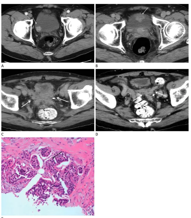
Fig. 1. Initial contrast-enhanced CT image in a 70-year-old man.
A. A contrast-enhanced CT image on early nephrographic phase shows an about 2.0 cm sized homogenous enhancing mass at right posterolat- eral wall of bladder (arrow).
B. A contrast-enhanced CT scan on excretory phase performed 8 years after first visit. An about 2.7 cm sized polypoid mass (arrow) is located in the lower anterior wall of bladder with no perivesical fatty infiltration nor abnormal lymphadenopathy.
C. Follow-up contrast-enhanced CT image on excretory phase, approximately four months after the final transurethral resection of bladder tu- mor. Ill-defined enhancing masses are developed in the perivesical space (arrows). This mass seemed to have invaded the bladder, seminal vesicle and adjacent peritoneum.
D. A coronal reconstructed CT image shows ill-defined, enhancing soft tissue lesion along the adjacent peritoneum (arrows).
E. Low power photomicrograph of the pathologic specimen obtained at extra-vesical mass around the bladder. Low power photomicrograph of the pathologic specimen obtained at extra-vesical mass around the bladder (arrow) shows low-grade infiltrating urothelial carcinoma (original magnification, × 200; hematoxylin-eosin stain).
CT = computed tomograph A
C
E
B
D
bladder with no evidence of perivesical fatty infiltration or ab- normal lymphadenopathy. Therefore he had second TURBT to remove this mass. Pathologic findings showed a low-grade by WHO, infiltrating urothelial carcinoma (stage T1), with only subepithelial connective tissue invasion. After that, TURBT was performed two times due to recurrence. Pathologic findings showed a low-grade, non-invasive papillary urothelial carcino- ma, with no evidence of bladder perforation or extravesical metastasis. Approximately four months after the final TURBT, the patient presented to the hospital after being unable to uri- nate for approximately 2 days. Contrast-enhanced CT revealed a newly developed ill-defined soft tissue mass in the perivesical space. The mass seemed to have invaded the bladder, seminal vesicle, adjacent peritoneum, and right distal ureter, resulting in the development of right hydronephrosis (Fig. 1C, D). CT guided biopsy was performed on the patient’s extravesical mass, and findings revealed metastatic, low-grade infiltrating urothe- lial carcinoma (Fig. 1E). Currently, the patient is receiving gem- citabine and carboplatin chemotherapy. He is undergoing fol- low-up, and has not shown any complications.
DISCUSSION
Bladder cancer accounts for 2% of all malignant neoplasms;
it is the second most common urological cancer (2, 3). Cystos- copy and TURBT are the primary methods of diagnosis and treatment of bladder cancer, and they cause fewer complica- tions and are less invasive compared to other treatment options.
According to recent research findings, the overall complication rate of TURBT is 5–6%, with common complications including urinary tract infection, hemorrhage requiring transfusion, and bladder perforation. Extravesical metastasis and intraperitoneal seeding of tumor cells following TURBT are very rarely report- ed, respectively (2, 3, 6).
There are three mechanisms explaining distant metastasis af- ter TURBT.
First, microperforation of bladder can cause peritoneal seed- ing. In rare cases, extravesical metastasis has been reported, that’s because microperforation of bladder occurred after TURBT (3).
Because TURBT is used to obtain a histologic diagnosis and to provide treatment through the resection of all visible disease, the scope of resection is usually deep and extensive, resulting in
an increased risk of bladder perforation (7). In cases with heav- ily pretreated, thin-walled bladders; and when the size of the tumor is large and located posteriorly or in the bladder dome, there is a high risk of perforation (8). Although such bladder perforation is rare, care needs to be taken; because it can lead to peritoneal or abdominal wall metastasis (3). Mydlo et al. (3) re- ported a case in which extensive peritoneal metastasis and liver metastasis occurred due to intraperitoneal perforation 4 months following TURBT, while Bus et al. (9) also reported a case in which seeding occurred in both adnexa due to intraperitoneal perforation following TURBT. Repeated TURBT caused thin- ning of the bladder wall, which is thought to have increased the risk of microperforation. This is also thought to have caused seeding to the adjacent peritoneum. However there is no defi- nite evidence of bladder perforation in our case on clinically and radiologically.
Second, increased internal pressure of the bladder after TURBT is the reason why the extravesical metastasis occurs (4). Engil- bertsson et al. (4) published a report on the seeding of cancer cells in the blood stream during TURBT. According to their study, the internal pressure of the bladder becomes greater than surrounding venous pressure due to fluid infusion during TURBT, which may lead to infusion of cancer cells into the sur- rounding venous system. A total of 16 patients took part in a test in which the number of cancer cells was measured in the inferior vena cava before and during TURBT. The results showed that the number of cancer cells increased significantly during TURBT. This study aimed to find an explanation for the occur- rence of distant metastasis following TURBT, but they could not present actual data on the subject.
Third, it is generally accepted that tumor seeding or distant metastasis are associated with histologic grade of tumor (5).
Papillary cancer of Ta grade 2 or 3 (high grade) tend to progress into the muscle-invasive bladder cancer and subsequent extra- vesical metastasis (10). However, the patient in our study ini- tially had stage Tis bladder cancer with invasion only extending into the mucosal layer. He underwent TURBT treatment, but owing to subsequent recurrence, TURBT was conducted 4 ad- ditional times. During this procedure, the histologic grade of the tumor was not exceeding low-grade, infiltrating urothelial carcinoma (stage T1).
According to the 2011 European Association of Urology (EAU)
guidelines on non-muscle-invasive bladder cancer, patients need to be follow-up by radiologic modality including chest X-ray, cystoscopy and CT because of the risk of recurrence and pro- gression; however, the frequency and duration of radiologic fol- low-up should reflect the degree of risk. Patients with small non-invasive (Ta), low-grade papillary tumors at low risk of re- currence and progression should have a cystoscopy at 3 month after TURBT. If negative, the following cystoscopy is advised 9 month later and then yearly for 5 years (1).
In summary, in patients who undergo TURBT to treat low- grade, Tis bladder cancer, and repeated TURBT due to recur- rence during follow-up, thorough radiologic follow-up should be performed due to the risk of subsequent extravesical metastasis.
REFERENCES
1. Babjuk M, Oosterlinck W, Sylvester R, Kaasinen E, Böhle A, Palou-Redorta J, et al. EAU guidelines on non-muscle-inva- sive urothelial carcinoma of the bladder, the 2011 update.
Eur Urol 2011;59:997-1008
2. Collado A, Chéchile GE, Salvador J, Vicente J. Early compli- cations of endoscopic treatment for superficial bladder tu- mors. J Urol 2000;164:1529-1532
3. Mydlo JH, Weinstein R, Shah S, Solliday M, Macchia RJ.
Long-term consequences from bladder perforation and/or violation in the presence of transitional cell carcinoma: re- sults of a small series and a review of the literature. J Urol
1999;161:1128-1132
4. Engilbertsson H, Aaltonen KE, Björnsson S, Kristmundsson T, Patschan O, Rydén L, et al. Transurethral bladder tumor re- section can cause seeding of cancer cells into the blood- stream. J Urol 2015;193:53-57
5. Gwynn ES, Clark PE. Bladder cancer. Curr Opin Oncol 2006;
18:277-283
6. Nieder AM, Meinbach DS, Kim SS, Soloway MS. Transure- thral bladder tumor resection: intraoperative and postop- erative complications in a residency setting. J Urol 2005;
174:2307-2309
7. Kim JH, Yang WJ. Delayed spontaneous perforation of uri- nary bladder with intraperitoneal seeding following radical transurethral resection of invasive urothelial cancer: a case report. BMC Res Notes 2014;7:167
8. Golan S, Baniel J, Lask D, Livne PM, Yossepowitch O. Trans- urethral resection of bladder tumour complicated by perfo- ration requiring open surgical repair - clinical characteris- tics and oncological outcomes. BJU Int 2011;107:1065-1068 9. Bus MT, Cordeiro ER, Anastasiadis A, Klioueva NM, de la Rosette JJ, de Reijke TM. Urothelial carcinoma in both ad- nexa following perforation during transurethral resection of a non-muscle-invasive bladder tumor: a case report and literature review. Expert Rev Anticancer Ther 2012;12:
1529-1536
10. Kitamura H, Tsukamoto T. Early bladder cancer: concept, di- agnosis, and management. Int J Clin Oncol 2006;11:28-37
비침습적 방광암의 경 요도 절제술 후 발생한 방광 외 전이에 대한 증례 보고
강가람
1· 손동완
2· 정동진
3*
비침습적 방광암의 경 요도 절제술은 방광암의 첫 번째 치료 방법으로 알려져 있다. 비침습적 방광암의 경 요도 절제술의 흔한 합병증으로는 요로감염, 수혈이 필요한 정도의 출혈, 방광 천공 등이 있다. 반면 시술 후 종양의 방광 외 전이 및 복막 으로의 파종 등은 매우 드물게 보고된다. 이에 저자들은 비침습적 방광암의 반복적인 경 요도 절제술 후 방광 외 전이가 발생한 증례를 보고하고자 한다.
1가톨릭대학교 의과대학 의정부성모병원 영상의학과, 가톨릭대학교 의과대학 여의도성모병원 2비뇨기과, 3영상의학과