Cystic duct closure during partial cholecystectomy:
ten years’ experience
Whanbong Lee
Department of Surgery, Sanbon Hospital, Wonkwang Univiversity, Kunpo, Korea
Backgrounds/Aims: When surgeons face difficulties in dissecting the Calot triangle during cholecystectomy due to se- vere inflammation or fibrosis, the proximal portion of the gallbladder is left in place to avoid injury to the bile duct;
this procedure is called partial cholecystectomy (PC), and it is associated with a much higher complication rate after the operation. Methods: We surveyed the clinical outcomes of 25 cases of PC by laparotomy during ten years from January 1998 to December 2007, for a total of 95 months of the mean follow-up period. Patients were separated in two groups for comparison: group I (n=15), in which cystic duct closure was tried from the intraluminal cystic ductal opening; and group II (n=10), in which cystic ductal circumferential ligation was possible. Results: Bile fistula occurred in 4 cases of group I, while no fistula occurred in group II. Postoperative peritonitis was observed in 4 cases from group I, with 3 of them caused by leakage of bile when the cystic duct could not be properly managed by stitches or staples. One of these peritonitis cases was fatal, but no case in group II showed peritonitis postoperatively. Wound infection, retained stone, and reoperations were also more frequent in group I, in 4, 2, and 5 cases. The mortality was 3 in group I and 1 in group II. Conclusions: When inevitable partial cholecystectomy is carried out, more attention should be focused on secure ligation of the cystic duct, with the expectation of an improved outcome of the operation on a large scale. Otherwise, patients should be clearly informed about the high risks of postoperative complications.
(Korean J Hepatobiliary Pancreat Surg 2013;17:176-180) Key Words: Cholecystitis; Partial cholecystectomy; Cystic duct
Received: October 5, 2013; Revised: October 11, 2013; Accepted: October 15, 2013 Corresponding author: Whanbong Lee
Department of Surgery, Sanbon Hospital, Wonkwang Univiversity, 1126-1, Sanbon-dong, Kunpo 435-040, Korea Tel: +82-31-390-2218, Fax: +82-31-390-2245, E-mail: [email protected]
Copyright Ⓒ 2013 by The Korean Association of Hepato-Biliary-Pancreatic Surgery Korean Journal of Hepato-Biliary-Pancreatic Surgery ∙ ISSN: 1738-6349
INTRODUCTION
Partial cholecystectomy (PC) is an inevitably or acci- dentally performed procedure in which the proximal por- tion of the gallbladder near the liver bed and Calot tri- angle is left in place. A deep-sited cystic duct with con- tinuous oozing on slight dissection or stony hard fibrous changes with collapse of the gallbladder hinders further dissection of the proximal gallbladder down to the cystic duct for its safe closure.
Safe closure of the cystic duct by cutting between liga- tions or by making a secure ligation without injuring other vital structures like the Rt. Hepatic duct or Rt. Hepatic artery or even the Rt. Portal vein would be an important factor in preventing bile leakage through the cystic duct.
The literature on the details of cystic duct treatment is still lacking. We reviewed our cases of PC from the view of cystic duct handling, the methods that were used, espe-
cially in the cases in which ligation was not possible, and related these with the outcomes to ascertain their importance.
METHODS
Patient selection
We retrospectively surveyed the clinical records of the patients’ profile and complications during the follow-up after the operation from January 1998 to December 2008, for 25 patients on whom PC was performed. Patient dem- ographics are illustrated in Table 1.
The patients were separated into two groups, I and II, for comparison of cystic ductal management in relation to whether the cystic duct was circumferentially dissected and ligated (group II) or not (group I), and the in- formation was reviewed or gathered through various ways.
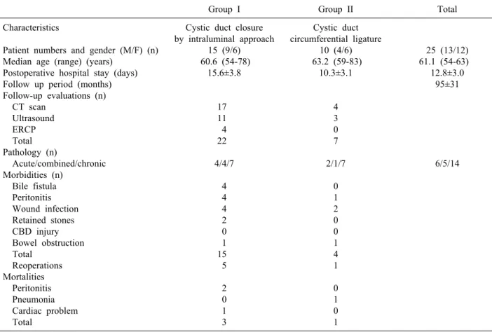
Table 1. Baseline demographics of patients and the clinical outcome
Group I Group II Total
Characteristics
Patient numbers and gender (M/F) (n) Median age (range) (years)
Postoperative hospital stay (days) Follow up period (months) Follow-up evaluations (n) CT scan
Ultrasound ERCP Total Pathology (n)
Acute/combined/chronic Morbidities (n)
Bile fistula Peritonitis Wound infection Retained stones CBD injury Bowel obstruction Total
Reoperations Mortalities Peritonitis Pneumonia Cardiac problem Total
Cystic duct closure by intraluminal approach
15 (9/6) 60.6 (54-78)
15.6±3.8
17 11 4 22 4/4/7
4 4 4 2 0 1 15 5 2 0 1 3
Cystic duct circumferential ligature
10 (4/6) 63.2 (59-83)
10.3±3.1
4 3 0 7 2/1/7
0 1 2 0 0 1 4 1 0 1 0 1
25 (13/12) 61.1 (54-63) 12.8±3.0
95±31
6/5/14
CT, computed tomography; ERCP, endoscopic retrograde cholangiopancreatography; CBD, common bile duct
Patients having serious associated or underlying dis- eases (n=4) or simultaneous biliary tract stones (n=2) or a previous upper abdominal major operative history (n=2) were excluded from the study, and patients with sepsis secondary to the cholecystitis were also excluded, as they had different risk rates of complications (n=3).
Operative methods
In the gallbladder approach, entry was made through the right paramedian transrectal vertical incision in all 25 patients by one surgeon, and converted procedures during the laparoscopic cholecystectomy were included (n=7).
In group II patients (n=10), in whom cystic duct liga- tion was possible but the proximal portion of Hartmann’s pouch was left, a guiding probe was used frequently to trace the course of the cystic duct and to remove possibly impacted stones. The tedious circumferential dissection of the cystic duct was complicated by the perforation of the cystic duct wall in 3 cases, and a further search for the cystic duct, and ligation were possible at last.
Cystic ductal ligation was performed in 7 cases, while 3 cases were ligated and cut between the ligations. In 3 cases, on the antemesenteric side, a large hole of the gall- bladder was torn right down to the cystic duct by cautious instrumental guiding and stitch closure of duct was performed.
In group I patients (n=15), cystic ductal circumferential dissection was not possible due to severe oozing from the surrounding structures of Calot triangle (n=6), or stony hard adhesions between the cystic duct and portal struc- tures (n=8) or too deep positioning of the cystic duct, with fibrotic adhesions (n=1). Probe guiding as well as other efforts were made but were of no use for isolating the cystic duct. But perforation of the cystic duct during dis- section did not occur, and perforation happened in 3 cases at the proximal part of Hartmann’s pouch. Attempts using various methods to close the cystic duct from Hartmann’s pouch were tried. Stitching of the cystic ductal opening at the exact site was possible in 4 cases. After cauteriza- tion of the opening, which was carried out in all the PC
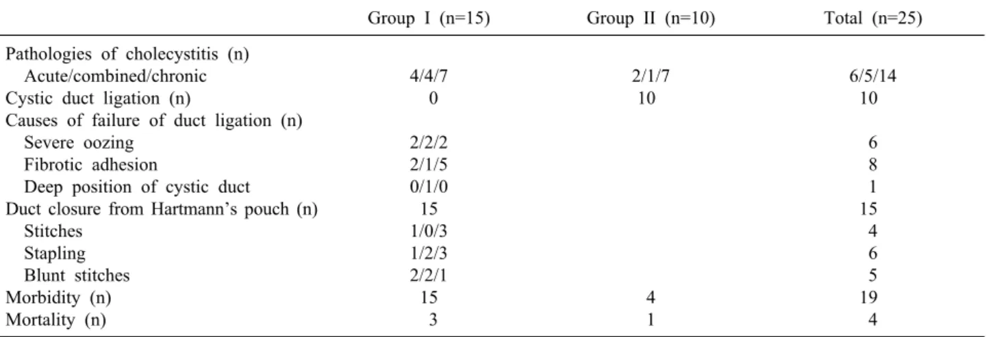
Table 2. Duct arrangement related with complications during partial cholecystectomy
Group I (n=15) Group II (n=10) Total (n=25) Pathologies of cholecystitis (n)
Acute/combined/chronic Cystic duct ligation (n)
Causes of failure of duct ligation (n) Severe oozing
Fibrotic adhesion
Deep position of cystic duct Duct closure from Hartmann’s pouch (n) Stitches
Stapling Blunt stitches Morbidity (n) Mortality (n)
4/4/7 0 2/2/2 2/1/5 0/1/0 15 1/0/3 1/2/3 2/2/1 15
3
2/1/7 10
4 1
6/5/14 10
6 8 1 15 4 6 5 19 4 cases (n=25), stapling was tried in 6 cases. Stapling was
rather a blunt procedure, so attempts were made to avoid injury of the bile duct nearby.
In 5 cases, neither stitches nor stapling were possible due to the poor operative field where even close con- frontation of the edges of the opening was nearly impossible. And therefore, blunt stitches around Hartmann’s pouch and plugging of the ductal opening by collagen fiber material were the only ways to close the ductal opening.
Additional blunt stitches or plugging after ductal liga- tion or closure of the ductal opening by stitches or sta- pling were not counted in this study.
A suction drain (Jackson-Pratt drain) was routinely left inside, running from the gallbladder through to the Rt.
flank, and it was usually removed a week after the oper- ation, depending on the drain’s output.
Statistic analysis
Descriptive analysis was carried out in detail for the complications, and chi-square tests for comparisons be- tween two variables were performed on outcomes for the complication, whose rates were high or for the clinical courses, assuming a normal distribution, with confidence intervals of 95%.
RESULTS
The patients’ ages were from 54 to 83 years, and the distribution of age between the two groups of PC was similar. And the follow-up period was also similar be-
tween the two groups, with a mean of 95 months. The hospital stay after the initial operation was longer in group I patients by about 5 days, showing a significant differ- ence (p<0.05). During the follow-up, 22 examinations were executed among group I patients than 7 imaging studies in group II patients, from postoperative symptoms and complaints from patients (p<0.01). A reoperation was necessary in 5 patients of group I, while 1 patient of group II needed reoperation, showing a prominent dif- ference even before the statistical analysis was performed.
Total morbidities were 15 cases in group I, and 4 cases in group II, showing a significant difference (p<0.05).
When the cystic duct could not be ligated in group I, bile fistula (n=4), peritonitis (n=4), wound infection (n=4), and retained stones (n=2) were more prevalent postoperatively, leading to more reoperations (n=5), as described above, than in group II cases where cystic ductal ligation was accomplished. Only one case of peritonitis, and 2 cases of wound infection occurred in group II. These results are summarized in Table 1 and Table 2.
Four cases of bile fistula in group I occurred after blunt closure of the cystic ductal opening (n=3), and after sta- pling of the cystic ductal opening (n=1).
Three of them had associated peritonitis, and one of them developed fatal sepsis. Bile fistula was managed by endoscopic stenting in 1 case, and supportive management in 2 cases, and one of the patients in the latter group suc- cumbed to fatal sepsis.
Considering the detailed conditions of the cystic duct where cystic duct ligation was not possible in group I, as shown in Table 2, fibrotic adhesions of structures (n=8)
and severe oozing from the field (n=6) in the Calot tri- angle were the most common conditions faced, occupying 14 out of 15 cases. A difficult position of the cystic duct was a rare cause of failure of ligation (n=1, 1 of 15, 6.7%). Failed cystic ductal ligation occurred when the acute inflammatory process spread, resulting in the com- bined pathologies of acute and chronic cholecystitis, which occurred in 8 out of 15 cases (53.3%).
In seven out of 11 cases (63.6%) of acute or combined pathology, the cystic duct could not be securely ligated, while in 7 out of 14 cases (50.0%) of chronic pathology, the duct could not be ligated, which was significantly dif- ferent (p<0.05). Among 15 cases of group I, stitches were possible in 4 cases and stapling at the opening of cystic duct was possible in 6 cases, which could be re- garded as a rather secure closing of the cystic duct. But in 5 cases, the cystic duct opening could not be closed properly, and electrocautery, blunt stitches, and collagen fiber plugging were the only ways to prevent post- operative bile fistula.
Mortality came from 1 case of bile fistula, 1 case of peritonitis without bile leakage and 1 cardiac problem in group I, and 1 pneumonic sepsis in group II. The repre- sentative mortality of the two groups was 4 of 15 (26.7%), and 1 of 10 (10.0%), respectively, which was not statisti- cally meaningful however (p>0.1).
DISCUSSION
PC itself generally imposes increased complications af- ter an operation; the incidence is well over 10%, as was proven in many publications,1-3 but reports separating PC cases according to whether the cystic duct was ligated or not, and according to the method of cystic ductal closure, are seldom seen.
Unsecure cystic duct closure (group I) was related to longer hospital stay, more frequent examinations after- wards the operation, and a higher reoperation rate, all of which were statistically meaningful, leading patients to experience prolonged discomfort from symptoms or com- plications, and higher financial expenditure.
When the cystic duct was closed after the patient com- plained of pain, taking cautions during the dissection of it and using a guiding probe through the cystic ductal opening in group II, the complications decreased, as we
would expect. No bile fistula, no retained stone, and no CBD injury were observed, except for the conventional complications of peritonitis (n=1), wound infection (n=2), and bowel adhesion related obstruction (n=1). Cystic duc- tal dissection was able to be performed more frequently when chronic pathology prevailed around gallbladder than in cases where the acute inflammatory process surrounded the gallbladder, as shown in Table 2.
It seemed that a friable edematous gallbladder and cyst- ic duct imposed more difficulty in dissection because of the higher incidence of troublesome bleeding and tearing or perforation during the process than cases of a fibrous planeless stiff structure in chronic cholecystitis.
Closure of the cystic ductal opening from Hartmann’s pouch was done in 4 cases by direct stitching across the opening in one case of acute cholecystitis and in 3 cases of chronic cholecystitis, where there was occurrence of bile leakage. When the gallbladder was edematous due to acute inflammation, secure stitches were more difficult to apply than in the case of a porcelain gallbladder.
In 6 cases where a stapling device was used for closure of the cystic ductal opening from Hartmann’s pouch, one case showed a bile fistula. Stapling was rather a blunt pro- cedure that was attempted with a poor operative view of the cystic ductal opening due to its deep position, and executed with concerns over potential tearing of the bili- ary duct or the causing of troublesome bleeding. During stapling, great care was taken to avoid perforation of the bile duct or a vessel, and stapling was kept away from them as much as possible. But when comparing bile leak- age after the placement of blunt stitches around Hartmann’s pouch and without placing stitches exactly at the ductal opening, bile leakage was less frequent in sta- pled patients (n=1) than in patients who received blunt stitches (n=3).
Through massive cauterization of the cystic ductal opening from Hartmann’s pouch, big bite blunt stitches including plugged collagen fiber materials were inevitable in 5 cases. Even cauterization around the cystic ductal opening was not easy due to the poor operative field caused by oozing and distortion of tissues secondary to inflammation. Four of these cases were acute or combined cholecystitis, and one case was chronic cholecystitis. In these patients, major morbidities and mortalities were seen such as bile leakage (n=3), wound infection (n=3), peri-
tonitis (n=3), retained stone (n=2), and need for a reopera- tion (n=3).
During the PC, gallbladder management was more chal- lenging in circumferential cystic duct dissection, and in performing stitching or stapling from Hartmann’s pouch, and in every other way, when acute inflammation was present than when there was chronic cholecystitis.
More advanced methods of secure closure of the cystic ductal opening from Hartmann’s pouch during PC should be sought rather than using temporary probing or stapling, as was shown in the review in this study. The timing of cholecystectomy might influence the success rate of clo- sure of the cystic ductal opening in the sense that when acute pathology is proceeding in the gallbladder, its man- agement is more difficult, so partial subsidence of the in- flammation should be attempted by delaying the operation while giving antibiotic coverage, as was recommended in some publications.4,5 Cystic ductal closure from the com- mon bile duct by choledochotomy and primary closure of the common bile duct without a T-tube as was recently proposed is another possible treatment.6-8
ACKNOWLEDGEMENTS
This paper was partially supported by Wonkwang University research foundation.
REFERENCES
1. Davis B, Castaneda G, Lopez J. Subtotal cholecystectomy versus total cholecystectomy in complicated cholecystitis. Am Surg 2012;78:814-817.
2. Sharp CF, Garza RZ, Mangram AJ, et al. Partial cholecystectomy in the setting of severe inflammation is an acceptable consid- eration with few long-term sequelae. Am Surg 2009;75:249-252.
3. Cakmak A, Genç V, Orozakunov E, et al. Partial chol- ecystectomy is a safe and efficient method. Chirurgia (Bucur) 2009;104:701-704.
4. Yamashita Y, Takada T, Kawarada Y, et al. Surgical treatment of patients with acute cholecystitis: Tokyo Guidelines. J Hepatobiliary Pancreat Surg 2007;14:91-97.
5. Catani M, De Milito R, Romagnoli F, et al. The best timing of surgery in laparoscopic cholecystectomy for acute cholecystitis:
when and how is to be performed. Hepatogastroenterology 2008;55:1993-1996.
6. Naraynsingh V, Hariharan S, Ramdass MJ, et al. Open common bile duct exploration without T-tube insertion- two decade expe- rience from a limited resource setting in the Caribbean. Indian J Surg 2010;72:185-188.
7. Yin Z, Xu K, Sun J, et al. Is the end of the T-tube drainage era in laparoscopic choledochotomy for common bile duct stones is coming? A systematic review and meta-analysis. Ann Surg 2013;257:54-66.
8. Cai H, Sun D, Sun Y, et al. Primary closure following laparo- scopic common bile duct exploration combined with intra- operative cholangiography and choledochoscopy. World J Surg 2012;36:164-170.