• 검색 결과가 없습니다.

Single Incision Thoracoscopic Left Lower Lobe Superior Segmentectomy for Non-Small Cell Lung Cancer

N/A
N/A
Protected

Academic year: 2021

Share "Single Incision Thoracoscopic Left Lower Lobe Superior Segmentectomy for Non-Small Cell Lung Cancer"

Copied!
4
0
0

로드 중.... (전체 텍스트 보기)

전체 글

(1)

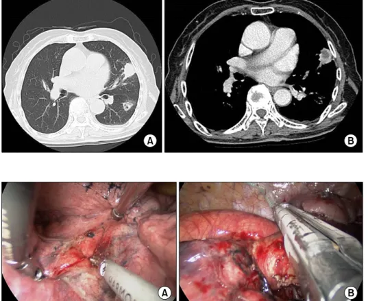
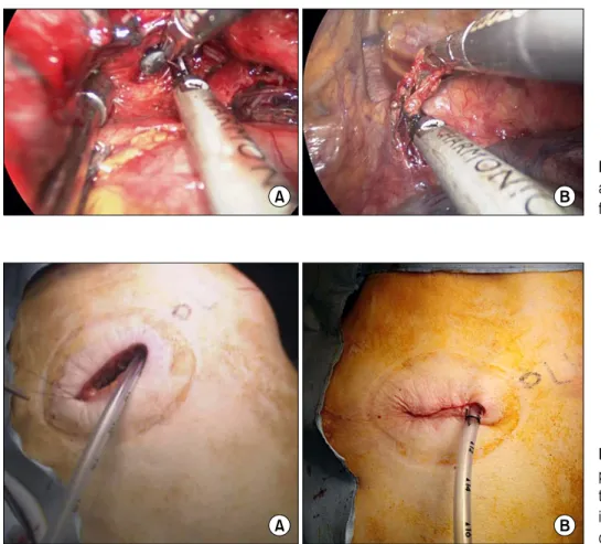
Korean J Thorac Cardiovasc Surg 2014;47:185-188 □ Case Report □ http://dx.doi.org/10.5090/kjtcs.2014.47.2.185 ISSN: 2233-601X (Print) ISSN: 2093-6516 (Online)

− 185 −

Department of Thoracic and Cardiovascular Surgery,

1

Bucheon St. Mary’s Hospital,

2

Seoul St. Mary’s Hospital,

3

St. Vincent’s Hospital, The Catholic University of Korea College of Medicine

Received: July 22, 2013, Revised: September 25, 2013, Accepted: October 1, 2013

Corresponding author: Young Pil Wang, Department of Thoracic and Cardiovascular Surgery, Seoul St. Mary’s Hospital, The Catholic University of Korea College of Medicine, 222 Banpo-daero, Seocho-gu, Seoul 137-701, Korea

(Tel) 82-2-2258-2858 (Fax) 82-2-594-8644 (E-mail) [email protected]

C

The Korean Society for Thoracic and Cardiovascular Surgery. 2014. All right reserved.

CC

This is an open access article distributed under the terms of the Creative Commons Attribution Non-Commercial License (http://creative- commons.org/licenses/by-nc/3.0) which permits unrestricted non-commercial use, distribution, and reproduction in any medium, provided the original work is properly cited.

Single Incision Thoracoscopic Left Lower Lobe Superior Segmentectomy for Non-Small Cell Lung Cancer

Hyun Woo Jeon, M.D.

1

, Soo Hwan Choi, M.D.

2

, Young Pil Wang, M.D., Ph.D.

2

, Kwan Yong Hyun, M.D.

3

Lobectomy with mediastinal node dissection has been standard treatment for non-small cell lung cancer (NSCLC).

Nowadays, video-assisted thoracoscopic surgery (VATS) is gaining acceptance as an alternative treatment option, given the quality-of-life benefits that it confers. For the VATS procedure, most surgeons create two or three ports with a utility incision of 3 to 5 cm. However, with acquired skill and instrumentation advances, single-incision thor- acoscopic surgery has emerged over time. Here, we report the case of an 86-year-old female with NSCLC treated by single-incision segmentectomy.

Key words: 1. Video-assisted thoracic surgery (VATS) 2. Minimally invasive surgical procedures 3. Lung neoplasms

4. Adenocarcinoma of lung 5. Segmentectomy

CASE REPORT

An 86-year-old female was referred to Seoul St. Mary's Hospital because of abnormal chest X-ray findings. The chief complaints were intermittent cough and mild fever ongoing for one month. Computed tomography (CT) revealed a mass (3.3×2.2 cm) in the lingula of the left upper lobe (LUL) and a second lesion (2.2×1.6 cm) in the superior segment of the left lower lobe (LLL), which were most likely malignant (Fig. 1A, B). Preoperative assessments included magnetic res- onance imaging of the brain, bone scan, whole-body positron emission tomography-CT, echocardiography, and broncho- scopy. Clinically, an abscess of LUL and a cancer of LLL (stage cT1bN0M0) were suspected. Preoperative forced ex- piratory volume in one second (FEV

1

) was 1.37 L. Echocar-

diography revealed normal ejection fraction (60%), but aki- nesia on the basal posterior wall and a ventricular premature complex were observed in the electrocardiography. For the complete resection, lingular segmentectomy and LLL lobec- tomy should be carried out. Postoperative FEV

1

was esti- mated to be 0.9 L. Due to advanced age and poor respiratory function, the surgical plan was wedge resection for the LUL lesion first. If the lesion was not malignant on the frozen re- port, the next step was the superior segmentectomy of LLL.

Under general anesthesia, a double-lumen endotracheal tube

was placed, and the patient was transitioned to the right later-

al decubitus position. Once selective lung ventilation was

achieved, a 4-cm incision was made in the fifth intercostal

space at the anterior axillary line with a wound protector

(Applied Medical, Rancho Santa Margarita, CA, USA). The

(2)

Hyun Woo Jeon, et al

− 186 −

Fig. 1. (A) Computed tomography revealed a mass measuring approx- imately 3.3×2.2 cm in the lingula and a nodule measuring 2.2×1.6 cm with speculation in the superior seg- ment of left lower lobe. (B) After contrast injection, the left upper lobe lesion showed peripheral enhance- ment with central low attenuation.

Fig. 2. (A) The first step is inter- lobar dissection using an ultrasonic device. (B) The superior segmental artery was identified and divided us- ing an endostapler.

Fig. 3. (A) The superior segmental bronchus was dissected and ob- structed using a right-angle clamp.

(B) After lung inflation, the segmen- tal plane was divided.

two distinct masses were digitally palpable, with a visible re- traction of the lower lobe visceral pleura. The cavitary lesion in LUL was sizable. Simple wedge resection was difficult for complete resection. First, the anterior portion of the fissure was dissected and divided, exposing the interlobar artery (Fig.

2A). The lingular segmental artery was then identified and divided, followed by the elevation and resection of the upper lobe mass. Then, complete resection was achieved, and the distance between the mass and the resection margin was 0.6 cm. After there was no evidence of malignancy on the frozen

section report, the superior segmental artery (encircled by a drain catheter) was identified and divided using an endo- stapler (TriStapler; Covidien, Norwalk, CT, USA) (Fig. 2B).

The inferior pulmonary ligament was divided to reveal the in-

ferior pulmonary vein. Eventually, the superior segmental

vein was divided using a Hem-o-lok (Teleflex Medical Inc.,

Research Triangle Park, NC, USA). After identifying the su-

perior segmental bronchus, the peribronchial tissue and lymph

nodes were dissected. Then, the bronchus was clamped, and

both lungs were ventilated (Fig. 3A), enabling the visual-

(3)

Single Incision Thoracosopic Segmentectomy

− 187 −

Fig. 4. (A) Subcarinal and (B) para- aortic lymph nodes were dissected for complete resection.

Fig. 5. (A) The chest tube was placed via the posterior portion of the utility incision, and (B) the skin incision was closed with the sub- cuticular running suture method.

ization of the segmental plane and stapler division (Fig. 3B).

Mediastinal lymph node dissection (para-aortic, subcarinal, in- ferior pulmonary ligament, and paraesophageal lymph nodes) was performed (Fig. 4A, B). A chest tube (24 Fr.) was placed in the pleural cavity through the utility incision, and the wound was closed (Fig. 5A, B). The total operative time was 135 minutes. Histopathology confirmed an abscess of LUL and an adenocarcinoma (1.6×1.4 cm) of LLL with the invasion of the visceral pleura (stage T2aN0M0), and a dis- tance of 3 cm was obtained between the tumor and the div- ided segmental plane. The patient was discharged on post- operative day 7 without complications.

DISCUSSION

Since its introduction in the 1990s, video-assisted thoraco- scopic surgery (VATS) has been an attractive treatment mo- dality for thoracic surgery, resulting in shorter recovery peri- ods and less postoperative pain than open procedures [1].

Although, generally, three or four incisions are used for VATS lobectomy or the segmentectomy of lung cancers, a report by Rocco et al. [2] on uniportal VATS wedge re- section, citing the potential to reduce postoperative pain and paresthesia, has caught the interest of thoracic surgeons. Since 2010, Gonzalez-Rivas et al. [3] have adopted this technique for lung cancer surgery, confirming its feasibility in a recent publication.

Recently, many centers have adopted the abovementioned

technique [4]. Furthermore, segmentectomy has also been per-

formed by single incision [5]. Although lobectomy is the

standard surgical management for early lung cancer, lobec-

tomy is not suitable for all patients. Most of these patients

are elderly and have poor cardiopulmonary reserve. Segmen-

tectomy is a reasonable alternative procedure. In a compar-

ison of segmentectomy with lobectomy for non-small cell

lung cancer <2 cm, Okada et al. [6] found that the 5-year

survival rates did not differ by procedure. In particular, in the

older patients, segmentectomy leads to less complications and

(4)

Hyun Woo Jeon, et al

− 188 − low comparable oncologic impact than lobectomy [7].

We extended our experience with VATS for lung cancer to include a single-incision approach using a 4-cm utility in- cision of the 5th intercostal space (at the anterior axillary line). A wound protector is employed to protect the camera lens from oozing by the intercostal muscle. The required in- strumentation does not otherwise differ from that of conven- tional VATS (endograsper, 10-mm camera with 30

o

angle, right-angle clamp, and ultrasonic device). The operative pro- cedure differs according to the affected lobe. During dis- section, we use a harmonic scalpel (Ethicon Endo-Surgery Inc., Cincinnati, OH, USA).

A harmonic scalpel is very useful and suitable for the dis- section and coagulation of microvessels because of its blunt tip and scissors-like action. We preferred a long right-angle clamp with a blunt tip during the tunneling and encircling of the vessels and the bronchus.

Single-incision VATS does have some limitations. Clearly, when the multiple views of conventional VATS are forfeited, obstruction of view and impingement of instruments com- monly occur. However, the surgical technique is not different, including the identification and division of the vessels and the bronchus, and lymph node dissection. We generally center the camera on the incision, inserting instruments on either side of it; further, for better visualization, the stapler is placed in the thoracic cavity beneath the camera. Conducting extensive surgery through a single, small incision is often er- gonomically problematic for surgeons and assistants. During an operation, the operator and the assistant are sometimes su- perimposed; therefore, it is important to prevent contami- nation.

We made a utility incision in the 5th intercostal space. The procedure through the 6th intercostal space allows the divi- sion of the inferior pulmonary ligament and the inferior pul- monary vein more easily. However, our first step is the dis- section of the interlobar space; this approach is easier through the 5th intercostal space. If a conversion to conventional VATS is required, a utility incision in the 5th intercostal space is more convenient because a utility incision in the 5th intercostal space is always used during conventional VATS.

In our experience, this approach is safe, feasible, and reproducible. Although segmentectomy is generally expected

to be more difficult than lobectomy, superior, basal, and lin- gula segmentectomies are more easily executed, owing to the readily defined segmental planes. However, without multiple views through a single incision, the division of segmental planes has to be performed in one direction. Division of seg- mental planes is slightly difficult during the division of seg- mental planes in single-incision segmentectomy.

In conclusion, single-incision VATS superior segmentec- tomy of LLL for early-stage lung cancer was both feasible and safe in this instance. However, its merits in cancer treat- ment require further study.

CONFLICT OF INTEREST

No potential conflict of interest relevant to this article was reported.

ACKNOWLEDGMENTS

This manuscript has been edited by the native English- speaking experts of BioMed Proofreading.

REFERENCES

1. Demmy TL, Nwogu C. Is video-assisted thoracic surgery lo- bectomy better?: quality of life considerations. Ann Thorac Surg 2008;85:S719-28.

2. Rocco G, Martin-Ucar A, Passera E. Uniportal VATS wedge pulmonary resections. Ann Thorac Surg 2004;77:726-8.

3. Gonzalez-Rivas D, Paradela M, Fernandez R, et al.

Uniportal video-assisted thoracoscopic lobectomy: two years of experience. Ann Thorac Surg 2013;95:426-32.

4. Kang DK, Min HK, Jun HJ, Hwang YH, Kang MK.

Single-port video-assisted thoracic surgery for lung cancer.

Korean J Thorac Cardiovasc Surg 2013;46:299-301.

5. Gonzalez-Rivas D, Fieira E, Mendez L, Garcia J. Single-port video-assisted thoracoscopic anatomic segmentectomy and right upper lobectomy. Eur J Cardiothorac Surg 2012;42:

e169-71.

6. Okada M, Koike T, Higashiyama M, Yamato Y, Kodama K, Tsubota N. Radical sublobar resection for small-sized non-small cell lung cancer: a multicenter study. J Thorac Cardiovasc Surg 2006;132:769-75.

7. Kilic A, Schuchert MJ, Pettiford BL, et al. Anatomic seg-

mentectomy for stage I non-small cell lung cancer in the

elderly. Ann Thorac Surg 2009;87:1662-6.

수치

Fig. 1. (A) Computed tomography  revealed a mass measuring  approx-imately 3.3×2.2 cm in the lingula  and a nodule measuring 2.2×1.6 cm  with speculation in the superior  seg-ment of left lower lobe
Fig. 4. (A) Subcarinal and (B) para-  aortic lymph nodes were dissected  for complete resection.

참조

관련 문서

Comparison of cumulative survival according to maspin expression in non-small cell lung cancer..

To our knowledge, this is the first case report of histopatholgically confirmed ILD following erlotinib therapy in a live patient with non-small cell lung cancer

Lung cancer (poorly differentiated adenocarcinoma) in right lower lobe shows iso signal intensity relative to muscle signal intensity on T1-weighted image (left) and shows high

We investigated the impact of genetic variants identified in GWASs for lung cancer susceptibility on the survival outcomes in patients with early stage non-small cell lung

Abbreviations: ADC = adenocarcinoma, EGFR = epidermal growth factor receptor, HPV = human papillomavirus, MCPyV = Merkel cell polyomavirus, NSCLC = nonsmall cell lung cancer,

Somatic mutations of the TP53 gene are among the most frequent genetic alterations detected in lung cancer and occur in -50% of patients with non-small cell lung cancer (NSCLC)

RET Fusion Genes in Korean Non-Small Cell Lung Cancer.. Recently, rearranged during transfection (RET) fusions have been

Conclusion : For non-small cell lung cancer, IMRT with restricted angle or VMAT could minimize the lung dose and lower the dose to spinal cord below the tolerance level.