ISSN: 2233-601X (Print) ISSN: 2093-6516 (Online)
Received: September 13, 2016, Revised: October 5, 2016, Accepted: October 17, 2016, Published online: August 5, 2017
Corresponding author: Jhingook Kim, Department of Thoracic and Cardiovascular Surgery, Samsung Medical Center, Sungkyunkwan University School of Medicine, 81 Irwon-ro, Gangnam-gu, Seoul 06351, Korea
(Tel) 82-2-3410-3483 (Fax) 82-2-3410-6986 (E-mail) [email protected]
© The Korean Society for Thoracic and Cardiovascular Surgery. 2017. All right reserved.
This is an open access article distributed under the terms of the Creative Commons Attribution Non-Commercial License (http://creativecommons.org/
licenses/by-nc/4.0) which permits unrestricted non-commercial use, distribution, and reproduction in any medium, provided the original work is properly cited.
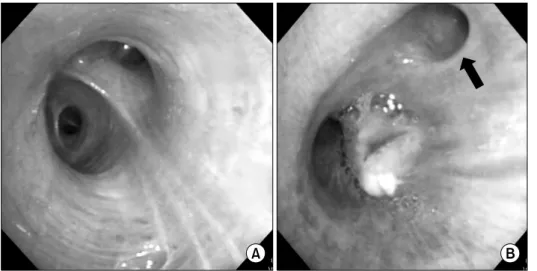
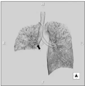
Right Lower Sleeve Bilobectomy for Lung Cancer with Posteparterial Tracheal Bronchus
Hongsun Kim, M.D. 1 , Jinsik Kim, M.D. 2 , Jong Ho Cho, M.D., Ph.D. 1 , Su Min Shin, M.D. 1 , Hong Kwan Kim, M.D., Ph.D. 1 , Jhingook Kim, M.D., Ph.D. 1
1