• 검색 결과가 없습니다.

A Large Epiphrenic Esophageal Diverticulum Communicating with the Left Lower Lobe

N/A
N/A
Protected

Academic year: 2021

Share "A Large Epiphrenic Esophageal Diverticulum Communicating with the Left Lower Lobe"

Copied!
4
0
0

로드 중.... (전체 텍스트 보기)

전체 글

(1)

ISSN: 2233-601X (Print) ISSN: 2093-6516 (Online)

− 40 −

Received: April 25, 2018, Revised: October 22, 2018, Accepted: October 22, 2018, Published online: February 5, 2019

Corresponding author: Jong Ho Cho, Department of Thoracic and Cardiovascular Surgery, Samsung Medical Center, Sungkyunkwan University School of Medicine, 81 Irwon-ro, Gangnam-gu, Seoul 06351, Korea

(Tel) 82-2-3410-2114 (Fax) 82-2-3410-6986 (E-mail) [email protected]

© The Korean Society for Thoracic and Cardiovascular Surgery. 2019. All right reserved.

This is an open access article distributed under the terms of the Creative Commons Attribution Non-Commercial License (http://creativecommons.org/

licenses/by-nc/4.0) which permits unrestricted non-commercial use, distribution, and reproduction in any medium, provided the original work is properly cited.

A Large Epiphrenic Esophageal Diverticulum Communicating with the Left Lower Lobe

Suk Kyung Lim, M.D., Jong Ho Cho, M.D., Ph.D.

Department of Thoracic and Cardiovascular Surgery, Samsung Medical Center, Sungkyunkwan University School of Medicine

Epiphrenic diverticula are known to cause a series of complications. We report the case of a 54-year-old woman who was diagnosed with an epiphrenic diverticulum at a regular checkup in November 2006. Ten years later, she presented with massive hematemesis. Imaging studies revealed an epiphrenic diverticulum measuring 7.8 cm in diameter and a large amount of bleeding inside the diverticulum. Computed tomography showed fistula formation between the diverticulum and the left lower lobe of the lung, leading to the devel- opment of a pulmonary abscess. Diverticulectomy and 180° posterior partial fundoplication were performed transabdominally. The pulmonary abscess was treated with antibiotics alone. She was discharged 16 days af- ter the operation without any complications over 7 months of follow-up.

Key words: 1. Esophageal diverticulum 2. Esophagopulmonary fistula

Case report

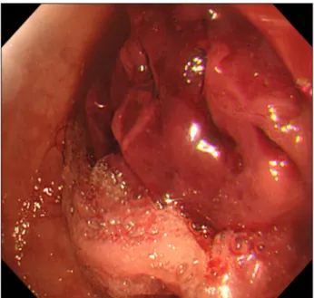
A 54-year-old woman was diagnosed with an esophageal diverticulum at a regular checkup in November 2006. No treatment was needed because the size of the diverticulum was small and she was asymptomatic. In September 2015, she presented with mild regurgitation and dysphagia. An esoph- agogastroduodenoscopic examination showed no spe- cific abnormalities, and she was treated conserva- tively. She returned to the emergency department 5 months later with massive hematemesis. An esoph- agogastroduodenoscopic examination revealed a Dieulafoy lesion within the diverticulum, in addition to a large hematoma (Fig. 1), and she underwent a clipping procedure. The esophagogram showed an epiphrenic diverticulum measuring 7.8 cm in diame- ter located 5 cm above the gastroesophageal junction (Fig. 2). Surgical treatment was indicated; however,

because of her poor nutritional condition and co- morbidities, surgery had to be put on hold. Her body mass index was 14.86 kg/m
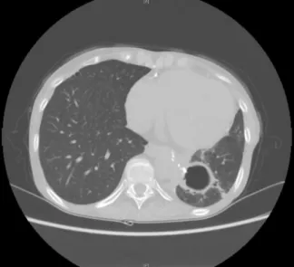
2

, and her weight was 33 kg and height was 149 cm. Her Eastern Cooperative Oncology Group performance status was only 3. The forced expiratory volume in 1 second was 58% and diffusing capacity of carbon monoxide was 38% of the expected value. She performed the 6-minute walk test but could only cover 156 m. She was diagnosed with end-stage renal disease and received hemo- dialysis. She had been administered long-term ste- roids to treat rheumatoid arthritis. In March 2017, her general condition improved to the point where she could use a wheelchair for ambulation, and sur- gery was therefore planned. Preoperative computed tomography of her chest revealed a concealed rup- ture of the diverticular sac, leading to the develop- ment of a fistula between the diverticulum and the left lower lobe of the lung and a consequent pulmo-

Korean J Thorac Cardiovasc Surg 2019;52:40-43 □ CASE REPORT □

https://doi.org/10.5090/kjtcs.2019.52.1.40

(2)

A Large Epiphrenic Esophageal Diverticulum

− 41 −

Fig. 1. An esophagogastroduodenoscopic examination revealed a large hematoma inside the diverticulum.

Fig. 2. An esophagographic examination showing an epiphrenic diverticulum on the left side of the esophagus.

nary abscess (Fig. 3A, B). However, considering her poor lung function and low performance status, we decided to treat the abscess conservatively. On May 16, 2017, diverticulectomy and 180° posterior partial fundoplication were performed via laparotomy. We made a hole with electrocautery in the diverticulum and drained the fluid inside, which provided us with a better view of the fistula. Then, we divided it with endo GIA staplers (Covidien, Seoul, Korea) from each side (the esophageal side and the left lower lobe) and added 3 or 4 reinforcement sutures below the GIA stapler line of each side. We could not proceed with left lower lobectomy due to the patient’s poor lung function, which was the reason for performing the reinforcement suture for the stapling line. We did not perform myotomy because there was no evi- dence of a motility disorder. Her postoperative course was uneventful (Fig. 4); she resumed a soft/liquid diet on May 21, 2017 and was discharged on June 1, 2017. Thus far, the patient has not shown any signs of recurrence or complications (Fig. 5).

Written informed consents were obtained from the patient.

Discussion

Epiphrenic diverticula are mucosal and submucosal

outpouchings of the distal third of the esophagus, which are commonly attributed to the action of pul- sion forces [1]. Most patients with epiphrenic diver- ticula are asymptomatic [2]; however, epiphrenic di- verticula can cause regurgitation or respiratory complaints. Fatal complications include diverticular perforation, progression to carcinoma [3], and rarely, fistula formation between a diverticulum and the res- piratory tract [4]. We describe a patient in whom a diverticulum communicated with the left lower lobe of the lung, leading to a pulmonary abscess. Fistula formation between an epiphrenic diverticulum and the respiratory tract, leading to the development of a pulmonary abscess, is a rare condition, and optimal surgical treatment has not been reported. A few pub- lished case reports have described resection of the affected parts of the lungs in patients presenting with severe complications such as respiratory failure or irreversible inflammatory lesions [5]. Notably, con- servative treatment for the inflamed lung lesion has shown benefits in a few patients [5]. In this case, the patient did not demonstrate serious respiratory symptoms or markedly elevated inflammatory marker levels. Therefore, considering the patient’s poor lung function, we decided to treat the pulmonary abscess conservatively. The surgical management of epiph- renic diverticula involves several different approaches.

The transthoracic approach has been conventionally

used, although a few surgeons have recently reported

(3)

Suk Kyung Lim and Jong Ho Cho

− 42 −

Fig. 3. (A) An image from a lung- window chest computed tomog- raphy scan shows an epiphrenic di- verticulum communicating with the left lower lobe of the lung. (B) An image from a mediastinal-window chest computed tomography scan of the epiphrenic diverticulum.

Fig. 5. An image from a mediastinal-window chest computed to- mography scan on postoperative day 65, showing only an air-con- taining cavity, corresponding to a change from the lung abscess.

Fig. 4. An image of esophagography on postoperative day 1, using gastrograffin.

using a transabdominal approach [2,6]. Based on our experience with successful transabdominal resection of diverticula in 12 patients over the past 20 years, in this patient, we dissected the diverticulum via a laparotomy. The transabdominal approach offers a better view of the gastroesophageal junction when performing a partial fundoplication, which represents a distinct advantage of this method [2]. Additionally, the transabdominal approach is associated with less postoperative pain and a shorter hospital stay. To summarize, we consider transabdominal epiphrenic diverticulectomy to be a feasible technique for the management of patients with this presentation. The optimal surgical technique for an epiphrenic divertic-

ulum remains controversial, particularly with regard

to whether myotomy is warranted in every case

[2,7]. A few authors maintain that myotomy should

be selectively performed in patients with a proven

motility disorder [5,6]. In this patient, a barium swal-

low study (esophagogram) did not show any abnor-

malities in the passage of barium. Additionally, intra-

operatively, no esophageal muscle hypertrophy was

identified. We did not perform esophageal man-

ometry because she already had undergone several

stressful examinations due to the presence of several

comorbid conditions, as mentioned above, so we

tried to minimize the number of tests required to

(4)

A Large Epiphrenic Esophageal Diverticulum

− 43 − confirm the diagnosis. In some cases, it is not possi- ble to differentiate the exact cause of esophageal di- verticulum even if comprehensive information is available from an esophageal motility study, including manometry results. This patient’s diverticulum had been diagnosed and followed by a gastroenterologist because she did not have significant symptoms, such as dysphagia, until the episode of hematemesis.

Therefore, we thought that she only required a diverticulectomy. Furthermore, it was crucial to re- duce the time and burden of the operation by sim- plifying the operative technique due to the patient’s comorbidities. For this reason, we did not perform myotomy, and choosing this simplified procedure led to favorable outcomes with a lower risk of com- plications.

Therefore, the patient underwent diverticulectomy and partial fundoplication. We recommend an in- dividualized approach to treat cases of a pulmonary abscess secondary to an esophagopulmonary fistula.

Myotomy is not always mandatory in patients with- out signs of motility disorders.

Conflict of interest

No potential conflict of interest relevant to this ar- ticle was reported.

ORCID

Suk Kyung Lim: https://orcid.org/0000-0002-2049-3709 Jong Ho Cho: https://orcid.org/0000-0003-3362-4621

References

1. Lee JM, Lee HS, Shin JK, Kim SH. Surgical treatment of ep- iphrenic esophageal diverticulum: one case report. Korean J Thorac Cardiovasc Surg 1990;23:584-7.

2. Zaninotto G, Portale G, Costantini M, Zanatta L, Salvador R, Ruol A. Therapeutic strategies for epiphrenic divertic- ula: systematic review. World J Surg 2011;35:1447-53.

3. Altorki NK, Sunagawa M, Skinner DB. Thoracic esophageal diverticula: why is operation necessary? J Thorac Cardiovasc Surg 1993;105:260-4.

4. Herbella FA, Del Grande JC. Benign esophagopulmonary fistula through an epiphrenic diverticulum and asympto- matic achalasia. Dig Dis Sci 2010;55:1177-8.

5. De Giacomo T, Francioni F, Venuta F, Rendina EA, Ricci C.

Benign esophageal-respiratory fistulae: the surgical treat- ment and results of 10 cases. Minerva Chir 1993;48:311-6.

6. Benacci JC, Deschamps C, Trastek VF, Allen MS, Daly RC, Pairolero PC. Epiphrenic diverticulum: results of surgical treatment. Ann Thorac Surg 1993;55:1109-13.

7. Soares R, Herbella FA, Prachand VN, Ferguson MK, Patti MG. Epiphrenic diverticulum of the esophagus: from path- ophysiology to treatment. J Gastrointest Surg 2010;14:

2009-15.

수치

Fig. 2. An esophagographic examination showing an epiphrenic  diverticulum on the left side of the esophagus.
Fig. 5. An image from a mediastinal-window chest computed to- to-mography scan on postoperative day 65, showing only an  air-con-taining cavity, corresponding to a change from the lung abscess.

참조

관련 문서

A source of light waves moving to the right with velocity 0.7c. The frequency is higher on the right, and lower

When two numbers are listed in a column, the number on the left reflects the higher-rate assumption and the number on the right reflects the lower-rate assumption. <

If both these adjustments are considered, the resulting approach is called a bootstrap-BC a -method (bias- corrected-accelerated). A description of this approach

Five days later, on 15 January 1975, the Portuguese government signed an agreement with the MPLA, FNLA and UNITA providing for Angola to receive its independence on 11

· 50% exemption from tuition fee Ⅱ for the student with a TOPIK score of level 3 or higher or completion of level 4 or higher class of the Korean language program at the

Since every classical or virtual knot is equivalent to the unknot via a sequence of the extended Reidmeister moves together with the forbidden moves, illustrated in Section 2,

웹 표준을 지원하는 플랫폼에서 큰 수정없이 실행 가능함 패키징을 통해 다양한 기기를 위한 앱을 작성할 수 있음 네이티브 앱과

• The supply and demand curves cross at the equilibrium price and quantity.. • You can read off approximate equilibrium values