ISSN: 2233-601X (Print) ISSN: 2093-6516 (Online)
†
This paper was presented in the 2015 Fall Conference of The Korean Society for Thoracic and Cardiovascular Surgery.
Received: July 5, 2017, Revised: September 13, 2017, Accepted: September 20, 2017, Published online: February 5, 2018
Corresponding author: Jun Sung Kim, Department of Thoracic and Cardiovascular Surgery, Seoul National University Bundang Hospital, 82 Gumi-ro 173beon-gil, Bundang-gu, Seongnam 13620, Korea
(Tel) 82-31-787-7140 (Fax) 82-31-787-4050 (E-mail) [email protected]
© The Korean Society for Thoracic and Cardiovascular Surgery. 2018. All right reserved.
This is an open access article distributed under the terms of the Creative Commons Attribution Non-Commercial License (http://creativecommons.org/
licenses/by-nc/4.0) which permits unrestricted non-commercial use, distribution, and reproduction in any medium, provided the original work is properly cited.
Mid-Term Results of Minimally Invasive Direct Coronary Artery Bypass Grafting
Dong Hyun Seo, M.D. 1 , Jun Sung Kim, M.D. 1,2 , Kay-Hyun Park, M.D., Ph.D. 1,2 , Cheong Lim, M.D., Ph.D. 1,2 , Su Ryeun Chung, M.D. 1 , Dong Jung Kim, M.D. 1
1
Department of Thoracic and Cardiovascular Surgery, Seoul National University Bundang Hospital,
2
Department of Thoracic and Cardiovascular Surgery, Seoul National University College of Medicine
Background: Minimally invasive direct coronary artery bypass grafting (MIDCAB) has the advantage of allow- ing arterial grafting on the left anterior descending artery without a sternotomy incision. We present our single-center clinical experience of 66 consecutive patients. Methods: All patients underwent MIDCAB through a left anterior small thoracotomy between August 2007 and July 2015. Preoperative, intraoperative, post- operative and follow-up data—including major adverse cardiovascular and cerebrovascular events (MACCE), graft patency, and the need for re-intervention—were collected. Results: The mean age of the patients was 69.4±11.1 years and 73% were male. There was no conversion to an on-pump procedure or a sternotomy incision. The 30-day mortality rate was 1.5%. There were no cases of stroke, although 2 patients had to be re-explored for bleeding, and 81.8% were extubated in the operating room or on the day of surgery. The median stay in the intensive care u nit and in the hospital were 1.5 and 9.6 days, respectively. The median follow-up period was 11 months, with a 5-year overall survival rate of 85.3%±0.09% and a 5-year MACCE-free survival rate of 72.8%±0.1%. Of the 66 patients, 32 patients with 36 grafts underwent a postoperative graft patency study with computed tomography angiography or coronary angiography, and 88.9% of the grafts were patent at 9.7±10.8 months postoperatively. Conclusion: MIDCAB is a safe procedure with low post- operative morbidity and mortality and favorable mid-term MACCE-free survival.
Key words: 1. Coronary artery disease 2. Coronary artery bypass 3. Minimally invasive surgery
Introduction
Since revascularization of the left anterior descend- ing artery (LAD) was first introduced in the 196 0s [1], the applications of coronary artery bypass graft- ing (CABG) in coronary artery disease have expanded to include minimally invasive direct coronary artery bypass grafting (MIDCAB). MIDCAB has 2 major ad-
vantages over other treatment interventions: one is
the use of a small incision through a left antero-
lateral thoracotomy without cardiopulmonary bypass,
and the other is the fact that it is the best option for
grafting the left internal thoracic artery (LITA) to the
LAD [2,3]. Those advantages result in better out-
comes, with fewer complications in terms of stroke,
mediastinitis, and bleeding, as well as lower medical
https://doi.org/10.5090/kjtcs.2018.51.1.8
Table 1. Preoperative data of 66 patients
Characteristic Value
Age (yr) 69.4±11.1
Male sex 50 (73)
Hypertension 45 (68)
Dyslipidemia 21 (32)
Diabetes mellitus 35 (53)
Chronic renal failure 11 (17)
Chronic obstructive pulmonary disease 4 (6)
Cerebrovascular accident 14 (21)
Smoking 35 (53)
Malignancy 4 (6)
EuroSCORE II (%) 1.99±2.05
(range, 0.5 –10.5)
Mean EF 53.7
Left ventricular dysfunction (EF <40%) 12 (18)
3VD 27 (41)
2VD 23 (35)
1VD 16 (24)
Previous percutaneous coronary intervention 26 (39) Peripheral arterial occlusive disease 6 (9)
Redo open heart surgery
a)4 (6)
Silent/stable angina 24 (37)
Unstable angina 29 (44)
STEMI/non-STEMI 13 (20)
Values are presented as mean±standard deviation or number of patients (%).
EF, ejection fraction; VD, vessel disease; STEMI, ST-segment elevation myocardial infarction.
a)
Coronary artery bypass graft surgery (n=3) and aortic valve replacement (n=1).
costs [2,4]. Recently, LITA-to-LAD MIDCAB has been considered to be the treatment of choice in sin- gle-vessel disease in the proximal LAD [5].
However, there are some obstacles hindering the universal use of MIDCAB in specific clinical situations.
The fact that thoracotomy is more painful than ster- notomy and the need for a learning curve, causing a longer operative time and target vessel misidentifi- cation, may be potential pitfalls of MIDCAB [6,7]. In this study, we evaluate the recent MIDCAB results from a single center and discuss the implications of our findings for this approach.
Methods
1) Patients
Between August 2007 and July 2015, a total of 66 patients underwent a MIDCAB operation through a left anterior minithoracotomy at Seoul National University Bundang Hospital, and the procedures were carried out by 3 cardiac surgeons who shared the same operative technique and strategy for MIDCAB.
Preoperative, intraoperative, postoperative, and follow- up data were collected, including major adverse car- diovascular and cerebrovascular events (MACCE), graft patency, and the need for re-intervention. The pre- operative clinical variables and demographics of the patients undergoing MIDCAB are summarized in Table 1.
The institutional review board of Seoul National University Bundang Hospital approved the present study (B-1511/322-109). And the requirement for in- formed patient consent was waived due to the retro- spective nature of the study.
2) Indications
We considered MIDCAB in the following circum- stances: (1) definitive MIDCAB, for single-vessel dis- ease of the proximal LAD, including restenosis of a previous LAD stent; (2) palliative MIDCAB, for multi- vessel disease with a significant underlying comor- bidity such as severe left ventricular (LV) dysfunction (n=12, 18%), stroke (n=14, 21%), malignancy (n=4, 6%), and the expectation of a major operation (n=5, 7.5%); and (3) hybrid MIDCAB, in cases of multi-ves- sel disease where MIDCAB is performed simulta- neously with a percutaneous coronary intervention (PCI) in another area (n=12, 18%). Hybrid MIDCAB
was considered for patients with multi-vessel disease who had high-grade proximal disease of the LAD along with favorable lesions for PCI in the left cir- cumflex (LCX) and right coronary artery (RCA) territories. There is no absolute contraindication for MIDCAB, but we generally did not perform MIDCAB in patients who were expected to have severe adhe- sions of the left hemithorax, poor pulmonary func- tion, or an intra-myocardial course of the LAD based on preoperative computed tomography.
3) Technique
We elevated the left hemithorax to 30
o, attached
external defibrillation pads, and prepared a dou-
ble-lumen endotracheal tube. MIDCAB was performed
through a 5- to 7-cm anterolateral minithoracotomy
in the fourth or fifth intercostal space. Harvesting of
the left internal mammary artery (LIMA) was done
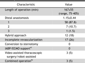
Table 2. Operative characteristics
Characteristic Value
Length of operation (min) 167±55
(range, 75 –405)
Distal anastomosis 1.15±0.44
1 58 (87.8)
2 7 (10.7)
3 1 (1.5)
Hybrid approach 12 (18)
Incomplete revascularization 17 (26)
Conversion to sternotomy 0
IABP/ECMO support
a)0
Video-assisted thoracoscopic surgery/robot-assisted
3 (5)
Combined operation
b)3 (5)
Values are presented as mean±standard deviation or number of patients (%).
a)
Intra-aortic balloon pump support and extracorporeal membrane oxygenation support.
b)Abdominal aortic aneurysm (n=2) and lung cancer (n=1).
under direct visualization with a specially designed retractor while deflating the left lung, and we usually harvested the LITA in a skeletonized fashion as far as the proximal origin of the left subclavian artery.
Heparin (1.5 mg/kg) was administered to maintain an activated clotting time over 300 seconds. A stand- ard stabilizer for off-pump CABG (Octopus; Medtronic Inc., Minneapolis, MN, USA) was used. Under a beat- ing heart, the anastomosis to the LAD was performed. The proximal LAD was snared with si- lastic loops. After the main procedure, we inserted a 24-Fr chest tube into the pleural space and the thor- acotomy was closed in layers. If there was no evi- dence of bleeding, aspirin was started on the day of the operation.
4) Statistical analysis
For statistical analyses, we used IBM SPSS ver.
22.0 (IBM Corp., Armonk, NY, USA). Continuous vari- ables are presented as mean±standard deviation or as median values with the range. Kaplan-Meier curves were used to analyze overall survival and freedom from MACCE.
Results
We identified 66 consecutive MIDCAB patients with a mean age of 69.4±11.1 years, 73% of whom were male and 53% of whom had diabetes. The pa- tients’ preoperative characteristics are shown in Table 1. Their mean EuroSCORE II was 1.99±2.05 (range, 0.5 to 10.5). Twenty-six patients had normal LV function (ejection fraction >60%), 28 patients had moderate LV dysfunction, and 12 patients had severe LV dysfunction (ejection fraction <40%).
Single- and multiple-vessel disease were present in 16 patients (24%) and 50 patients (76%), respecti- vely. Twenty-six patients (39%) had a prior PCI, 3 patients (4.5%) had a prior CABG, and 1 patient (1.5%) had a prior aortic valve replacement. Thirteen patients (20%) had a history of an acute myocardial infarction.
The operative characteristics are shown in Table 2.
The mean total operative time was 16 7 minutes (range, 75 to 405 minutes). There were no iatrogenic injuries to the LITA. Isolated LAD-only revasculariza- tion was performed in 58 patients (87.8%). A re- constructed composite graft with the saphenous vein
was used for the anastomosis of additional branches or lengthening of the LITA graft in 8 patients.
Twelve patients underwent a hybrid approach for suitable lesions of the LCX and/or RCA using a standard commercial stent immediately after surgery in the hybrid operating room or preoperatively.
There were no conversions to an on-pump proce- dure, sternotomy, or intra-aortic balloon pump or ex- tracorporeal membrane oxygenation support. Robot- assisted MIDCAB and combined operations required longer operative times. If we consider anterolateral minithoracotomy MIDCAB for isolated LAD revascula- rization (LITA to LAD), the actual mean operative time was 144 minutes.
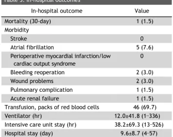
The in-hospital outcomes are shown in Table 3.
The 30-day mortality rate was 1.5% (1 of 66). One 81-year-old patient died due to idiopathic massive gastric bleeding on day 4 after surgery. There were no cerebrovascular events. Atrial fibrillation was ob- served in 5 patients (7.6%), but there were no cases of perioperative myocardial infarction or low cardiac output syndrome. Two patients (3.0%) had to be re-explored for bleeding. Two patients (3.0%) had a superficial wound problem associated with the thoracotomy. There was 1 case (1.5%) of pulmonary complication and 1 case (1.5%) of acute renal failure.
Eighty-one percent of the patients were extubated in
the operating room or on the day of surgery. The
Table 3. In-hospital outcomes
In-hospital outcome Value
Mortality (30-day) 1 (1.5)
Morbidity
Stroke 0
Atrial fibrillation 5 (7.6)
Perioperative myocardial infarction/low cardiac output syndrome
0
Bleeding reoperation 2 (3.0)
Wound problems 2 (3.0)
Pulmonary complication 1 (1.5)
Acute renal failure 1 (1.5)
Transfusion, packs of red blood cells 46 (69.7)
Ventilator (hr) 12.0±41.8 (1 –336)
Intensive care unit stay (hr) 38.2±69.3 (13 –526)
Hospital stay (day) 9.6±8.7 (4 –57)
Values are presented as number of patients (%) or mean±standard deviation (range).
Fig. 1. Kaplan-Meier curve showing mid-term outcomes with a survival rate of 85.3%±0.09%.
Table 4. Follow-up data of 65 patients
Follow-up data No. of patients (%)
Mortality 7 (10.8)
Cardiac 5 (7.7)
Non-cardiac 2 (3.1)
Morbidity
Stroke 1 (1.5)
Myocardial infarction 2 (3.1)
Target vessel revascularization 1 (1.5) Non-target vessel revascularization 3 (4.6)
Fig. 2. MACCE-free survival curve: mid-term outcomes with a MACCE-free survival rate of 72.8%±0.1%. MACCE, major adverse cardiovascular and cerebrovascular events.
median stay in the intensive care unit and in the hospital were 23 hours (range, 13 to 526 hours) and 7 days (range, 4 to 57 days), respectively. Twenty patients (30.3%) received no transfusion.
The follow-up data are shown in Table 4. The me- dian follow-up period was 11 months (range, 0 to 90 months). Except for in-hospital mortality, regular fol- low-up of the 65 survivors was completed in 100%
of the patients, and their data were included in the outcome analysis. Among these patients, there were 7 late deaths (10.8%), among which 5 were due to cardiac causes (congestive heart failure). Fig. 1 shows the Kaplan-Meier survival curve of cumulative surviv- al: the calculated 5-year overall survival rate was 85.3%±0.09%. One cerebrovascular event was obser- ved. Two patients (3.1%) had a myocardial infarction due to an occluded LIMA graft and 1 patient was successfully treated with re-intervention on the tar-
get vessel. Another 3 patients (4.6%) had a re-inter- vention due to progressive disease of a non-target vessel during the follow-up period. Fig. 2 shows the calculated cumulative 5-year MACCE-free survival rate of 72.8%±0.1%. Forty-eight percent of the pa- tients (32 of 6 6 ), with a total of 36 grafts, under- went a postoperative graft patency study with com- puted tomography angiography or coronary angio- graphy. Eighty-nine percent of these grafts (32 of 36) were patent at 9.7±10.8 months postoperatively (Table 5).
Discussion
In recent years, MIDCAB is increasingly becoming
Table 5. Results of postoperative computed tomography angiography or coronary angiography in 32 patients with a total of 36 grafts