• 검색 결과가 없습니다.

Assessment of the Thickness of the Roof of the Glenoid Fossa Using Cone Beam Computed Tomography in Orthognathic Surgery Patients: A Preliminary Study JOMP

N/A
N/A
Protected

Academic year: 2021

Share "Assessment of the Thickness of the Roof of the Glenoid Fossa Using Cone Beam Computed Tomography in Orthognathic Surgery Patients: A Preliminary Study JOMP"

Copied!
6
0
0

로드 중.... (전체 텍스트 보기)

전체 글

(1)

pISSN 2288-9272 eISSN 2383-8493 J Oral Med Pain 2019;44(4):154-159 https://doi.org/10.14476/jomp.2019.44.4.154

Assessment of the Thickness of the Roof of the Glenoid Fossa Using Cone Beam Computed Tomography in Orthognathic Surgery Patients:

A Preliminary Study

Hyun-Jeong Park 1 , Yo-Seob Seo 2 , Sung-Hoon Lim 3 , Ji-Won Ryu 4

1 Department of Oral Medicine, Chosun University Dental Hospital, Gwangju, Korea

2 Department of Oral and Maxillofacial Radiology, School of Dentistry, Chosun University, Gwangju, Korea

3 Department of Orthodontics, School of Dentistry, Chosun University, Gwangju, Korea

4 Department of Oral Medicine, School of Dentistry, Chosun University, Gwangju, Korea

Received November 24, 2019 Revised December 16, 2019 Accepted December 17, 2019

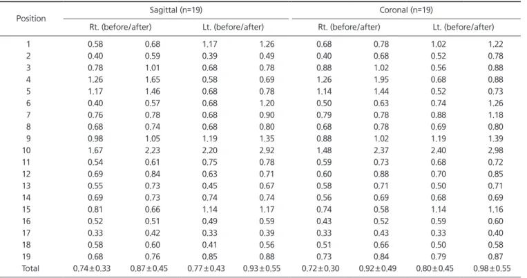
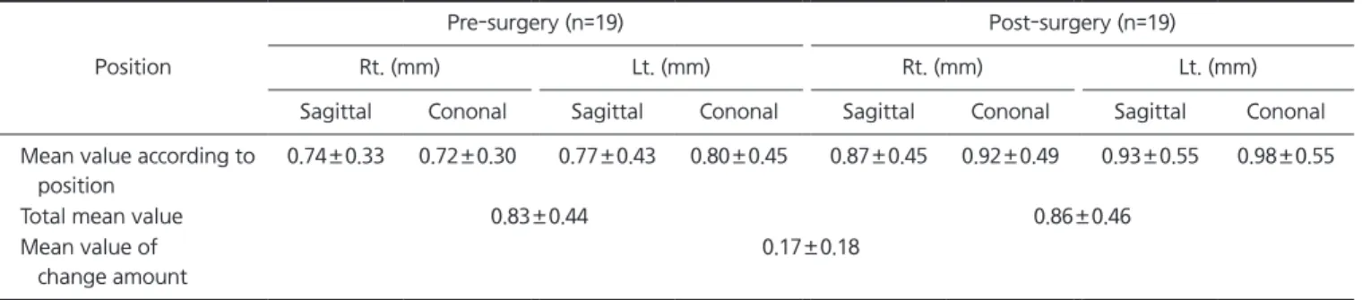
Purpose: The aim of this study was to assess the change in thickness of the roof of the gle- noid fossa (RGF) in patients undergoing orthognathic surgery using cone-beam computed tomography (CBCT) images.

Methods: This retrospective study measured the thickness of the RGF in 19 patients (10 males, 9 females) who underwent orthognathic surgery at Chosun University Dental Hospi- tal. The thickness of the RGF was measured perpendicularly between the ‘glenoid fossa line’

and ‘middle cranial fossa line’ on parasagittal and paracoronal reconstructions.

Results: The mean RGF thickness increased from 0.83±0.44 mm to 0.86±0.46 mm after sur- gery. The average change in thickness of the RGF was 0.17±0.18 mm. The thickness of the RGF in the temporomandibular joint (TMJ) showed no significant difference by sex, and the change in thickness of the TMJ did not vary by surgical method.

Conclusions: We found that the thickness of the RGF increased after orthognathic surgery, as revealed by CBCT. Further studies including larger numbers of subjects and long-term follow-up are needed to confirm the results of this study.

Key Words: Cone-beam computed tomography; Orthognathic surgery; Roof of glenoid fossa; Temporomandibular joint

Correspondence to:

Ji-Won Ryu

Department of Oral Medicine, School of Dentistry, Chosun University, 309 Pilmun- daero, Dong-gu, Gwangju 61452, Korea Tel: +82-62-220-3897

Fax: +82-62-234-2119 E-mail: [email protected]

https://orcid.org/0000-0002-5586-8195 This study was supported by research fund from Chosun University, 2016.

JOMP Journal of Oral Medicine and Pain

Copyright Ⓒ 2019 Korean Academy of Orofacial Pain and Oral Medicine. All rights reserved.

CC

This is an open-access article distributed under the terms of the Creative Commons Attribution Non-Commercial License (http://creativecommons.org/licenses/by-nc/4.0/), which permits unrestricted non-commercial use, distribution, and reproduction in any medium, provided the original work is properly cited.

INTRODUCTION

The temporomandibular joint (TMJ), unlike other joints of the body, is very complicated, being composed of osse- ous components (condyles, glenoid fossa, and articular emi- nences) and soft tissues (articular discs and joint capsules) [1].

The marked thickness of the roof of the glenoid fossa (RGF) of the TMJ is important to withstand the various types of stimulation associated with facial trauma involving superi- or displacement of the mandibular condyle into the middle cranial fossa, as well as the effects of TMJ arthroplasty and

joint reconstruction [2].

In general, orthognathic surgery is performed in patients with skeletal class II and III dentofacial deformities, and in patients with asymmetry. Orthognathic surgery includes one-jaw surgery (mandibular advancement or mandibular setback) and two-jaw surgery (bimaxillary advancement or setback). Orthognathic surgery affects the relationships of the soft tissues, as well as the osseous components of the TMJs [3,4].

Many studies have evaluated changes in condyle posi-

tion and condyle shape after orthognathic surgery [3,5-

7]. Cevidanes et al. [3] studied changes of the condyle of

(2)

the TMJs after orthognathic surgery, and reported surgical displacement and adaptive responses relative to adjacent structures in the craniofacial complex [3]. Skeletal stabil- ity differs depending on the orthognathic surgical method used. Busby et al. [6] and Proffit et al. [7] found that man- dibular setback alone was more stable over the long term than when combined with maxillary surgery.

We suspect that not only changes in the condyle, but also in the morphology of the glenoid fossa, could be caused by orthognathic surgery. A previous study determined that the thickness of the RGF is clinically important [8]. This study evaluated changes in the glenoid fossa following orthogna- thic surgery using cone beam computed tomography (CBCT).

CBCT images, which can be obtained relatively economical- ly and involve lower radiation doses, have proven remark- ably useful in craniofacial investigations [3]. The aim of this study was to assess the change in thickness of the RGF in patients undergoing orthognathic surgery.

MATERIALS AND METHODS

1. Subjects

This study included patients who underwent orthognathic surgery at the Oral and Maxillofacial Surgery Department of Chosun University Dental Hospital. Patients were diagnosed with mandibular prognathism, retrognathism, or asymme- try underwent one-jaw surgery (mandibular setback or ad- vancement) or two-jaw surgery (maxillomandibular setback or advancement). The total number of patients undergoing

orthognathic surgery from January 2015 to September 2019 was 104. However, only 19 patients (10 males, 9 females;

mean age, 26.5±6.7 years) were selected, due to the exclu- sion of those without postoperative CBCT data, a truncat- ed TMJ on CBCT images, or temporomandibular disorder prior to surgery. This retrospective study was approved by the Institutional Review Committee of Chosun University Dental Hospital (CUDH) (IRB no. 1901/013).

2. Image Analysis

Images of the glenoid fossa of the temporal bone were edited using three-dimensional imaging software (OnDemand3D; CyberMed Co., Seoul, Korea). As in a previ- ous study [8], the images were reconstructed based on the angle of the condylar head. Paracoronal sections were ob- tained parallel to the horizontal axis of the condylar head, and parasagittal sections were reconstructed parallel to the line connecting the center point of the condylar head with the coronoid process (1 mm slice thickness, 1 mm slice in- terval). The reconstructed data sets were exported as Digital Imaging and Communications in Medicine image stacks.

3. Measurement of the Thickness of the Roof of the Glenoid Fossa

The method used for measuring RGF thickness was the same as in previous studies [8]. Measurements were ob- tained on images of the central region of the glenoid fossa in parasagittal and paracoronal sections using G3 software (Infinitt Healthcare, Seoul, Korea). The images were assessed

RGF

A

B C

D

Fig. 1. Parasagittal reconstruction of

cone beam computed tomography

images. (A) Roof of the glenoid fossa

(RGF), thickness of the Roof of glenoid

fossa. (B) Thickness of the RFG before

orthognathic surgery. (C) Thickness of

the RGF after orthognathic surgery. (D)

A super-imposition of images before

and after orthognathic surgery. Pre-

operative image for yellow color, post-

op image for grey color.

(3)

in a darkened room on a personal computer with a 32-inch LCD monitor. The contrast and brightness of the images were adjusted using the image processing tool of G3 to en- sure optimal visualization. The thickness of the RGF was determined as the perpendicular distance between the ‘gle- noid fossa line’ and ‘middle cranial fossa line’ on parasagit- tal (Fig. 1) and paracoronal reconstructions. RGF thickness was measured three times, by one investigator, at the thin- nest part of the glenoid fossa among multiple slices, with the mean value calculated for statistical analysis. The same examiner measured the thickness of the RGF, in the same way as in a previous study [8], therefore, interexaminer agreement was not assessed in this study.

4. Statistical Analysis

Statistical analyses were performed using PASW Statistics for Windows, Version 18.0 (SPSS Inc., Chicago, IL, USA).

The Shapiro-Wilk test was used to test the normality of the data distribution. A p-value <0.05 was regarded as statisti- cally significant. All quantitative variables are described as means±standard deviations. In a previous study [8], intra- and interexaminer agreement with respect to RGF thickness

was evaluated using the intraclass correlation coefficient.

Comparison of the RGF thickness of patients pre-and post- surgery, and analyses of RGF thickness according to sex and surgical method, were performed using t-tests.

RESULTS

This study involved 19 patients (10 males and 9 females) who underwent corrective surgery (Table 1). The mean age was 26.5±6.7 years (Table 1). Increases in RGF thickness were observed in all 19 patients (Table 2). The mean RGF thickness increased from 0.83±0.44 mm to 0.86±0.46 mm

Table 1. Patients demographic data

Variable Value

Age 26.5±6.7

Sex

Male 10

Female 9

Surgical method

1-Jaw surgery 11

2-Jaw surgery 8

Values are presented as mean±standard deviation or number only.

Table 2. The thickness of the RGF on pre- and post-surgery (unit: mm)

Position Sagittal (n=19) Coronal (n=19)

Rt. (before/after) Lt. (before/after) Rt. (before/after) Lt. (before/after)

1 0.58 0.68 1.17 1.26 0.68 0.78 1.02 1.22

2 0.40 0.59 0.39 0.49 0.40 0.68 0.52 0.78

3 0.78 1.01 0.68 0.78 0.88 1.02 0.56 0.88

4 1.26 1.65 0.58 0.69 1.26 1.95 0.68 0.88

5 1.17 1.46 0.68 0.78 1.14 1.44 0.52 0.73

6 0.40 0.57 0.68 1.20 0.50 0.63 0.74 1.26

7 0.76 0.78 0.68 0.90 0.79 0.78 0.88 1.18

8 0.68 0.74 0.68 0.80 0.68 0.78 0.69 0.80

9 0.98 1.05 1.19 1.35 0.88 1.02 1.19 1.39

10 1.67 2.23 2.20 2.92 1.48 2.37 2.40 2.98

11 0.54 0.61 0.75 0.78 0.59 0.73 0.68 0.72

12 0.69 0.84 0.63 0.71 0.60 0.88 0.70 0.85

13 0.55 0.73 0.45 0.67 0.58 0.71 0.50 0.71

14 0.69 0.73 0.74 0.74 0.56 0.69 0.68 0.69

15 0.81 0.66 1.14 1.17 0.74 0.58 1.14 1.16

16 0.52 0.51 0.49 0.59 0.43 0.52 0.59 0.60

17 0.33 0.42 0.33 0.39 0.33 0.43 0.33 0.40

18 0.58 0.60 0.41 0.56 0.51 0.66 0.50 0.58

19 0.68 0.76 0.85 0.88 0.73 0.84 0.79 0.87

Total 0.74±0.33 0.87±0.45 0.77±0.43 0.93±0.55 0.72±0.30 0.92±0.49 0.80±0.45 0.98±0.55 RGF, roof of glenoid fossa; Rt., right ; Lt., left.

Values are presented as distance or mean±standard deviation only.

(4)

after surgery (Table 3). The average change in thickness of the RGF was 0.17±0.18 mm (Table 4).

The change in thickness of the RGF on parasagittal and paracoronal images showed no significant difference by sex (Table 5).

In total, 8 of 19 patients underwent two-jaw surgery (7 patients underwent maxillary LeFort 1 surgery and mandib- ular setback, and 1 underwent maxillary and mandibular advancement), and 11 patients underwent one-jaw surgery (mandibular setback) (Table 1). There was no significant dif- ference in the change in thickness of the TMJ according to surgical method (one- vs. two-jaw surgery) (Table 6).

DISCUSSION

The TMJ is affected by the surrounding environment [9].

Factors contributing to changes in the structure of the TMJ include disc derangement, degenerative arthritis and or- thognathic surgery [9].

CBCT is a highly efficient and low-cost imaging modality suitable for observing bone changes [10]. In particular, since the radiation dose is lower than that of conventional CT, CBCT can be performed several times over the follow-up period [10]. In general, CBCT is used to accurately evaluate skeletal structure, devise a surgical plan before orthognathic surgery, and evaluate skeletal changes during follow-up [3,10]. We analyzed before- and after-surgery CBCT images using three-dimensional imaging software in our evalua- tion of the skeletal structure of the TMJ.

In this study, the thickness of the RGF was not significantly different from the value for Koreans obtained in a previous study [8]. We evaluated the change in thickness (pre- vs.

post-surgery) of the RGF on CBCT images in patients who underwent orthognathic surgery. The average thickness of the RGF increased from 0.83±0.44 mm to 0.86±0.46 mm (Table 3). There have been many studies of changes in the condyle and glenoid fossa of TMJs after orthognathic sur- gery [11-15]. Cottrell et al. [11] reported minimal long-term changes in healthy patients after orthognathic surgery. An et al. [12] noted that surgical correction of skeletal deformi- ties requires a soft-tissue response and condylar remodel- ing, to adapt to the new environment. They noted condylar

Table 3. The mean of the thickness of RGF on pre- and post surgery

Position

Pre-surgery (n=19) Post-surgery (n=19)

Rt. (mm) Lt. (mm) Rt. (mm) Lt. (mm)

Sagittal Cononal Sagittal Cononal Sagittal Cononal Sagittal Cononal

Mean value according to position

0.74±0.33 0.72±0.30 0.77±0.43 0.80±0.45 0.87±0.45 0.92±0.49 0.93±0.55 0.98±0.55

Total mean value 0.83±0.44 0.86±0.46

Mean value of change amount

0.17±0.18

RGF, roof of glenoid fossa; Rt., right ; Lt., left.

Values are presented as mean±standard deviation.

Table 4. The change amount of the thickness of the RGF on pre- and post-surgery

Position Sagittal (n=19) Coronal (n=19) Rt. (mm) Lt. (mm) Rt. (mm) Lt. (mm)

1 0.10 0.09 0.10 0.20

2 0.19 0.10 0.28 0.26

3 0.23 0.10 0.14 0.32

4 0.39 0.11 0.69 0.20

5 0.29 0.10 0.30 0.21

6 0.17 0.52 0.13 0.52

7 0.02 0.22 –0.01 0.30

8 0.06 0.12 0.10 0.11

9 0.07 0.16 0.14 0.20

10 0.56 0.72 0.89 0.58

11 0.07 0.03 0.14 0.04

12 0.15 0.08 0.28 0.15

13 0.18 0.22 0.13 0.21

14 0.04 0.00 0.13 0.01

15 –0.15 0.03 –0.16 0.02

16 –0.01 0.10 0.09 0.01

17 0.09 0.06 0.10 0.07

18 0.02 0.15 0.15 0.08

19 0.08 0.03 0.11 0.08

Total 0.13±0.16 0.15±0.18 0.20±0.23 0.18±0.16 RGF, roof of glenoid fossa; Rt., right ; Lt., left.

Values are presented as distance or mean±standard deviation only.

(5)

surface changes, and reported that condylar inward rota- tion was closely related to changes in the condylar surface [12]. Kim et al. [13] reported that orthognathic surgery was able to correct dentofacial deformities and alter the neu- romuscular environment. They noted that surgical correc- tion led to a physiological adaptation process, according to changes in the position of the condyle [13]. Owtad et al. [14]

reported that the glenoid fossa was remodeled according to changes in the surrounding environment, likely involving the condyle. Kai et al. [15] showed that the RGF was thicker in patients with versus without degenerative joint changes and disc displacement. They suggested that this may be at- tributable to remodeling, or might occur in response to me- chanical stress [15].

Many studies have reported bone changes, such as flat- tening and rotation of the condyle, after orthognathic sur- gery [16,17]. We suggest that these changes in the surface and location of the condyle also affect the glenoid fossa of the TMJ. The increase in thickness of the RFG seen in this study showed that structural changes and mechanical stress after surgery affected the glenoid fossa of the TMJ [17].

There was no significant difference in the thickness of the RGF according to sex (Table 5). These results are consistent with previous studies showing that the thickness of the RGF is not affected by age or sex [2,18].

In this study, 11 of 19 patients underwent one-jaw sur- gery (mandibular setback), while 8 patients underwent two-jaw (maxillary) surgery (Table 6). There was no sig- nificant difference in the variation in thickness of the TMJ

according to the surgical method (Table 6). An et al. [12] re- ported no significant difference in the extent of remodeling of the constant bone between one- and two-jaw surgeries.

However, Wolford et al. [17] reported greater bone remod- eling in two- versus one-jaw surgery. In one-jaw surgery, there is a difference in the position of the condyle, and the stress applied to the glenoid fossa, according to whether mandibular setback or advancement is performed [3,5,17].

Wolford et al. [17] reported that physiological adaptation is difficult when surgical correction is performed in the coun- terclockwise direction.

There were several limitations to our study. First, the small sample size makes it difficult to generalize the results.

Also, Koreans typically have a class III skeletal structure [19,20]. In this study, only one patient had a class II skeletal structure (Table 6). In addition, many studies have shown that bone to remodeling after surgery can take a relatively long time [21,22]; however, in our study, CT was performed only about 1 year after surgery. Therefore, long-term fol- low-up should be implemented in future studies to assess bone remodeling.

In conclusion, we found that the thickness of the RGF increased after orthognathic surgery, as revealed by CBCT.

Further studies including larger numbers of subjects and long-term follow-up are needed to confirm the results of this study.

CONFLICT OF INTEREST

No potential conflict of interest relevant to this article was reported.

ORCID

Hyun-Jeong Park

http://orcid.org/0000-0002-5237-005X

Table 5. Differences between male and female about orthognathic surgery patients

Sex Rt. coronal Lt. coronal Rt. sagittal Lt. sagittal

Male 0.26±0.29 0.21±0.20 0.15±0.18 0.22±0.23

Female 0.13±0.13 0.16±0.11 0.11±0.13 0.09±0.06

RGF, roof of glenoid fossa; Rt., right; Lt., left.

Statistically significant difference (p<0.05) (t-test).

Table 6. Differences according to surgical methods (1-jaw/2-jaw surgery)

Surgical method 1-jaw surgery (0.17±0.18)

2-jaw surgery (0.15±0.18)

1- jaw surgery (0.17±0.18) - -

2- jaw surgery (0.15±0.18) 0.496* -

*Statistically significant difference (p<0.05) (t-test).

(6)

Yo-Seob Seo

http://orcid.org/0000-0003-1804-5648 Sung-Hoon Lim

http://orcid.org/0000-0003-4528-8514 Ji-Won Ryu

http://orcid.org/0000-0002-5586-8195

REFERENCES

1. Bag AK, Gaddikeri S, Singhal A, et al. Imaging of the temporo- mandibular joint: an update. World J Radiol 2014;6:567-582.

2. Honda K, Kawashima S, Kashima M, Sawada K, Shinoda K, Sugi- saki M. Relationship between sex, age, and the minimum thick- ness of the roof of the glenoid fossa in normal temporomandibu- lar joints. Clin Anat 2005;18:23-26.

3. Cevidanes LH, Bailey LJ, Tucker SF, et al. Three-dimensional cone-beam computed tomography for assessment of mandibular changes after orthognathic surgery. Am J Orthod Dentofacial Or- thop 2007;131:44-50.

4. Motta AT, Cevidanes LH, Carvalho FA, Almeida MA, Phillips C. Three-dimensional regional displacements after mandibular advancement surgery: one year of follow-up. J Oral Maxillofac Surg 2011;69:1447-1457.

5. Vandeput AS, Verhelst PJ, Jacobs R, Shaheen E, Swennen G, Politis C. Condylar changes after orthognathic surgery for class III dentofacial deformity: a systematic review. Int J Oral Maxillofac Surg 2019;48:193-202.

6. Busby BR, Bailey LJ, Proffit WR, Phillips C, White RP Jr. Long- term stability of surgical class III treatment: a study of 5-year postsurgical results. Int J Adult Orthodon Orthognath Surg 2002;17:159-170.

7. Proffit WR, Bailey LJ, Phillips C, Turvey TA. Long-term stability of surgical open-bite correction by Le Fort I osteotomy. Angle Orthod 2000;70:112-117.

8. Park HJ, Seo YS, Yoon A, Kim JH, Ryu JW. Assessment of the thickness of the roof of the glenoid fossa using cone beam com- puted tomography in asymptomatic Korean adult patients. J Oral Med Pain 2019;44:112-117.

9. Wadhwa S, Kapila S. TMJ disorders: future innovations in diag- nostics and therapeutics. J Dent Educ 2008;72:930-947.

10. Sukovic P. Cone beam computed tomography in craniofacial imaging. Orthod Craniofac Res 2003;6 Suppl 1:31-36; discussion 179-182.

11. Cottrell DA, Suguimoto RM, Wolford LM, Sachdeva R, Guo IY. Condylar change after upward and forward rotation of the

maxillomandibular complex. Am J Orthod Dentofacial Orthop 1997;111:156-162.

12. An SB, Park SB, Kim YI, Son WS. Effect of post-orthognathic surgery condylar axis changes on condylar morphology as de- termined by 3-dimensional surface reconstruction. Angle Orthod 2014;84:316-321.

13. Kim YI, Cho BH, Jung YH, Son WS, Park SB. Cone-beam com- puterized tomography evaluation of condylar changes and stabil- ity following two-jaw surgery: Le Fort I osteotomy and mandibu- lar setback surgery with rigid fixation. Oral Surg Oral Med Oral Pathol Oral Radiol Endod 2011;111:681-687.

14. Owtad P, Park JH, Shen G, Potres Z, Darendeliler MA. The biology of TMJ growth modification: a review. J Dent Res 2013;92:315- 321.

15. Kai Y, Matsumoto K, Ejima K, Araki M, Yonehara Y, Honda K.

Evaluation of the usefulness of magnetic resonance imaging in the assessment of the thickness of the roof of the glenoid fossa of the temporomandibular joint. Oral Surg Oral Med Oral Pathol Oral Radiol Endod 2011;112:508-514.

16. Yamada K, Hanada K, Hayashi T, Ito J. Condylar bony change, disk displacement, and signs and symptoms of TMJ disorders in orthognathic surgery patients. Oral Surg Oral Med Oral Pathol Oral Radiol Endod 2001;91:603-610.

17. Wolford LM, Reiche-Fischel O, Mehra P. Changes in temporo- mandibular joint dysfunction after orthognathic surgery. J Oral Maxillofac Surg 2003;61:655-660; discussion 661.

18. Lobo F, Tolentino ES, Iwaki LCV, Walewski LÂ, Takeshita WM, Chicarelli M. Imaginology tridimensional study of temporoman- dibular joint osseous components according to sagittal skeletal relationship, sex, and age. J Craniofac Surg 2019;30:1462-1465.

19. Ong MA. Spectrum of dentofacial deformities: a retrospective survey. Ann Acad Med Singapore 2004;33:239-242.

20. Ruslin M, Forouzanfar T, Astuti IA, Soemantri ES, Tuinzing DB.

The anthropological aspects of dentofacial deformities: a compar- ison between Indonesian and Dutch cohorts (aspek antropologi kelainan bentuk dentofasial: sebuah perbandingan antara kelom- pok Indonesia dan Belanda). J Dentomaxillofac Sci 2014;13:48- 54.

21. Hoppenreijs TJ, Stoelinga PJ, Grace KL, Robben CM. Long- term evaluation of patients with progressive condylar resorp- tion following orthognathic surgery. Int J Oral Maxillofac Surg 1999;28:411-418.

22. Hwang SJ, Haers PE, Zimmermann A, Oechslin C, Seifert B, Sailer

HF. Surgical risk factors for condylar resorption after orthogna-

thic surgery. Oral Surg Oral Med Oral Pathol Oral Radiol Endod

2000;89:542-552.

수치

Fig.  1.  Parasagittal  reconstruction  of  cone  beam  computed  tomography  images. (A) Roof of the glenoid fossa  (RGF), thickness of the Roof of glenoid  fossa
Table 2. The thickness of the RGF on pre- and post-surgery (unit: mm)
Table 4. The change amount of the thickness of the RGF on pre-  and post-surgery
Table 5. Differences between male and female about orthognathic surgery patients

참조

관련 문서

After choosing the type of bike, the next step is the right bike size. the right size for you from

44 글의 첫 번째 문장인 The most important thing in the Boat Race is harmony and teamwork.을 통해 Boat Race에서 가장 중요한 것은 조 화와 팀워크임을

Now that you have the right bike for you, it is important to learn the right riding position.. A poor riding position can lead to injuries

44 글의 첫 번째 문장인 The most important thing in the Boat Race is harmony and teamwork.을 통해 Boat Race에서 가장 중요한 것은 조 화와 팀워크임을

12 that절 내의 주어를 문장의 주어로 쓰고 that절 내의 동사를 to부정사로 써 서 수동태 문장을 만들 수 있다... 13 반복되는 the reason을 관계대명사 which로 바꿔주고

Now that you have the right bike for you, it is important to learn the right riding position.. A poor riding position can lead to

웹 표준을 지원하는 플랫폼에서 큰 수정없이 실행 가능함 패키징을 통해 다양한 기기를 위한 앱을 작성할 수 있음 네이티브 앱과

_____ culture appears to be attractive (도시의) to the