대흉외지 2007;40:802-804 □ 증례보고 □
− 802 −
*울산대학교 의과대학 강릉아산병원 흉부외과학교실
Department of Thoracic and Cardiovascular Surgery, Gangneung Asan Hospital, University of Ulsan College of Medicine
**울산대학교 의과대학 서울아산병원 흉부외과학교실
Department of Thoracic and Cardiovascular Surgery, Asan Medical Center, University of Ulsan College of Medicine
***울산대학교 의과대학 강릉아산병원 병리학교실
Department of Pathology, Gangneung Asan Hospital, University of Ulsan College of Medicine
****울산대학교 의과대학 강릉아산병원 마취과학교실
Department of Anesthesiology, Gangneung Asan Hospital, University of Ulsan College of Medicine 논문접수일:2007년 9월 4일, 심사통과일:2007년 9월 29일
책임저자:김종욱 (210-711) 강원도 강릉시 사천면 방동리 415번지, 강릉아산병원 흉부외과 (Tel) 033-610-3259, (Fax) 033-641-8070, E-mail: [email protected]
흉강내 이소성 간
1예 보고
유동곤*ㆍ김종욱*ㆍ박종빈*ㆍ장용진**ㆍ엄대운***ㆍ김영기****
Intrathoracic Ectopic Liver
A case report
Dong Gon Yoo, M.D.*, Chong Wook Kim, M.D., Ph.D.*, Chong Bin Park, M.D., Ph.D.*, Yong Jin Chang, M.D.**, Dae-Woon Eom, M.D.***, Young Ki Kim, M.D., Ph.D.****
Although intrathoracic liver in association with a congenital diaphragmatic hernia has been well documented, the finding of intrathoracic ectopic liver tissue in the presence of an intact diaphragm is an extremely rare congenital anomaly. We have experienced a case of intrathoracic ectopic liver without any diaphragmatic hernia. A 37-year-old woman was admitted for the treatment of an incidentally detected right lung mass. A chest computed tomography scan revealed a right lower lobe lung mass close to the diaphragm, and this was suspicious for bronchial carci- noid tumor. Upon surgery, 2 round solid masses 3.5x3.5 cm and a 2.0x2.0 cm in size were noted, with their bot- toms attached to the diaphragm dome. The masses were completely resected. Histologically, they were confirmed to be intrathoracic ectopic livers. The patient had an uncomplicated postoperative course.
(Korean J Thorac Cardiovasc Surg 2007;40:802-804) Key words: 1. Pleural space
2. Diaphragm 3. Ectopic tissue 4. Liver
증 례
평소 건강하던 37세 여자 환자가 건강검진에서 우연히 발견된 폐종양에 대한 치료를 위해 본원 흉부외과에 입 원하였다. 환자는 간헐적인 기침 외에 호소하는 특이 증 상이 없었으며, 이학적 검사상 특이 소견도 없었다. 말초 혈액 검사, 심전도 모두 정상이었다. Chest X-ray에서 우
측 폐하엽에 우측 횡격막에 연하여 3.0×2.0 cm 정도의 종양처럼 보이는 병변이 있었다(Fig. 1). 흉부 컴퓨터단층 촬영 스캔상 우측 폐하엽에 경계가 명확한 3.0×3.5 cm 정도의 종양이 확인되었으며 이는 횡격막과 연해 있었 다(Fig. 2). 세침흡입술 검사상 정상 간조직이 나와 검사 실패로 생각하고, 기관지 유암종(bronchial carcinoid tu- mor) 의심 하에 수술을 시행하였다.
유동곤 외 흉강내 이소성 간
− 803 − Fig. 1. Chest X-ray shows round mass (arrow) in right lower lung field, close to right diaphragm.
Fig. 2. (A) Axial chest CT scan shows a well defined mass in right lower lobe (arrow), suggesting benign lung tumor. (B) Coronal reformation chest CT scan reveals right lower lung mass (arrow), but it cannot be differentiated with a diaphragmatic mass.
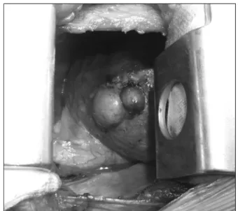
Fig. 3. Upon surgical finding, two round masses are noted and they look like liver tissues.
수술은 우측 후측방 개흉술을 시행하였다. 수술 소견 상, 흉막유착이나 흉막삼출액은 없었으며, 폐도 정상으로 보였다. 우측 횡격막의 중심 부위에 3.5×3.5 cm와 2.0×
2.0 cm 정도의 둥글고 매끈한 간 조직처럼 보이는 2개의 종양이 있었으며, 이 종양들은 횡격막에 부착되어 있었 다(Fig. 3). 정상적인 간과 연결되는 혈관 등을 포함한 경 구조물(pedicle)은 없었고, 횡격막 탈장이나 누공도 보이 지 않았다. 횡격막의 일부를 포함한 종양의 절제를 시행 하고 횡격막은 단순봉합하였다. 병리학적 검사에서 흉강 내 이소성 간으로 확진되었다. 환자는 특별한 문제 없이
수술 후 7일째 퇴원하였다.
고 찰
이소성 간 조직은 흔하지 않으며, 대부분 흉강보다는 복강내에 발생한다[1]. 특히 횡격막 탈장이 동반되지 않 은 흉강내 이소성 간은 매우 희귀하여, 문헌상 15예 정 도 보고되어 있는 실정이다[1]. 복강내 이소성 간 조직은 장간막(mesentery) 등에 의해 정상 간에 연결되어 있는 경우가 많으며, 비장, 췌장, 부신, 담낭, 후복강, 복망
대흉외지 2007;40:802-804
− 804 − (omentum) 등의 다양한 복강 내 구조물에 위치할 수 있 다[1,2]. 복강내 이소성 간은 복강내 종양으로 발견되는 경우가 많으나, 임상적으로 의미가 없는 경우가 많다. 그 러나 드물게 이소성 간에 간경변과 간암이 발생하였다 는 보고들이 있다[1].
흉강내 이소성 간은 일반적으로 횡격막 탈장과 동반되 며, 좌측 흉강, 폐, 그리고 심낭에서 발생한 보고도 있지 만, 주로 우측 흉강에 발생한다[1,3,4]. 이소성 간은 경구 조물에 의해 정상간에 연결되어 있는 경우가 많으나, 독 립된 종괴로 존재하기도 한다[1].
Collan 등[5]은 이소성 간을 4가지 종류로 분류하였다. 그 들은 상당한 크기의 간 부엽이 정상간과의 연결이 있을 경 우(accessory lobe of liver of considerable size and with a con- necting stalk to the liver)를 제I군, 작은 간 부엽이 정상간에 연결된 경우(small accessory lobe of the liver attached to the liver)를 제II군, 정상간과의 연결이 있거나 없는 이소성 간 (ectopic liver located with or without connection to the liver) 을 제III군, 그리고 현미경적 이소성 간 조직(microscopic ec- topic liver)을 제IV군으로 나누었다. 본 증례는 그들의 분류 에 따르면 제 III군에 해당한다고 볼 수 있다.
발생기전이 명확하지는 않지만, 횡격막 상부의 이소성 간은 발생학적으로 매우 연관이 있는 간과 횡격막의 발 생학적 이상에서 초래될 것으로 생각한다[6]. 간배아(hep- atic bud)는 전장(foregut)에서 발생하여 횡중격(septum trans- versum)으로 자라며, 큰 두부(cradial portion)와 작은 미부 (caudal portion)로 나누어진다. 두부는 횡중격의 간충조직 (mesenchymal tissue)으로 분화되는데, 횡격막의 중심건 (central tendon)도 횡중격에서 발생한다. 횡격막이 태아의
다른 부위에 비해 부적절하게 빠른 분화가 일어날 경우 횡격막 상부에 이소성 간이 발생하게 된다[6].
흉강내 이소성 간은 호흡곤란 또는 폐렴을 동반할 수 있으나, 일반적으로 무증상인 경우가 많다[2,7]. 흉부 하 측에 종양이 있을 경우 한번쯤 의심하는 것이 중요하며, HIDA 스캔(hepatobiliary iminodiacetic acid scan)이 진단에 도움이 된다[2]. 폐격리증과의 감별에 혈관조영술이 도움 이 되나, 최종 진단은 주로 수술 중에 이루어진다[1,2].
본 증례는 국내 최초의 횡격막 탈장이 동반되지 않은 흉강내 이소성 간으로 여겨지며, 흉강내 이소성 간의 발 생학적 발병기전을 이해하는 데 도움이 되리라 생각한다.
참 고 문 헌
1. Chen F, Heller DS, Bethel C, Faye-Petersen O. Intra- thoracic ectopic lobe of liver presenting as pulmonary sequestration. Fetal Pediatr Pathol 2005;24:155-9.
2. Babu R, Van der Avoirt A. Ectopic intrathoracic liver.
Pediatr Surg Int 2001;17:461-2.
3. Salman AB. Left-sided congenital diaphragmatic hernia associated with intrathoracic ectopic liver lobule. Eur J Cardiothorac Surg 2002;21:558-60.
4. Kinnunen P, Kulmala P, Kaarteenaho-Wiik R, Vuopala K. Ectopic liver in the human pericardium. Histopath- ology 1997;30:277-9.
5. Collan Y, Hakkiluoto A, Hastbacka J. Ectopic liver. Ann Chir Gynecol 1978;67:27-9.
6. Le Roux BT. Heterotopic intrathoracic liver. Thorax 1961;16:68-71.
7. Iber T, Rintala R. Inrapulmonary ectopic liver. J Pediatr Surg 1999;34:1425-6.
=국문 초록=
선천성 횡격막 탈장이 동반된 흉강내 이소성 간은 드물지 않게 보고되고 있으나 횡격막 탈장이 동반 되지 않은 흉강내 이소성 간은 매우 희귀한 선천성 질환이다. 횡격막 탈장이 동반되지 않은 흉강내 이소성 간을 체험하였기에 증례를 보고한다. 환자는 37세 여자로, 우연히 발견된 폐종양의 치료를 위 해 내원하였다. 흉부 컴퓨터단층촬영 스캔상 우측 폐하엽에 횡격막에 인접해 있는 폐종양이 확인되 었으며, 기관지 유암종을 의심하고 수술을 시행하였다. 수술 소견상, 우측 횡격막 중심부에 지름 3.5 cm 와 2.0 cm 정도의 2개의 둥근 종양이 위치하고 있었으며, 종양의 절제를 시행하였다. 병리학적으 로 흉강내 이소성 간으로 확진되었고, 환자는 수술 후 특별한 문제 없이 퇴원하였다.
중심 단어:1. 흉막강 2. 횡격막 3. 이소성 조직 4. 간