INTRODUCTION
Lemierre syndrome, known as postanginal sepsis or necrobacillosis, is an uncommon but have potentially life-threatening complication of acute pharyngotonsillitis.
Anaerobic oropharyngeal infection may result in septic thrombophlebitis of the internal jugular vein (IJV) with subsequent septicemia and septic embolization, which cause metastatic abscesses, commonly in the lungs. It was well depicted by Dr. Andr Lemierre (1875-1956) in 1936.é 1)In the preantibiotic era, Lemierre syndrome was common and often followed a fulminant course, with a mortality rate of 90%. Substantial decrease of mortality and morbidity with the introduction of antibiotics has made this syndrome into status that is frequently forgotten or overlooked when it appears.2,3)We report here on a case of Lemierre syndrome to increase the awareness of this disorder.
CASE REPORT
A 54-year-old male patient, suffering from a painful right-sided neck mass and painful swelling of right upper arm, was admitted. He had a history of sore throat that started 10 days before admission, followed two days later by an enlarging mass on the right side of his neck. He also complained of dysphagia, subjective fever and painful swelling of right upper arm. He was known patient with diabetes mellitus. He had no alcohol, smoking, or illegal drug use.
On physical examination, the patient appeared acutely ill but in no respiratory distress. He had mild erythema of the oropharynx, with right sided oropharyngeal swelling. The neck showed a large tender mass adjacent to the anterior border of the sternocleiomastoid muscle, measuring approximately 4×5 . And the right upper arm showed㎝ swelling, erythematous skin with tenderness.
Laboratory studies revealed a white blood cell count of 15,000 with 84% neutrophils, 10.5% lymphocytes, 5.4%
monocytes, and 0.1% eosinophils. Hemoglobin was 12.8 g/dL and hematocrit, 37.5%. Platelet count was 210×103/ L. Fasting capillary blood glucose level was 350 mg/dL.
μ
Erythrocyte sedimentation rate was 62mm/h, and C-reactive protein level was 17.86 ng/mL.
고신대학교 의과대학 학술지 제 권 제 호24 1 Kosin Medical Journal
Vol. 24. No. 1, pp. 193 196, 2009∼
Kyung-Soon Jeong ․ Mi-Hee Jung
Department of Radiology, Gospel Hospital, Kosin University College of Medicine, Busan, Korea
――― Abstract ――――――――――――――――――――――――――――――――――――――――
We report a 54-year-old man who had anaerobic pulmonary sepsis associated with internal jugular venous thrombosis secondary to pharyngotonsillitis. The patient had classic clinical presentations and the computed tomography (CT) scan of the neck and chest showed an irregularly enhanced cystic mass with right jugular vein thrombosis and multiple lung nodules with cavity, which are characteristics of Lemierre syndrome. The CT and clinical implications are described.
―――――――――――――――――――――――――――――――――――――――――――――――――
Key words : Lung abscess, Peritonsillar abscess
교신저자 Mi Hee Jung:
Add : Department of Radiology, Gospel Hospital, Kosin University College of Medicine,
34 Amnam dong, Seo-Gu, Busan 602-902, Korea TEL : +82.51-990-6341, FAX : +82.51-255-2764 E-mail : [email protected]
고신대학교 의과대학 학술지 제 권 호24 1 , 2009
Plain chest film demonstrated multiple patch increased opacities with cavitation scattered in right lung, measured about 3cm in largest diameter in right middle lung zone.
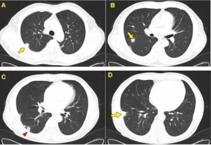
Computer tomography(CT) scans of the neck and chest were obtained with Somatome Plus-4 system (Siemens medical solution, Erlangen, Germamy) after the intravenous administration of 100mL of non-ionic contrast (ultravistⓇ, Schering, Germany). CT scan of the neck revealed thrombosis of the right internal jugular vein. There was an extensive abscess with extension to adjacent sternocleidomastoid muscle (Fig. 1). CT scan of the chest showed numerous pulmonary nodules with variable degrees of cavitation that were situated predominantly in the periphery of the lung.
There were some nodules with evidence of feeding vessels, indicated septic pulmonary emboli in both lungs (Fig. 2).
Fig. 1. A 54-year-old man with a painful right-sided neck mass and painful swelling of the right upper arm. The plain chest posteroanterior view shows multiple patch and nodular opacities with cavitations at the right middle and lower lung zones (arrowheads).
Fig. 2. Transverse ultrasound image of the right internal jugular vein (calipers) shows occlusion by an echogenic thrombus. A normal right carotid artery (arrowhead) is also shown. Note that multiseptated abscess formations (*) surround the right internal jugular vein.
Fig. 3. Both non-enhanced (A) and enhanced (B) axial CT scans of the neck at the level of the hyoid bone show lobulating cystic mass with irregular enhancing wall in the right carotid space. Note thrombosis within the right internal jugular vein (arrow).
Fig. 4. Conventional transverse CT scans (A-D) show multiple peripheral lung nodules(arrow) with cavitation (arrowhead) in some nodules.
On the basis of these findings, a diagnosis of Lemierre syndrome was made. Blood and aspiration cultures were Klebsiella pneumoniae. The patient was started on intravenous antibiotics, such as cefoperazone 2.0 g every 24 hours, isepamicin 400 mg every 12 hours, clindamycin 600 mg every 8 hours.
Despite appropriate antimicrobial therapy, the patient remained unwell, with painful swelling of right neck and upper arm. Urgent decompression and excessive debridement of the neck and right upper arm and excision of right IJV were performed. Surgical findings were shown as right internal jugular vein thrombosis with adjacent abscess formation and adhesion to sternocleiodomastoid muscle.
DISCUSSION
Lemierre syndrome consists of septic thrombophlebitis of the internal jugular vein secondary to oropharyngeal infection, which is an uncommon but potentially life-threatening complication of acute phryngotonsilitis.4) According to series review by Sinave and colleagues,5) Fusobacteriumnecrophorum, an obligate Gram-negative anaero bicbacillus, is most frequently isolated organism for causing Lemierre syndrome , accounting for 81% of cases, followed by other Fusobacterium species, such as Fusobacteriumnucleatum for 11%, and other Gram negative organisms like Enterococcus spp. for 8%. Of 12.8% of documented cased, the culture is inconclusive.2,5) In this case, Klebsiella pneumoniae of Enterococcus spp. was cultured.
Infection of the parapharyngeal space, which may occur secondary to direct spread of oropharyngeal infection or by lymphatic or tonsillar venous dissemination, is central to the development of Lemierre syndrome.2) Infection at this site may affect not only the carotid sheath and its contents but also cranial nervesⅨ Ⅻ- , with consequent neurogenic signs and symptoms. Internal jugular venous thrombosis that results from the adjacent inflammatory process or extension from the tonsillar veins acts as a nidus of infection that may spread hematogenously and results in septicemia and septic embolization.4,5) Pulmonary emboli are found in most untreated patients, however empyema is rare. Seeding of other sites occurs, mostly to the joints. Other potential sites that are involved are the liver causing 'bacteremic jaundice'.6)
Patient with Lemierre syndrome typically presents with acute pharyngitis followed 3-10 days later by the development of a high fever, rigors and malaise with onset of septicemia.4)Internal jugular venous thrombosis typically causes pain, tenderness, swelling along the line of the vein, and trismus; involvement of the lower cranial nerves may result in hoarseness and dysphagia. However, these findings may be minor and readily overlooked.4) Dyspnea and pleuric chest pain are seen with pleuropulmonary disease.
The radiologic findings in Lemierre syndrome are integral to its diagnosis. Referal to the radiology department
may be for the evaluation of symptoms caused directly by internal jugular venous clot or for imaging of the septic embolic complications.4)Awareness of this syndrome by the radiologist should lead to the suggestion of Lemierre syndrome whether by demonstration of internal jugular venous thrombosis at CT or ultrasonogram (US) or through chest CT findings suggestive of septic emboli.2,4)On doppler sonography, which is sensitive and an excellent serial examination, thrombus appears homogenously echogenic or as a complex solid and cystic mass. Acute thrombus may be of low echogenicity and difficult to detect without doppler analysis.7) But there are limits to full visualization of the internal jugular vein due to cardiac pulsatility and respiration or inaccessible areas to US (eg. behind the clavicle or the skull base). In contrast, CT and magnetic resonance (MR) imaging enable visualization of the entire internal jugular vein, as well as assessment of the thorax for proximal compressive cause of venous obstruction.4)
Contrast enhanced CT is the most common modality used, with excellent differentiation of lymphadenopathy, infectious loculations, and thrombus. Thrombosed vein are distended, with enhancing walls and low attenuation filling defects, whereas adjacent tissue may be edematous.7) MR imaging, with its flow sensitivity and excellent soft-tissue contrast, has been shown to be an effective modality for the identification and characterization of thrombi, although cost and availability currently limit its use.4)
Differential diagnostic consideration with Lemierre syndrome are primarily the cause of IJV thrombosis, including intravenous drug abuse, hypercoagulable state, and radiation therapy, as well as endocarditis.7)
The mainstay of treatment of Lemierre syndrome remains high dose intravenous antibiotics, which should cover both anaerobes and β-lactamase production. Surgical drainage of any abscess that develops is recommended.
However, ligation of internal jugular vein or anticoagulation remains controversial and is typically reserved for cases that are refractory to treatment with antibiotics alone .2,8)
고신대학교 의과대학 학술지 제 권 호24 1 , 2009
CONCLUSION
Lemierre syndrome, although very rare, still exists and remains potentially life threatening. Early diagnosis can expedite the treatment and decrease the unwanted complications. Therefore, CT absolutely stand high as the key modality in being reminiscent of the forgotten disease.
The radiologist should be aware of Lemierre syndrome, particularly in a young, previously well patient without a history of drug abuse, and should suggest this diagnosis when radiologic and clinical features are compatible with septic emboli.
국문초록
폐혈성 폐 색전증을 동반한 르미에르 증후군 증례 보고:
르미에르 증후군은 드문 질환으로 구인두 감염이 있었던 환자에서 내경정맥 혈전과 폐혈성 폐 색전증을 보이는
것이 특징이다. 54세 남자환자가 목의 우측에 만져지는
종괴와 열감을 주소로 내원하였다 시행한 목과 흉부 컴.
퓨터 단층 촬영에서 우측 내경정맥 내부와 주변부에 저 음영의 조영증강을 보이는 종괴가 보였고 우폐야에 내부
공동을 보이는 경계가 불분명한 결절들이 보였다 저자.
들은 르미에르 증후군의 임상 소견과 영상학적 소견을 보고하고자 한다.
REFERENCE
1) Lemierre A : On certain septicaemias due to anaerobic organisms. Lancet 1:701-703, 1936
2) Yen-Jun Lai, Jiing-Feng Lirng, Feng-Chi Chang, Chao-Bao Lou, Micheal Mu-Huo Teng, Cheng-Yen Chang : Computed tomographic finding in Lemierre syndrome. J Chin Med Assoc 67:419-421, 2004
3) Julio A Chirinos, Daniel M Lichtstein, Javier Garcia, Leonardo J Tamariz : The evolution of Lemierre syndorme. Medicine 81:458-65, 2002
4) Screaton NJ, Ravenel JG, Lehner PJ, Heitzman ER, Flower CDR : Lemierre syndrome: forgotten but not extinct-report of 4 cases. Radiology 213:369-374, 1999
5) Sinave CP, Hardy GJ, Fardy PW : The Lemierre syndrome : suppurative thrombophlebitis of the internal jugular vein secondary to oropharyngeal infection. Medicine 68:85-94, 1989
6) Itzhak Brook : Microbiology and management of deep facial infection and Lemierre Syndrome ORL 65:117-120, 2003 7) Andrew E Auber, Patricia A Mancuso : Lemierre syndrome:
magnetic resonance imaging and computed tomographic appearance. Military Medicine 165(8):638-640, 2000 8) Andrea Williams, Mark Nagy, Jennifer Wingate, Luna Bailey,
Mark Wax : Lemierre syndrome: a complication of acute pharyngitis. Int J Pediatr Otorhinolaryngol 45:51-55, 1998