• 검색 결과가 없습니다.

Ruptured Abdominal Aortic Aneurysm after Endovascular Aortic Aneurysm Repair

N/A
N/A
Protected

Academic year: 2021

Share "Ruptured Abdominal Aortic Aneurysm after Endovascular Aortic Aneurysm Repair"

Copied!
4
0
0

로드 중.... (전체 텍스트 보기)

전체 글

(1)

Korean J Thorac Cardiovasc Surg 2011;44:68-71 □ Case Report □ DOI:10.5090/kjtcs.2011.44.1.68 ISSN: 2233-601X (Print) ISSN: 2093-6516 (Online)

− 68 −

*Department of Thoracic and Cardiovascular Surgery, School of Medicine, Pusan National University

**Department of Radiology, School of Medicine, Pusan National University

†This work was supported for two years by Pusan National University Research Grant.

Received: August 4, 2010, Revised: September 15, 2010, Accepted: September 26, 2010

Corresponding author: Sung Woon Chung, Department of Thoracic and Cardiovascular Surgery, School of Medicine, Pusan National University, 10, 1-ga, Ami-dong, Seo-gu, Busan 602-739, Korea

(Tel) 82-51-240-7263 (Fax) 82-51-243-9389 (E-mail) chungsungwoon@hanmail.net

C

The Korean Society for Thoracic and Cardiovascular Surgery. 2011. All right reserved.

CC
This is an open access article distributed under the terms of the Creative Commons Attribution Non-Commercial License (http://creative- commons.org/licenses/by-nc/3.0) which permits unrestricted non-commercial use, distribution, and reproduction in any medium, provided the original work is properly cited.

Ruptured Abdominal Aortic Aneurysm after Endovascular Aortic Aneurysm Repair

Chung Won Lee, M.D.*, Sung Woon Chung, M.D.*, Jong Won Kim, M.D.*, Sangpil Kim, M.D.*, Mi Ju Bae, M.D.*, Chang Won Kim, M.D.**

In treating uncomplicated abdominal aortic aenurysm, endovascular aortic aneurysm repair (EVAR) has been em- ployed as a good alternative to open repair with low perioperative morbidity and mortality. However, the aneurysm can enlarge or rupture even after EVAR as a result of device failure, endoleak, or graft migration. We experienced two cases of aneurismal rupture after EVAR, which were successfully treated by surgical extra-anatomic bypass.

Key words: 1. Aneurysm 2. Aorta, abdominal 3. Rupture

4. Endovascular surgery

CASE REPORT 1) Case 1

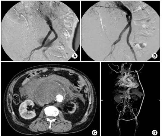
A sixty-six-year-old male patient visited the emergency room with severe abdominal pain and distension. The patient had received endovascular aortic aneurysm repair (EVAR) us- ing bifurcated stent graft (S&G Biotech inc., Seongman, Korea), about five years before. The patient had a history of the second endovascular intervention for endoleak due to the separation of the left iliac limb one year after EVAR (Fig.

1A, B). On arrival at the emergency room, his blood pressure was 70/40 mmHg. As the abdominal computed tomography (CT) showed an enlarged abdominal aortic aneurysm with ret- roperitoneal hematoma (Fig. 1C), an emergency operation was performed. Through median laparotomy the abdominal aorta was dissected, but surgical exposure was difficult because of

the massive hematoma. After Supra-celiac aortic clamping

was done, removal of the implanted stent graft was

attempted. However, the proximal bare part of the stent,

which was firmly incorporated into the aortic wall by neo-

intimalization, could not be retrieved. Because bleeding from

the aorta was so severe on the attempt for stent retrieval and

bowl ischemia was suspected due to supraceliac clamping of

the aorta, extra-anatomic bypass grafting was contemplated as

a salvage procedure. After the stent was cut inside the aorta,

infrarenal portion of the aorta were oversewn and supraceliac

aortic clamp was released for bowl reperfusion. Two prox-

imal portions of the common iliac arteries were also over-

sewn, and left axillo-femoral and femorofemoral bypass was

performed using a ring reinforced 8mm polytetrafluoro-

ethylene (PTFE) grafts (Gore-Tex, USA). After adequate cir-

culation to the both feet was checked, he was transferred to

(2)

Ruptured Abdominal Aortic Aneurysm after Endovascular Aortic Aneurysm Repair

− 69 −

Fig. 1. Case 1: (A) Contrast extra- vasation of the left limb of stent graft (Type III endoleak). (B) A co- mpletion angiogram after additional stent graft deployment. (C) Abdomi- nal CT showing the enlarged abdo- minal aortic aneurysm and retroperi- toneal hematoma. (D) CT angiog- raphy showing patent axillo-bifemoral bypass graft one month after the operation. CT=Computerized tomogr- aphy.

Fig. 2. Case 2: (A) An angiogram after endovascular aortic aneurysm repair. (B) Abdominal CT showing the ruptured abdominal aortic aneurysm after EVAR. (C) CT angiography showing patent axillo-bifemoral bypass graft one month after the operation. CT=Computerized tomography; EVAR=Endovascular aortic aneurysm repair.

the intensive care unit (ICU) in a critical condition. During the postoperative course, he developed acute renal failure and continuous renal replacement therapy was performed. He stayed at the ICU for eight days, and he could be discharged without any further complications at postoperative day 41.

The graft patency is has been well maintained for 24 months of follow-up (Fig. 1D).

2) Case 2

A seventy-three-year-old male patient presented with severe

(3)

Chung Won Lee, et al

− 70 − abdominal pain and hypotension. He underwent EVAR about four years ago using a straight stent graft (S&G Biotech inc., Seongman, Korea) (Fig. 2A). He had histories of hyper- tension and coronary artery disease. Because his blood pres- sure was 60/40 mmHg and abdominal CT showed ruptured abdominal aortic aneurysm (Fig. 2B), an emergency operation was performed. When he arrived in the operating room, his systolic blood pressure was checked at 40 mmHg. To main- tain the cerebral and systemic perfusion, cardiopulmonary by- pass was used commenced by cannulating the right femoral vein and the right axillary artery. After median laparotomy incision was made under the cardiopulmonary bypass, the aorta was dissected and clamped below the renal arteries.

Because the stent graft could not be completely removed and collapsed remnant of the stent graft hindered the inflow anas- tomosis to a prosthetic graft, the infrarenal aorta and open- ings of both common iliac arteries were oversewn for ex- tra-anatomic bypass grafting. After cardiopulmonary bypass was discontinued and laparotomy wound was closed, axil- lo-bifemoral bypass was performed using a ring reinforced 8mm PTFE graft (Gore-Tex, USA). He was transferred to the intensive care unit in a critical condition. He was supported by a mechanical ventilator for 12 days and transferred to the general ward in a stable state at postoperative day 15. CT an- giography checked one month after the surgery showed good flow through the bypass graft (Fig. 2C).

DISCUSSION

Since the first report of endovascular aortic aneurysm re- pair (EVAR) by Parodi in 1991 [1], EVAR has been replac- ing the open repair in most of the patients with favorable anatomic features because of lower perioperative morbidity and mortality than the latter [2,3].

However, the complications of EVAR such as device fail- ure, endoleak, and graft migration may require a conversion to open repair or lead to aneurysm enlargement or rupture.

Verzini et al. [4] reported that 4.5% patients required open repair during a median follow-up of 38 months. Harris et al.

[5] reported an annual cumulative risk of 2.1% for conversion to open repair in the EUROSTAR registry.

Furthermore, open surgical repair after EVAR usually turns

out to be more risky than the primary open repair. In the lit- eratures, the mortality rates were reported to be as high as 20% [5,6]. The authors speculated that high mortality and morbidity may be related to the presence of the endograft which makes dissection difficult, and the need for suprarenal or supraceliac aortic cross clamping.

We performed axillo-bifemoral bypass in two cases. This procedure is much easier to perform than abdominal aortic surgery, and can be applied to patients with risk factors such as severe heart disease, chronic renal failure, severe pulmo- nary dysfunction, and severe obesity. In our cases, one pa- tient showed left kidney atrophy on preoperative CT, and the other patient had a history of coronary artery disease. We think that the extra-anatomic bypass alleviated the risk of postoperative morbidity and mortality thanks to the shortening of aortic cross clamping time. Because the collapsed stent graft made it difficult to perform proximal inflow anasto- mosis, extra-anatomic bypass was thought to be a safer op- tion than time-consuming conventional aortoiliac graft interposition. Long-term patency of the axillofemoral grafts, however, is to be closely followed up.

Halpern et al. [7] reported that loss of consciousness is a preceding sign of mortality in patients with ruptured abdomi- nal aortic aneurysm (AAA). When AAA ruptures, an in- dependent risk factor associated with immediate mortality is the hemodynamic instability of the patients [8]. Because pre- operative vital signs of our second patient were very unstable, we decided to use cardiopulmonary bypass (CPB) to maintain the cerebral perfusion and systemic circulation. The employ- ment of CPB might be helpful as a life-saving measure for the patient who shows ruptured AAA with unstable vital signs.

In conclusion, we could successfully treat the patients with

EVAR-related AAA rupture by extra-anatomic bypass graft-

ing, with or without the aid of cardiopulmonary bypass. We

think that the options for post-EVAR aneurysm rupture

should include a conversion to extra-anatomic bypass, be-

cause performing a salvage procedure is safer than sticking to

anatomic repair. Using CPB also could be helpful in case of

critically ill patients with ruptured AAA.

(4)

Ruptured Abdominal Aortic Aneurysm after Endovascular Aortic Aneurysm Repair

− 71 − REFERENCES

1. Parodi JC, Palmaz JC, Barone HD. Transfemoral intra- luminal graft interposition for abdominal aortic aneurysm.

Ann Vasc Surg 1991;5:491-9.

2. Cao P, Verzini F, Parlani G, et al. Clinical effect of abdomi- nal aortic aneurysm endografting: 7-year concurrent com- parison with open repair. J Vasc Surg 2004;40:841-8.

3. Schermerhorn ML, O’Malley AJ, Jhaveri A, Cotterill P, Pomposelli F, Landon BE. Endovascular vs. open repair of abdominal aortic aneurysm in the medicare population. N Engl J Med 2008;358:464-74.

4. Verzini F, Cao P, De Rango P, et al. Conversion to open re- pair after endogrfting for abdominal aortic aneurysm: cause, incidence and results. Eur J Vasc Endovasc Surg 2006;31:

136-42.

5. Harris PL, Vallabhaneni SR, Desgranges P, Becquemin JP, van Marrewijk C, Laheij RJ. Incidence and risk factors of late rupture, conversion, and death after endovascular repair of infrarenal aortic aneurysms: the EUROSTAR experience. J Vasc Surg 2000;32:739-49.

6. May J, White GH, Yu W, et al. Conversion from endolumi- nal to open repair of abdominal aortic aneurysm. A hazard- ous procedure. Eur J Vasc Endovasc Surg 1997;14:4-11.

7. Halpern VJ, Kline RG, D’Angelo AJ, Cohen JR. Factors that affect the survival rate of patients with ruptured ab- dominal aortic aneurysms. J Vasc Surg 1997;26:939-48.

8. Coppi G, Silingardi R, Gennai S, Saitta G, Ciardullo AV. A single-center experience in open and endovascular treatment of hemodynamically unstable and stable patients with rup- tured abdominal aortic aneurysms. J Vasc Surg 2006;44:

1140-7.

수치

Fig. 1. Case 1: (A) Contrast extra- extra-vasation of the left limb of stent  graft (Type III endoleak)

참조

관련 문서

- leaves the abdominal cavity lateral to the inferior epigastric vessels and enters the deep inguinal ring. - traverses the entire inguinal canal - exits through

Therefore depending on the size of the fracture in the veneering porcelain, intraoral repair methods using surface treatment and composite resins may

Construction New construction sites, road repair sites, demolition of  buildings.

In this study we will survey the changes in DNA repair factors during aging in rat, including mismatch repair (MMR), base excision repair (BER), and double-strand break

To investigate the levels of mismatch repair proteins, such as MSH2, MSH6, MSH3, MHL1, and PMS2, in young, intermediate and senescent CCD-18Co and IMR90 cells,

- 소유 광산 : Ust-Kamenogorsk Metallurgical Combine(구 UstKa- menogorsk Industrial Complex), Riddersky Repair and Mechanical Plant, Bukhtarminsky Hydro Energy

(A) Before repair of RCT shows fatty degeneration of supraspinatus (stage 2) and infraspinatus (stage 3) muscle and atrophy (Grade 2) (B) After 18months later

treat wide wide wide wide necked necked necked necked aneurysms aneurysms aneurysms aneurysms that that that that selection selection selection selection of of