8
1)
이 연구는 계명대학교 대학원 학생 학술연구장학금에 의한 것임.
접수 : 2002년 8월 25일, 수정 : 2002년 12월 6일, 승인 : 2002년 12월 7일 책임저자 : 하정숙, 계명대학교 의과대학 진단검사의학교실
Tel : 053)250-7266, Fax : 053)250-7275, E-mail : [email protected]
백혈병환자에서의 세포유전학 검사; 515례에 대한 분석
계명대학교 의과대학 진단검사의학교실1, 성균관의대 마산삼성병원 진단검사의학과2
하정숙1․류남희1․전동석1․김재룡1․김영재2
The Cytogenetic Analysis of Leukemia Patients;
A Study of 515 Cases
Jung-Sook Ha, M.D.1, Nam-Hee Ryoo, M.D.1, Dong-Seok Jeon, M.D.1, Jae-Ryong Kim, M.D.1, and Young-Jae Kim, M.D.2
Department of Laboratory Medicine1, University of Keimyung College of Medicine, Daegu,
Department of Laboratory Medicine2, Sungkyunkwan University School of Medicine, Masan Samsung Hospital, Masan, Korea
Background : Cytogenetic study is important in prediction of prognosis and evaluation of treatment effect in leukemia. The cytogenetic aberrations of leukemia are nonrandom, but uneven geographic distribution of specific abnormalities have been reported in a few studies. So we analyzed cytogenetic study to find these uneven distribution patterns.
Methods : The conventional cytogenetic study was performed for 515 cases with acute and chronic leukemia on initial diagnosis. The results were analysed in each subtypes classified according to FAB criteria.
Results : The aberration rate was 62.0% in acute myelogenous leukemia (AML), 72.0% in acute lymphoblastic leukemia (ALL), 92.8% in chronic myelogenous leukemia (CML), 37.5% in chronic lymphocytic leukemia (CLL), 56.9% in myelodysplastic syndrome (MDS) and 36.4% in acute undetermined leukemia. The frequent anomalies were t(8;21)(q22;q22), t(15;17)(q22;q11), -Y,
+8, +21 in AML, t(9;22)(q34;q11), del(6q), +8, t(1;19)(q23;p13), +21, -20 in ALL, -7/del(7q), +8, del(12p), +11 in MDS. Philadelphia chromosome was found in 94.8% of CML and +22q-, +8 was frequent secondary changes. The incidence of t(8;21)(q22;q22) in M2, t(15;17)(q22;q11) in M3, t(9;22)(q34;q11) in ALL and -5/del5q, -7/del7q in MDS were 54.9%, 95.2%, 23.6%, 4.0% and 40.0%, respectively.
Conclusion : There were no marked differences in distribution pattern of common aberrations compared to previous reports. But the frequency of some anomalies showed specific findings. The incidence of t(8;21) in M2 subtype and t(9;22)(q34;q11) in ALL were higher in oriental countries including our results than in western countries. The incidence of -5/del(5q) in MDS was lower in oriental countries. These findings suggest the geographic heterogeneity which may give some help to investigate the genetic and environmental influence on the karyology of tumors. (Korean J He- matol 2003;38:8~14)
Key Words : Leukemia, Cytogenetic study, Geographic heterogeneity, t(8;21), t(9;22)(q34;q11), -5, del(5q)
서 론
세포유전학 검사는 종양의 발생 연구뿐 아니라 진단, 분 류, 예후 추정, 치료효과 평가에 중요한 역할을 한다. 그 중 혈액 종양은 염색체 이상이 가장 많이 연구된 영역으로, 주요 이상들은 최근 WHO에서 제시한 혈액 종양 분류에서 새로운 분류기준으로 제시되었다.1) 최근에는 분자유전기 법을 이용하여 좀더 빠른 진단이 가능해졌으나, 이차 이상 의 검출이나 빈도가 낮은 새로운 이상의 발견이라는 측면 에서 전형적인 세포유전학 검사는 아직까지 중요한 위치를 차지하고 있다.
염색체 이상 분포는 보고마다 조금씩 다르며, 몇몇 주요 이상의 빈도는 지역에 따른 차이가 관찰되는데 이는 유전 적, 환경적 요인이 영향을 미치리라는 것을 암시한다.2∼5) 따라서 저자들은 1992년 1월에서 2001년 12월까지 11년 간 본원에 의뢰된 급,만성 백혈병 환자를 대상으로 배양성 적과 이상핵형의 분포를 살펴보고, 주요 이상에 대해서는 지역에 따른 차이점을 살펴보고자 하였다.
대상 및 방법 1. 대 상
1992년 1월부터 2001년 12월까지 본원을 방문하여 조혈 기 질환으로 염색체 검사를 의뢰한 1086례 중, 골수천자도 말 및 말초혈액도말검사상 백혈병으로 첫 진단을 받은 515 례를 대상으로 분석하였고, 추적 검사는 분석에서 제외하 였다.
FAB (French-American-British, 이하 FAB) 분류 기준 에 따라 급성골수성백혈병(acute myelogenous leukemia;
이하 AML), 급성림프아구성백혈병(acute lymphoblastic leukemia; 이하 ALL), 만성골수성백혈병(chronic myelo- genous leukemia; 이하 CML), 만성림프구성백혈병 (chronic lymphocytic leukemia; 이하 CLL), 골수이형성 증후군(myelodysplastic syndrome; 이하 MDS) 및 각 아 형으로 구분하였으며, 형태 관찰과 면역화학염색으로 골수 구나 림프구계열로 구분할 수 없고, 면역표지자검사를 실 시하지 못했던 경우는 미분류 급성 백혈병(acute leukemia undetermined)으로 따로 분류하여 관찰하였다.
2. 방 법
대부분의 검체는 헤파린 처리한 골수로 검사를 실시하였
으며, CML, CLL의 경우나, 골수검체를 얻을 수 없으면서 말초혈액에서 종양 세포가 10% 이상일 경우 말초혈액으로 도 검사를 실시하였다. 배양방법은 직접법, 단기배양법, methotrexate 또는 thymidine 동기화고정도분염법, 장기 배양법의 방법을 각 검체당 질병 유형별로 2∼3가지 방법 을 조합하여 실시하였다. 검체가 도착한 즉시 배양 용기에 배지 9mL (RPMI 1640 7.5mL, FCS 1.5mL, penicillin/
streptomycin 0.1mL)를 분주하여 37℃에서 가온한 후 107/mL의 농도로 맞춘 검체 1mL를 접종하였다. 직접법과 단기, 장기 배양법은 각각 1시간, 24시간, 48∼72시간을 배양한 후 10µg/mL colcemid를 0.01mL 첨가하고 10분 배양한 후 수획하였고, 동기화고정도분염법은 검체 접종 후 3∼5시간 항온을 하고 105M methotrexate를 0.1mL 첨 가하여 14∼17시간 추가 배양 후 상층액을 버리고 103M thymidine을 0.1mL 첨가하여 5∼6시간 배양 후 같은 방법 으로 colcemid를 첨가하여 세포를 수획하였다. 수획한 세 포는 0.075M KCL 저장 용액으로 10분 처리 후 고정 (methanol:acetic acid=3:1) 단계를 거쳐 슬라이드를 제조 하였다. 염색 방법은 trypsin 처리없이 Wright 염색액으로 G-분염을 실시하였으며, 클론의 인정과 염색체 명명은 ISCN6)의 기준에 따랐다.
이상핵형은 FAB 아형에 따라 각 유형별 관련이상7)으로 알려져 있는 것은 표에 나열하였고, 여기에 속하지 않는 이 상은 기타 이상으로 분류하여 염색체 번호 순서대로 나열 하였다.
결 과 1. 배양성적
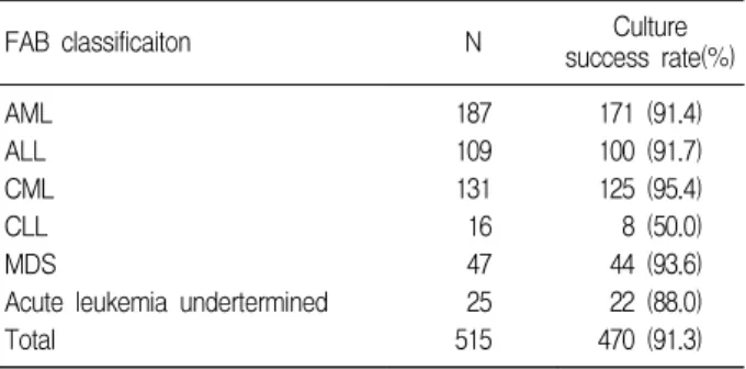
총 515례 중 470례(91.3%)에서 배양에 성공하였으며,
Table 1. The culture success rates according to FAB classification
FAB classificaiton N Culture
success rate(%) AML
ALL CML CLL MDS
Acute leukemia undertermined Total
187 109 131 16 47 25 515
171 (91.4) 100 (91.7) 125 (95.4) 8 (50.0) 44 (93.6) 22 (88.0) 470 (91.3) Abrreviations : AML, acute myelogenous leukemia; ALL, acute lym- phoblastic leukemia; CML, chronic myelogenous leukemia; CLL, chronic lymphocytic leukemia; MDS, myelodysplastic syndrome
유형별로 AML 91.4%, ALL 91.7%, CML 95.4%, CLL 50.0%, MDS 93.6%, 미분류 급성백혈병 88.5%의 성공 률을 보였다(Table 1). 330례(70.2%)에서 이상핵형을 관 찰할 수 있었고, AML 62.0%, ALL 72.0%, CML 92.8
%, CLL 37.5%, MDS 56.9%, 미분류 급성백혈병 36.4%에서 관찰되었다(Table 2∼6).
2. 유형별 이상핵형
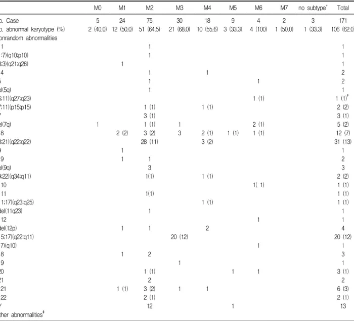
AML은 106례(62.0%)에서 이상핵형이 관찰되었고, 아
형별로 M6 100%, M3 68.0%, M2 64.5%, M4 55.6%, M7 50.0%, M1 50.0%, M0 40.0%, M5 33.3%순이었 다. 이 중 가장 빈도가 높은 이상핵형은 t(8;21)(q22;q22) 로 31례(29.2%)에서 관찰되었고, t(15;17)(q22;q11) 20 례(18.9%), -Y 13례(12.3%), +8 12례(11.3%), +21 6례(5.7%), del(7q) 5례(4.7%)순으로 나타났다. 단일이 상으로는 t(8;21)(q22;q22) 13례, t(15;17)(q22;q11) 12 례, +8 7례, +21 3례 순이며, -Y는 13례 모두 t(8;
21)(q22;q22)와 동반된 형태였다(Table 2).
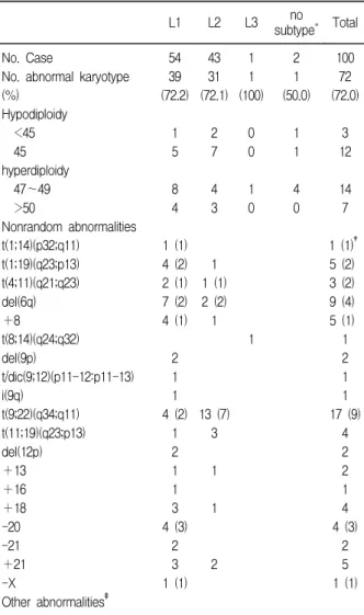
ALL은 72례(72.0%)에서 이상핵형이 관찰되었고, t(9;
Table 2. The distribution pattern of chromosome aberrations in acute myelogenous leukemia
M0 M1 M2 M3 M4 M5 M6 M7 no subtype* Total
No. Case
No. abnormal karyotype (%) Nonrandom abnormalities
+1 t(1:7)(q10;p10) t(3;3)(q21;q26)
+4 -5 del(5q) t(6;11)(q27;q23) t(7;11)(p15;p15) -7
del(7q)
+8
t(8;21)(q22;q22) -9
+9 del(9q) t(9;22)(q34;q11)
+10
+11
t(11;17)(q23;q25) t/del(11q23)
+12 t/del(12p) t(15;17)(q22;q11) i(17)(q10) -18 -19 -20 -21
+21
+22 -Y
Other abnormalities‡
5 2 (40.0)
1
24 12 (50.0)
1
2 (2)
1 1
1
1
1 (1) 75 51 (64.5)
1 1
1 1 1
1 (1) 3 (1) 1 (1) 3 (2) 28 (11)
1 3 1(1)
1(1)
1
1
2
1 (1) 2 3 (2) 2 (1) 12
30 21 (68.0)
1 3
20 (12)
1
1
18 10 (55.6)
1
1 (1)
2 (1) 3 (2)
1 (1)
1 (1)
2
1 9 3 (33.3)
1 (1)
1
1
4 4 (100)
1
1 (1)
2 (1) 1 (1)
1( 1)
1
1
1
2 1 (50.0)
3 1 (33.3)
171 106 (62.0)
1 1 1 2 2 1 1 (1)†
2 (2) 3 (1) 5 (2) 12 (7) 31 (13)
1 2 3 2 (2) 1 (1) 1 (1) 1 (1) 1 1 4 20 (12)
1 3 1 3 (1)
2 6 (3) 2 (1) 13
*unclassifiable cases by FAB classification; †number of solitary change; ‡-1, del(1)(p13;p32), t(1;X)(p31.2;p22.1), t(1;?), del(2)t(2;?), t(2;12)(q33;p12.1)
§, t(3;19)(q21.3;q13.4), t(4;8), t(4;13)(pter;q21), t(5;7;12;18)(p13;q22;q13;pter), t(5;12)(q22;q15), -6, r(6)(p25;q27), t(6;10)(q21;p15), +7q, del(7)(p11), t(7;9)(q22;q22), t(7;12) t(7;21)(q32;q22), t(7;?), i(9)(p10), 9p+, t(9;17), -10, -11, del(11)(q14;q12), t(11;22)(p11.2;q11.2), t(12;21),t(12;22), del(13q), t(14;?), del(14)(q24), -15, t(15;18), -16, -17, t(17;20)(q21;pter), t(19;?), t(22;?)(q13.2;?), t(22;?)(q13.3;?), -X, t(Y;9)(q12;q13); §detected as solitary change
22)(q34;q11) 17례(23.6%), del(6q) 9례(12.5%), t(1;
19)(q23;p13) 5례(6.9%), +8 5례(6.9%), +21 5례 (6.9%), -20 4례(5.6%)순으로, 단일이상은 t(9;22) (q34;q11) 9례, del(6q) 4례, -20 3례 순으로 흔히 관찰 되었다. L1은 고이배체 12례(30.8%), 저이배체 6례(15.4
%)로, L2는 고이배체 7례(22.6%), 저이배체 9례(29.0
%)로 나타나, L1에서는 고이배체가, L2에서는 저이배체 의 비율이 상대적으로 높았다(Table 3).
CML은 116례(92.8%)에서 이상핵형이 관찰되었고, 그 중 110례에서 philadelphia 염색체(이하; Ph)가 관찰되었 다. 6례는 변이형으로, 단순변이형 2례와 3가지 이상의 염 색체가 연관된 복합변이형 4례이며 모두 절단점이 9q34와 22q11을 포함하였다. 18례에서 Ph에 동반된 추가이상이 관찰되었는데 +22q-와 +8이 가장 흔히 동반되었다. Ph 가 관찰되지 않은 15례 중 9례는 정상핵형이, 6례는 다른 이상이 관찰되었다(Table 4).
MDS는 25례(56.9%)에서 이상핵형이 관찰되었고, RA (refractory anemia) 37.5%, RARS (refractory anemia with ringed sideroblasts) 33.3%에 비해 RAEB (re- fractory anemia with excess blasts) 69.2%, RAEB-T (refractory anemia with excess blasts in transformation) 68.8%로 이상핵형의 비율이 높았다. 각 이상별 빈도는 -7/del(7q), +8, del(12p), +11순이었고, -5/del(5q)는 1례에서만 관찰되었다(Table 5).
CLL은 3례(37.5%)에서만 이상핵형이 관찰되고 정상핵 형이 관찰된 비율이 높았다. 미분류 급성백혈병에서 계열 특 이성이 높은 이상들10,12)이 관찰된 경우는 재분류되었고, 나 머지 8례에서는 다양한 이상핵형이 관찰되었다(Table 6).
고 찰
염색체의 배양성공률은 대부분 90% 이상이고, 이상핵 형의 발견율은 보고문헌에 따라 다양하나 세포유전학기법 Table 3. The distribution pattern of chromosome
aberrations in acute lymphoblastic leukemia
L1 L2 L3 no
subtype* Total No. Case
No. abnormal karyotype (%)
Hypodiploidy <45
45
hyperdiploidy 47∼49
>50
Nonrandom abnormalities t(1;14)(p32;q11)
t(1;19)(q23;p13) t(4;11)(q21;q23) del(6q)
+8
t(8;14)(q24;q32) del(9p)
t/dic(9;12)(p11-12:p11-13) i(9q)
t(9;22)(q34;q11) t(11;19)(q23;p13) del(12p)
+13
+16
+18 -20 -21
+21 -X
Other abnormalities‡
54 39 (72.2)
1 5
8 4
1 (1) 4 (2) 2 (1) 7 (2) 4 (1)
2 1 1 4 (2)
1 2 1 1 3 4 (3)
2 3 1 (1)
43 31 (72.1)
2 7
4 3
1 1 (1) 2 (2) 1
13 (7) 3
1
1
2 1 1 (100)
0 0
1 0
1 2 1 (50.0)
1 1
4 0
100 72 (72.0)
3 12
14 7
1 (1)† 5 (2) 3 (2) 9 (4) 5 (1) 1 2 1 1 17 (9)
4 2 2 1 4 4 (3)
2 5 1 (1)
*unclassifiable cases by FAB classification; †number of solitary change; ‡+1, del(1)(p22), del(1q), inv(1)(q25;q42), i(1q),t(1;3)(q36.1;
q26), t(1;3)(q44;q27), t(1;6)(p34.1;q13), del(1q), t(1;13)(p13;q32), t(1;13) (q12;q32),t(1;16)(q23;q22)§, t(1;16)(q23;p13.3), t(1;5;2;?)(q32;q31;q31;?), t(2;8)(p21;qter), t(2;9), t(2;12)(p16.3-21;p13.3), t(2;12)(p23;p13), t(2;9;14) (q13q21;q12q13;q24), +3, t(3;9)(q21;p24), t(3;13)(p23;q34), +4, t(4;?), t(4;17;8), -5, +5, del(5)(p14), del(5)(q13), t(5; 7;8)(p15.2;p13;q11.2),
+6, t(6;13;14)(q23;q22;q32.2), t(6;21)(q16;q22), i(6p), -7, +7, del (7q32), t(7;?)(p22;?), t(8;21)(qter;q11.2), t(8;14;20), -9, +9, del(9)(q22), t(9;11)(p24;p11), t(9;12) (q10;q10), t(9;15)(q11;p11), t(9;18)(p24;q11),
+10, t(10;14)(q11;qter), +11, del(11)(q13q14), -12, +12, -14, +14, t(14;17), -15, +15, del(16q), i(16q), inv(16)(p13;q22), -17, +17, t(17;?), -18, i(18q), -19, der(19)t(19;?), +22, +der(22)t(9;22)(q34;q11) ,+X, del(Xq22), r(?), t(?,20); §detected as solitary change
Table 4. The chromosome aberrations in chronic myelogenous leukemia
N No. Case
No. abnormal karyotype (%) philadelphia positive standard variant*
additional change† philadelphia negative normal karyotype other‡
125 116 (92.8) 110 104 6 18 15 9 6
*t(17;22)(p13;q11) 2cases, t(3;9;22)(q14;q34;q11), t(7;9;9;22)(q36;q34;p24;
q11), t(9;12;22) (q34;q13;q11), t(9;13;22)(q34;q14;q11); †del(1) (p22), ins(1;2)(q14;p23pter), t(1;2;5;22), t(1;4;17)(p36.3;q23;p13), t(2;3) (p22;
q28), t(2;7), t(3;21)(q26.2;q22.1), t(7;16), -8, +8, t(8;17;21), t(8;21)(q22;
q22), ider(9)(q10)t(9;22)(q34;q11), inv(16), del(17)(p11.2), -21, +22, der(22)t(9;22)(q34;q11); ‡t(4;22)(q23-25;q22), del(5)(q13q33), del(5) (q22), -7, t(8;13)(p23;q14), +9, i(17q)
의 발달로 높아져 AML 55∼78%,8) ALL 60∼85%9,10)로 평균 2/3정도에서 관찰이 가능하고, MDS는 38.8∼
60.4%9,11)로 AML, ALL에 비해서는 낮다. CLL의 경우 분열지수가 낮아 이상을 관찰하기가 쉽지 않은데, B-CLL 의 경우 B-cell 분열촉진제를 쓴 후 40∼100%9)까지 이상 핵형을 발견할 수 있으나, 본 연구에서는 37.5%에서만 이 상이 발견되고 정상 핵형이 많이 관찰되어 적절한 분열촉 진제의 사용을 비롯해 배양 방법의 개선이 필요할 것으로 생각된다.
각 유형별로 흔한 이상은 AML에서 t(8;21)(q22;q22),
t(15;17)(q22;q11), +8, -Y, ALL에서 t(9;22)(q34;
q11), t(4;11), del(6q), +8, t(8;14), t(1;19), +21, MDS에서 -5/del(5q), -7/del(7q), +8 등으로8∼13) 본 연 구의 결과와 유사하였다.
그러나, 각 주요 이상들의 빈도는 지역별로 차이를 보이 는데,4,5) M2에서 t(8;21)의 빈도는 유럽 18.8∼53.8%, 미국 12.5∼27.3%에 비해 중국 74.1%, 일본 58.3%로 동양인에게 특히 높다. 김영숙 등14)과 본 연구에서도 49.5%, 54.9%로 높은 빈도를 보여, 동양인에서 특히 t(8;21)의 빈도가 높음을 확인할 수 있었다. M3에서 t(15;17)의 빈도는 본 연구의 95.2%를 비롯해 유럽 58∼
89.7%, 일본 96%, 미국 88.9∼100%로 모두 높은 빈도 를 보이며 지역별 차이가 없었다. 그러나, 본 연구에서 t(15; 17)의 발견율은 66.7%로 낮아, 분자유전학적 검사 를 실시하지 못했던 9례의 정상핵형과 다른 이상핵형만이 관찰된 1례에 대해서는 내재된 t(15;17)의 가능성을 배제 할 수 없다.
ALL에서 del(6q)와 +8, +21의 빈도는 국가별로 차이 가 없었고, t(9;22)(q34;q11)은 유럽 5.4∼19%, 미국 1.3∼3.6%에 비해 홍콩3) 50%, 한국14) 26.4%, 본 연구 23.6%로 동양국가에서 더 높은 빈도를 보였다.
MDS에서는 -5/del(5q)의 빈도가 미국 15.8∼21.6%, 독일 24∼32.6%에 비해 일본 4%, 대만 14.7%, 한국 13%11)로 동양 국가에서 낮은 경향을 보이는데, 본 연구에 서도 4%로 매우 낮아 인종별 차이의 가능성이 있다.
-7/del(7q)는 미국 10.5∼25%, 독일 12∼23.9%, 일본 Table 6. Chromosome aberrations in chronic lym-
phocytic leukemia and acute leukemia undeter- mined
CLL Acute leukemia undetermined No. Case
No. abnormal karyotype (%) Nonrandom abnormalities
+3 del(6q)
+8
+13 inv(14)(q11q32) -17
Other abnormalities†
8 3 (37.5)
1 (1)*
1
22 8 (36.4)
1 1 1 (1)
2
*number of solitary change; †dup(1)(q21.3-q31.3), ins(1;?)(q21;?), t(2;8)(p21;q12), t(2;12)(p23;p13), t(4;8),t(5;11)(q31.1;pter), i(8q), t(8;13) (p23;q14), -17, t(19;Y)(p13;q11), del(17)(p12), -Y
Table 5. The distribution pattern of chromosome aberrations in myelodysplstic syndrome subtypes
RA RARS RAEB RAEBT CMMoL no subtype* Total
No. Case
No. abnormal karyotype (%) Nonrandom abnormalities t(3;3)(q21;q26)
del(5q) -7 del(7q)
+8
t(8;21)(q22;q22)
+11 del(11q) del(12p) -21 -Y
Other abnormalities‡
8 3 (37.5)
1
1(1)
1
3 1 (33.3)
13 9 (69.2)
1 (1) 1 4 (1) 2 (1) 4 (2) 1
1 4 1 2
16 11 (68.8)
3 (1)
1 (1) 2 (2) 1
3 1 (33.3)
1 (1)
1 0
44 25 (56.9)
1 (1)† 1 8 (2) 2 (1) 5 (3) 2 (1) 3 (3) 2 4 2 2
*unclassifiable cases by FAB classification; †number of solitary change; ‡t(1;6)(q22;p21.1), t(1;20)(q41;qter), del(2)(q33), t(3;7;17)(p22;q29;p15-21;p15.3), t(2;9)§, del(3)(p22-24),del(3)(q26), t(3;6;17)(p22;q21;q25), r(4), -6, -9, -10, t(11;12)(pter;p12.3), t(11;12)(q22;q21.3), t(11;19), i(12q), -14, -18, -20; § detected as solitary change
4.5%, 대만 32.6%, 한국11) 11.9%의 결과에 비해 본 연 구에서는 40.0%로 매우 높게 관찰되었고, 한국에서 특징 적으로 많이 관찰되는 것으로 보고된11) trisomy 1q는 본 연 구에서는 관찰되지 않고, +11이 3례(12.0%)로 상대적으 로 자주 관찰되어 앞으로 더 많은 증례를 대상으로한 검토 가 필요하다.
CML은 90%이상에서 Ph를 관찰할 수 있으며,14∼18) Ph 가 관찰되지 않는 5∼10%정도의 CML에서는 bcr-abl 유 전자재배열을 검출하여 진단할 수 있다.18) 본 연구에서 Ph 를 관찰할 수 없었던 15례는 임상적으로나 형태학적으로 CML이 의심된 경우로 분자유전학적 재검사가 이루어진다 면 유전자재배열 음성인 비전형 CML이나 형태학적 오진의 가능성을 배제할 수 있을 것이다. Ph에 동반되는 이차적 이상 중에는 +8, +22q-, i(17q), +19, -Y, +21, + 17, -7이 가장 흔한데,19) 본 연구에서도 +22q- 와 +8이 각각 6례(33%)와 2례(11%)로 자주 관찰되었고, AML이 나 ALL로 전환된 형에 따른 이차적 이상에서는 관찰 증례 가 많지 않아 특이할 만한 차이점은 없었다.
이상핵형이 관찰되지 않는 경우는 더 잘 분열하는 정상 세포의 핵형이거나 미세현미경적 이상으로 발견할 수 없는 경우로, 후자의 경우 통상적인 배양 검사에 비교했을 때 더 높은 민감도와 예민도를 보이고 있는2,20,21) 분자유전학적 방법으로 검출률을 높이고 있다. 하지만 이런 기법들은 새 로운 이상의 발견에는 아직까지 제한적이며, 이차적 이상 이나 빈도가 낮아 아직까지 잘 알려져있지 않은 이상을 발 견하기 위해서는 통상적인 배양 검사가 필수적이다. 본 연 구에서도 알려진 이상 이외에 다양한 기타 이상핵형들을 관찰할 수 있었는데 이러한 이상들이 새로운 종양관련 이 상으로 밝혀지고, 지역적 분포에 대한 비교를 위해서는 더 많은 세포유전학 검사에 대한 증례 보고 및 검사결과 분석 이 필요할 것으로 생각된다.
요 약
배 경 : 혈액종양의 세포유전학 검사는 진단, 예후 추정, 치료 효과 평가에 중요한 구실을 하며, 매년 새로운 이상이 보고되고 있다. 종양의 종류 및 아형별로 특징적인 이상들 이 나타나나 각각의 이상별 빈도는 보고마다 다양하며, 지 리적으로 특징적인 분포를 보인다. 이에 저자들은 세포유 전학 검사 결과와 이상핵형 분포를 분석하고, 지리적 분포 의 이질성에 해당하는 소견이 있는지 알아보고자 하였다.
방 법 : 1992년 1월부터 2001년 12월까지 11년간 본원을 방문하여 급,만성 백혈병으로 첫 진단을 받은 515례를 대 상으로, 통상적인 세포유전학 검사를 실시하여, FAB 각
아형별로 이상핵형의 분포를 살펴보았다.
결 과 : 이상핵형은 급성골수성백혈병(AML) 62.0%, 급 성림프구성백혈병(ALL) 72.0%, 만성골수성백혈병(CML) 92.8%, 만성림프구성백혈병(CLL) 37.5%, 골수이형성증 후군(MDS) 56.9%, 미분류 급성백혈병 36.4%에서 관찰되 었고, 흔한 이상은 AML t(8;21)(q22;q22), t(15;17) (q22;q11), -Y, +8, +21, ALL t(9;22)(q34; q11), del(6q), +8, t(1;19)(q23;p13), +21, -20, MDS-7/
del(7q), +8, del(12p), +11순이었다. CML에서는 94.8
%에서 Philadelphia 염색체가 관찰되었으며, 22q-와 +8 이 흔한 이차이상이었다. M2에서 t(8;21)(q22;q22)은 54.9%, M3에서 t(15;17)(q22;q11)은 95.2%, ALL에서 t(9;22)(q34;q11)은 23.6%, MDS에서 -5/del5q와 -7/
del7q는 각각 4.0%, 40.0%로 관찰되었다.
결 론 : 각 종양별로 흔한 이상은 이전 보고와 유사하였고, M2에서 t(8;21)(q22;q22)과 ALL의 t(9;22)(q34;q11)의 빈도는 다른 동양국가와 같이 서양에 비해 빈도가 높았고, MDS에서 -5/del(5q)는 오히려 동양에서 낮은 경향을 보였 다. 이러한 지리적 분포의 차이는 종양 발생에 있어 유전적, 환경적 인자가 미치는 영향에 대한 연구에 중요한 자료가 될 수 있을 것으로 생각된다.
참 고 문 헌
1) Bennett JM : World health organization classification of the acute leukemias and myelodysplastic syn- drome. Int J Hematol 72:131-133, 2000
2) Koo SH, Kwon GC, Chun HJ, Park JW : Cyto- genetic and fluorescence in situ hybridization analyses of hematologic malignancies in Korea.
Cancer Genet Cytogenet 101:1-5, 1998
3) Chan LC, Kwong YL, Liu HW, Chan TK, Todd D, Ching LM : Cytogenetic analysis of hematologic malignancies in Hong-Kong. Cancer Genet Cyto- genet 62:154-159, 1992
4) Mitelman F : Geographic heterogeneity of chromo- some aberrations in hematologic disorders. Cancer Genet Cytogenet 20:203-208, 1986
5) Johansson B, Mertens F, Mitelman F : Geographic heterogeneity of neoplasia-associated chromosome aberrations. Genes Chromosomes Cancer 3:1-7, 1991
6) Mitelman F(ed) : ISCN(1995), An international sys- tem for human cytogenetic nomenclature. Basel, S Karger, 1995
7) Mitelman F, Kaneko Y, Trent J : Report of the committee on chromosome changes in neoplasia.
Cytogenet Cell Genet 58:1053-1079, 1991
8) Mrozekk, Heinonen K, Chapelle A, Bloomfield C : Clinical significance of cytogenetics in acute myeloid leukemia. Semin Oncol 24:17-31, 1997 9) Heim S, Mitelman F : Cancer cytogenetics. 2nd ed,
New York, Wiley-Liss, 1995, pp69-236
10) Faderl S, Kantarjian HM, Talpaz M, Estrov Z : Clinical significance of cytogenetic abnormalities in adult acute lymphoblastic leukemia. Blood 91:3995- 4019, 1998
11) Lee DS, Kim SH, Seo EJ, Park CJ, Chi HS, Ko EK, Yoon BH, Kim WH, Cho HI : Predominance of trisomy 1q in myelodysplastic syndromes in Korea:
Is there an ethnic difference? A 3-year multicenter study. Cancer Genet cytogenet 132:97-101, 2002 12) Kim KY : Chromosome studies in hematopoietic
disorders. Korean J Hematol 23:335-343, 1988 13) Mitelman F, Heim S : Quantitative acute leukemia
cytogenetics. Genes Chromosomes Cancer 5:57-66, 1992
14) Kim YS, Kim GS, Lee CH, Choi SI, Rhang DW, Cho HC : The cytogenetic study of acute and chronic leukemic patients in Korea. Korean J Clin Pathol 17:898-911, 1997
15) Lee MH, Chun HJ, Jeon DS, Kim JR : Cytogenetics studies in chronic myelogenous leukemia. Korean J Clin Pathol 13:689-698, 1993
16) Shin JS, Choi IY, MS Lyu, YK Baik : The Phila- delphia chromosome in chronic myelogenous leuke- mia. Korean J Hematol 17:159-165, 1982
17) Estop AM, Sherer C, Cieply K, Groft D, Burcoglu A, Jhanwar S, Thomas J : A ph-negative chronic myeloid leukemia patient with a non-classical BCR-ABL rearrangement characterized by fluores- cence in situ hybridization. Cancer Genet Cyto- genet 96:174-176, 1997
18) Reddy KS, Grove B : A Philadelphia-negative chron- ic myeloid leukemia with a BCR/ABL fusion gene on chromosome 9. Cancer Genet Cytogenet 107:48- 50, 1998
19) Johansson B. Fioretos T. Mitelman F : Cytogenetic and molecular genetic evolution of chronic myeloid leukemia. Acta Haematologica 107:76-94, 2002 20) Frohling S, Skelin S, Liebisch C, Scholl C, Schlenk
RF, Dohner H, Dohner K : Comparison of cytoge- netic and molecular cytogenetic detection of chro- mosome abnormalities in 240 consecutive adult patients with acute myeloid leukemia. J Clin Oncol 20:2480-2485, 2002
21) Kim KH, Han JY : Simultaneous reverse transcrip- tion-polymerase chain reaction for detection of 7 gene rearrangements in acute leukemia. Korean J Clin Pathol 21:24-33, 2001