J Korean Soc Coloproctol Vol. 18, No. 4, 2002
205
G enetic Polymorphism of Xenobiotics Me- tabolizing Enzymes and Individual Sus- ceptible Genes to Colorectal Cancer Pa- tients in Korea
H eung W oo Lee, M.D1
.,
M in Soo K im , M.D.1,
Pa Jong Jung, M.D1.,
H yun Jun K im , B.S.2,
G u K ong, M.D.2,
H oK yung C hun, M.D.3Departments of 1General Surgery and 2Pathology, College of Medicine, Hanyang University, 3Department of Surgery, Sung- kyunkwan University School of Medicine, Samsung Medical Center, Seoul, Korea
Individual susceptibility to cancers may result from several factors including differences in xenobiotics metabolism, DNA repair, altered oncogenes and suppressor genes, and environmental carcinogen exposures. To determine the frequencies of the genotypes of phase I (CYP1A1 and CYP2E1) and phase II (GSTM1 and NAT2) metabolizing enzymes and to identify the high-risk genotypes of these metabolic enzymes to colon cancer in Korean, we have analyzed 113 colorectal cancer patients and corresponding age and sex matched healthy controls using polymerase chain reaction-restriction fragment length polymorphism (PCR-RFLP). In analysis of phase I enzymes, m1/m2, m2/m2 and Val/Val genotypes in CYP1A1 enzyme polymorphisms and C1/C2 genotype in CYP2E1 polymorphism were as- sociated with high relative risks to colorectal cancers (Odds ratio; 1.51, 1.59, 1.76 and 1.38, respectively). Among the phase II enzymes polymorphisms, GSTM (-) genotype of GSTM1 enzyme and slow acetylator (S/S) of NAT2 enzyme had 1.48 and 1.34 times of relative risks to colorectal cancers, respectively. In combined genotyping of phase I enzymes and GSTM1 polymorphisms, the patients with m1/m2 and GSTM (-), Val/Val and GSTM (-), and C1/C2 and GSTM (-) combined genotypes had higher relative risk than the patients with each baseline of combined genotypes (Odds ratio; 2.15, 5.81 and 2.20, respectively).
In combined genotyping of phase I enzyme and NAT2
polymorphisms, the combined genotypes of m1/m2 with slow acetylator and C1/C2 with slow acetylator were more susceptible to colorectal cancer (Odds ratio; 3.5 and 4.5, respectively). These results suggest that the combined genotypes of Val/Val and GSTM (-), m1/m2 and slow acetylator, and C1/C2 and slow acetylator were more susceptible to colorectal cancer in Korean. And genotyping of xenobiotics metabolizing enzymes could be useful for predicting an individual susceptibility to colorectal cancer.
J K orean S oc C oloproctol 2002;18:205-215
K ey W ords: Susceptible gene, Phase I and II metabolizing en- sym es, G enotyping, PC R-RFLP, C olorectal cancer
감수성 유전자, Phase I과 Phase II 대사효소, 유전자분류, PCR-RFLP, 대장암
ꠏꠏꠏꠏꠏꠏꠏꠏꠏꠏꠏꠏꠏꠏꠏꠏꠏꠏꠏꠏꠏꠏꠏꠏꠏꠏꠏꠏꠏꠏꠏꠏꠏꠏꠏꠏꠏꠏꠏꠏꠏꠏꠏꠏꠏꠏꠏꠏꠏ
서 론
암의 발생에 대한 개개인의 감수성(individual cancer susceptibility)은, 발암 원인 물질의 노출 정도, 발암 물 질과 관련된 xenobiotics 대사 효소 활성도, DNA 수복 계의 활성도, 여러 종양 관련 유전자의 변이율 등, 여 러 발암 요인의 개인적 차이에 의해 결정된다. 이들 중 xenobiotics 대사 효소의 활성도는 유전적으로 결정되 며, 대부분의 외부 및 내부 발암 물질이 DNA와 결합 하여 변이를 일으키기 위해서 이들 효소에 의해 활성 화되기 때문에, 암 발생에 대한 개개인의 감수성과 밀 접한 관계가 있는 것으로 알려져 있다.1-3 특히, 흡연자 에서 발생빈도가 높은 폐암, 후두암 등에서는 암의 발 생 빈도와 개인의 유전적 감수성(individual genetic susceptibility)과의 상관관계를 밝히려는 연구가 활발 히 진행되고 있다.4-8 이러한 개인의 유전적 감수성을 결정하는 데는 발암물질의 대사에 관여하는 효소, 즉 phase I 과정의 cytochrome P450 family (CYPs)와 phase II 과정의 glutathione S-transferase (GST)와 N-acetyl- transferase (NAT) 등이 중요 효소들로 알려져 있다. 외
Xenobiotics 대사 효소의 다형성과 한국인 대장암 환자의 감수성 유전자에 대한 연구
한양대학교 의과대학 1외과학교실 및 2병리학교실, 3성균관대학교 의과대학 삼성서울병원 외과학교실
이흥우1․김민수1․정파종1․김현준2․공 구2․전호경3
책임저자: 전호경, 서울시 강남구 일원동 50번지 성균관대학교 의과대학 삼성서울병원 외과 (우편번호: 135-710)
Tel: 02-3410-3465, Fax: 02-3410-0040 E-mail: [email protected]
부 또는 내부의 발암물질은 phase I 대사에 의해 활성 화되고 활성화된 발암물질은 DNA와 결합하여 DNA adduct를 형성하며, 한편으로 활성화된 발암물질은 phase II 대사에 의해 비활성화된다. 인체 DNA의 손상 및 변이는 발암물질과 결합된 DNA adduct 형성에 의 해 발생되므로 phase I 과 phase II 대사 효소의 활성도 에 따른 DNA adduct 양에 의해 암 발생의 위험도가 결정된다.8-11
흡연과 관련된 폐암, 후두암, 방광암 등에서 xen- obiotics 대사 효소의 다형성 및 유형과 상대적 위험도 에 대한 많은 연구가 진행되어 왔지만, 대장암과 xen- obiotics 대사 효소의 다형성 및 유형에 대한 연구는 GSTM1, GSTT1 및 NAT2 등의 phase II 대사 효소에 대하여 소수에서 이루어져 있으며, phase I 대사 효소 에 대한 유전적 감수성 연구나 phase I이나 phase II 대 사 효소 간의 조합 유형에 대한 연구 등은 이루어져 있지 않다.
본 연구에서는 정상 한국인과 대장암 환자군에서 xenobiotics 대사와 관련된 phase I 및 phase II 대사의 대표적 효소인 CYP1A1, CYP2E1, GSTM1 및 NAT2에 대한 유형 빈도의 분포를 PCR-RFLP (polymerase chain reaction-restriction fragment length polymorphism) 방법 을 이용하여 분석하고, 각 유전자 유형 및 phase I과 phase II 대사 효소의 유전자 조합 유형 간의 상대적 위험도를 측정하므로써, 상대적 위험도가 가장 높은 집단을 분류하여 한국인에서 발생한 대장암 환자에서 의 개인적 감수성 결정에 연관성을 규명하고자 한다.
또한 이들 유전자 다형성에 의한 고위험도군을 예방 적 차원에서 중요한 지표(marker)로 이용할 수 있는 가 능성을 제시하고자 한다.
방 법
1) 대상 집단의 설정
(1) 대장암 실험군의 설정: 생검을 통하여 병리조직 학적으로 대장암으로 진단 받은 113명의 환자를 실험 군의 대상으로 하였다.
(2) 대조군의 설정: 한국인의 일반 집단에서 대장암 의 발생과 관련이 있는 각 유전인자의 빈도를 밝히기 위해서, 암으로 진단 받은 경험이 없는 113명의 정상 인에서 유전자 빈도 분석을 시행하였으며, 정상 대조 군으로 신뢰성을 높이기 위해 실험군의 성 및 연령에 따른 분포 빈도를 일치하게 하였다. 실험군 및 대조군 으로부터 각각 말초 혈액을 채취하여 실험재료로 사
용하였다. 말초 혈액으로부터의 DNA 추출은 WizardTM Genomic DNA purification system (Promega, Madison, WI, USA)에 의해 시행하였다.
2) Xenobiotics 대사 효소 다형성에 대한 유전자 부위 결정 방법
유전자의 다형성 분석을 PCR-RFLP 방법에 의해 분 석하였으며, 다형성을 위한 중합 효소 반응(PCR)을 위 한 primer의 염기서열은 Table 1에 요약하였다. 중합효 소 반응(이하 PCR)은 1xPCR buffer (50 mM KCl, 10 mM Tris-Cl (pH=8.0, 0.01% gelatin, 0.1% Triton x-100), 1.5 mM Mgcl2), 200μM dNTPs, 1 unit의 Taq DNA polymerase (Takara, Tokyo, Japan)의 혼합액에 각각의 primer 20 pmol 및 0.2μg의 DNA를 혼합하여 총 20μl 용량을 맞춘 후 DNA thermal cycler를 이용하여 PCR 반응을 시켰다.
(1) CYP1A1 다형성에 대한 PCR 반응 및 결과분석 ① CYP1A1의 Msp I 다형성에 대한 PCR 반응 및 결과 분석; CYP1A1의 Msp I 다형성에 대한 PCR 반응 은 denaturation은 95oC에서 1분 및 annealing은 60oC에 서 1분간 연장 반응하였다. PCR 산물은 5μl를 2%
agarose gel에 전기영동하여 PCR 유무를 확인하였다.
PCR 반응을 확인 후 PCR 산물 15μl, 1x enzyme buffer,
Table 1. Primers sequences for genotyping of xenobiotic metabolizing enzyme
ꠚꠚꠚꠚꠚꠚꠚꠚꠚꠚꠚꠚꠚꠚꠚꠚꠚꠚꠚꠚꠚꠚꠚꠚꠚꠚꠚꠚꠚꠚꠚꠚꠚꠚꠚꠚꠚꠚꠚꠚꠚꠚꠚꠚꠚꠚꠚꠚꠚ
∙CYP1A1 Msp I polymorphism (Reference: 5)
Forward: 5'-CAG TGA AGA GGT GTA GCC GCT-3' Backward: 5'-TAG GAG TCT TGT CTC ATG CC-3'
∙CYP1A1 exon 7 polymorphism (Reference: 18) Forward: CYP1A1 A (Ile)
5'-CGG AAG TGT ATC GGT GAG ACCA-3' CYP1A1 G (Val)
5'-CGG AAG TGT ATC GGT GAG ACCG-3' Backward: 5'-GTA GAC AGA TCT AGG CCT CA-3'
∙CYP2E1 Pst I polymorphism (Reference: 17)
Forward: 5'-CCA GTC GAG TCT ACA TTG TCA-3' Backward: 5'-TTC ATT CTG TCT TCT AAC TGG-3'
∙GSTM1 deletion polymorphism (Reference: 47) Forward: 5'-CTG CCC TAC TTG ATT GAT GGG-3' Backward: 5'-CTG GAT TGT AGC AGA TCA TGC-3'
∙NAT2 polymorphism (Reference: 39)
Forward: 5'-GAC ATT GAA GCA TAT TTT GAA AG-3' Backward: 5'-GAT GAA AGT ATT TGA TGT TTA GG-3' ꠏꠏꠏꠏꠏꠏꠏꠏꠏꠏꠏꠏꠏꠏꠏꠏꠏꠏꠏꠏꠏꠏꠏꠏꠏꠏꠏꠏꠏꠏꠏꠏꠏꠏꠏꠏꠏꠏꠏꠏꠏꠏꠏꠏꠏꠏꠏꠏꠏ
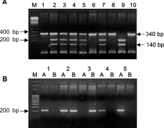
Msp I 효소(5 unit/μl) 0.5μl을 총 20μl에 맞춘 후에 37oC에서 1일간 반응시켰다. 반응 후 60∼70oC에서 5 분간 가열하여 제한효소를 불활성화시킨 후 2% aga- rose gel에서 전기 영동하여 제한효소에 의한 인식 부 위 유무에 따라 CYP1A1의 Msp I 다형성을 결정하였 다. 제한효소 처리 전에 PCR 산물의 염기 크기는 340 bp이며 두 개의 allele 모두에 제한효소의 인식 부위가 있으면 140 bp, 200 bp 크기의 band가 나타나며 (m2/m2 type), 한 개의 allele만 있으면 340 bp, 200 bp, 140 bp가 나타난다(m1/m2 type). 두 개의 allele 모두가 제한효소 의 인식 부위가 없을 때는 340 bp의 band만이 나타난 다(m1/m1 type)(Fig. 1A).
② CYP1A1의 exon 7 다형성에 대한 PCR 반응 및 결과분석; CYP1A1의 exon 7 다형성은 대립인자-특이 중합반응(allelic specific PCR) 방법을 이용하였다. 즉 forward primer의 3'-말단 부위에 다형성을 나타내는 염기 배열의 차이로 primer를 제작하고 backward prim- er를 공통으로 사용하여 두 쌍의 반응을 동시에 일으 켜 PCR 반응 유무에 따라 다형성을 결정하였다. PCR 반응 조건은 denaturation은 95oC에서 1분, annealing은 60oC에서 1분간 총 35 cycle 반응시켰다. 두 개의 PCR
반응 유무에 따라 AA (Ile/Ile), AG (Ile/Val), GG (Val/
Val) 유전자 유형으로 나누었다. 중합효소 반응 산물 의 염기 크기는 200 bp이며 A (Ile) primer를 이용한 중 합효소 반응에서만 band가 나타나면 Ile/Ile 유형이고, B (Val) primer를 이용한 중합효소 반응에서만 band가 나타나면 Val/Val 유형이며, A (Ile), B (Val) 모두에서 band가 나타나면 Ile/Val 유형이 된다(Fig. 1B).
(2) CYP2E1의 Pst I 다형성에 대한 PCR 반응 및 결 과분석: CYP2E1의 Pst I 다형성에 대한 PCR 반응은 denaturation은 95oC에서 1분, annealing은 55oC에서 1분, 그리고 extension은 72oC에서 1분을 35 cycle 시행한 후 마지막으로 72oC에서 10분간 연장반응하였다. PCR 반 응 확인 후 PCR 산물 15μl, 1x enzyme buffer, Pst I 효 소(5 unit/μl) 0.5μl을 총 20μl에 맞춘 후 37oC에서 1일 간 부양하였다. 부양 후 70oC에서 5분간 가열 후 2%
agarose gel에 전기 영동하여 제한효소에 의한 인식 부 위 유무에 따라 CYP2E1의 Pst I 다형성을 결정하였다.
제한효소 처리 전에 PCR 산물의 염기 크기는 410 bp이 며, 두 개의 allele 모두가 제한효소의 인식 부위가 있 으면, 290 bp, 120 bp 크기의 band가 나타나며(C2/C2 type), 한 개만 있으면 410 bp, 290 bp, 120 bp의 band가 나타나며(C1/C2 type), 두 개의 allele 모두가 제한효소 의 인식 부위가 없을 때에는 410 bp만 나타난다(C1/C1 type)(Fig. 2).
(3) GSTM1의 결손 부위 다형성에 대한 PCR 반응 및 결과분석: GSTM1의 결손 부위 다형성을 위한 PCR 반응은 denaturation이 95oC에서 1분, annealing이 55oC 에서 1분, 그리고 extension이 72oC에서 1분으로 35 cycle 시행한 후 마지막으로 72oC에서 10분 연장 반응 하였다. PCR 반응물을 2% agarose gel에 전기영동하여 PCR 유무를 확인한 후 273 bp band가 있으면 GSTM (+)로, band가 없으면 GSTM1 (-)로 분류하였다(Fig. 3).
Fig. 1. Picture shows a genotyping of the CYP1A1 gene polymorphism. A. Genotyping of the CYP1A1 gene in Msp I polymorphism. Lane 9 = homozygotes with (+) restriction site of Msp I (m2/m2); Lane 2, 3, 4, 5, 7 = heterozygote (m2/m1); Lane 1, 6, 8, 10 = homozygotes with (-) restriction site of Msp I (m1/m1) (M: molecular marker). B.
Genotyping of the CYP1A1 gene in Val-Ile polymorphism.
Lane 1, 4, 5 = predominant homozygotes for isoleucine (Ile/Ile); Lane 2 = heterozygotes of Ile/Val polymorphism (Ile/Val); Lane 3 = rare homozygotes for valine (Val/Val) (M: molecular marker).
200 bp
M A B A B A B A B A B1 2 3 4 5
B
400 bp
200 bp 340 bp
140 bp M 1 2 3 4 5 6 7 8 9 10
A
200 bp
M A B A B A B A B A B1 2 3 4 5
B
400 bp
200 bp 340 bp
140 bp M 1 2 3 4 5 6 7 8 9 10
A
Fig. 2. Picture shows a genotyping of the CYP2E1 gene.
Lane 1, 3, 4, 6 = homozygotes with (-) restriction site of Pst I (C1/C1); Lane 5, 7 = heterozygotes of Pst I poly- morphism (C1/C2); Lane 2, 8 = hemozygotes with (+) restriction site of Pst I (C2/C2) (M: molecular marker).
410 bp 290 bp
M 1 2 3 4 5 6 7 8
120 bp 410 bp
290 bp
M 1 2 3 4 5 6 7 8
120 bp 400 bp
200 bp
200 bp
(4) NAT2 다형성에 대한 PCR 반응 및 결과분석:
PCR 반응 조건은 94oC에서 5분간 denaturation, 그리고 마지막으로 extension 반응을 72oC에서 10분간 시켰다.
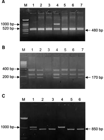
PCR 산물을 2% agarose gel에서 전기영동하여 1,000 bp의 산물을 확인한 후에 제한효소 결핍 부위에 따라 S1, S2, 그리고 S3의 slow acetylator 유전자 유형을 결 정하였다.
① S1 유형의 결정; S1 유형은 1,000 bp의 PCR 산물 10μl을 Kpn I 제한효소(Boehringer Mannheim, Man- nheim, Germany) 9 unit를 가지고 37oC에서 3시간 이상 반응시켰다. 반응 후 2% agarose gel에 전기영동하였 다. 완전히 1,000 bp의 PCR 산물이 분해된 경우는 즉, 520 bp과 480 bp만 보이는 경우 S1 allele가 없는 homo- zygote로 분류하였으며, 분해가 전혀 없이 1,000 bp만 보이는 경우를 S1 allele의 homozygote로 하였으며, 1,000 bp와 520 bp, 480 bp가 보이는 경우는 S1 allele의 he- terozygote로 하였다(Fig. 4A).
② S2 유형의 결정; S2 유형은 ①에서와 마찬가지로 PCR 산물을 Taq I 제한효소(Boehringer Mannheim, Mannheim, Germany)로 전기영동하였다. 400 bp의 band 와 400 bp의 분해 크기인 230 bp과 170 bp의 유무에 따라 S2의 유형을 결정하였다. 즉, 400 bp의 band만 있 으면 S2 homozygote로, 230 bp과 170 bp의 band만 있으 면 S2가 없는 homozygote로 분류하였다(Fig. 4B).
③ S3 유형의 결정; S3의 유형은 ①, ②에서 마찬가 지로 PCR 산물을 BamH I 제한효소(Boehringer Man- nheim, Mannheim, Germany) 11 unit를 가지고 37oC에서 3시간 이상 부양하였다. 전기영동상에서 1,000 bp만 보 이면 S3 allele의 homozygote로, 150 bp과 850 bp만 보이 면 S3 allele가 없는 homozygote로 분류하였다(Fig. 4C).
④ 유전자 유형과 표현형 유형 결정; 이상의 3가지 유형을 각 실험대상 표본에서 결정한 후 S1, S2, S3 유 형 중 두 allele 모두 있는 경우 slow acetylator 유전자
유형 S/S로, 두 allele 모두 없는 경우 fast acetylator 유 전자 유형 F/F로, 한 개만이 있는 경우 intermediate 유 전자 유형 S/F로 분류하였다. 또한 S/F와 F/F 유전자 유형을 fast acetylator 표현형 유형으로 분류하였고, S/S 유전자 유형은 slow acetylator 표현형 유형으로 분 류하였다.
3) 통계처리
각각 xenobiotics 대사 효소들의 다형성에 의한 유형 분포 차이에 대한 평가 및 상대 위험도 차이의 평가 를 위해 Chi-square test를 사용하였다. 각 유전자 유형 및 각 유전자 유형 간의 조합에 따른 상대적 위험도 측정을 위해 Odds ratio가 사용되었다. 모든 통계 처리 는 SPSS program (ver. 10.0, SPSS Inc, Chicago, IL, USA)를 이용하였다.
Fig. 3. Picture shows a genotyping of the GSTM1 gene.
Lane 2, 3, 8 = homozygous and/or heterzygous present alleles, GSTM (+); Lane 1, 4, 5, 6, 7 = homozygous null GSTM (-) (M: molecular marker).
M 1 2 3 4 5 6 7 8
273 bp
M 1 2 3 4 5 6 7 8
273 bp Fig. 4. Picture shows a genotyping of the NAT2 gene. A.
Determination of S1 allele with Kpn I digestion. S1X = Lane 4; XX = Lane 1, 2, 3, 5, 6, 7. B. Determination of S2 allele with Taq I digestion. S2S2 = Lane 3, 5; S2X = Lane 1, 6;
XX = Lane 2, 4, 7. C. Determination of S3 allele with BamH I digestion. S3S3 = Lane 4; S3X = Lane 1; XX = Lane 2, 3, 5, 6 (X: not determined, M: molecular marker).
480 bp 1000 bp
520 bp
M 1 2 3 4 5 6 7
M 1 2 3 4 5 6 7
M 1 2 3 4 5 6
A
400 bp
200 bp 170 bp
B
C
1000 bp 850 bp
480 bp 1000 bp
520 bp
M 1 2 3 4 5 6 7
M 1 2 3 4 5 6 7
M 1 2 3 4 5 6
A
400 bp
200 bp 170 bp
B
C
1000 bp 850 bp
결 과
1) 정상인에서 xenobiotics 대사 효소들의 다형성 유형 분포
113명의 대조군에서 phase I 대사 효소 중 CYP1A1 Msp I 다형성 유형 분포는 m1/m1, m1/m2, m2/m2 유형 이 각각 41.6%, 46.0% 그리고 12.4%였으며 exon 7 다 형성에 의해서는 Ile/Ile, Ile/Val, Val/Val 유형이 각각 59.3%, 36.3%, 그리고 4.4%였다. CYP2E1 Pst I 다형성 에 대한 유형은 C1/C1, C1/C2, C2/C2 유형이 각각
61.1%, 32.7% 그리고 6.2%였다(Table 2).
Phase II 대사 효소 중 GSTM1 결핍유무에 따른 다형 성 유형에서는 GSTM (+) 및 GSTM (-)가 각각 53.9%
및 46.1%였다. NAT2 효소 유형의 분포는 F/F, F/S, S/S 유형이 각각 43.4%, 48.7% 및 8.0%였다(Table 3).
2) 대장암 환자에서 xenobiotics 대사 효소들의 다 형성의 유형 분포 및 유전자 유형에 따른 상대 적 위험도
113명의 대장암 환자에서 phase I 대사 효소의 유형 분포는 CYP1A1의 Msp I 다형성 유형 분포는 m1/m1,
Table 3. Frequencies of phase II enzyme polymorphism in healthy control and colon cancer patients
ꠚꠚꠚꠚꠚꠚꠚꠚꠚꠚꠚꠚꠚꠚꠚꠚꠚꠚꠚꠚꠚꠚꠚꠚꠚꠚꠚꠚꠚꠚꠚꠚꠚꠚꠚꠚꠚꠚꠚꠚꠚꠚꠚꠚꠚꠚꠚꠚꠚꠚꠚꠚꠚꠚꠚꠚꠚꠚꠚꠚꠚꠚꠚꠚꠚꠚꠚꠚꠚꠚꠚꠚꠚꠚꠚꠚꠚꠚꠚꠚꠚꠚꠚꠚꠚꠚꠚꠚꠚꠚꠚꠚꠚꠚꠚꠚꠚꠚꠚꠚꠚꠚꠚꠚ
Genotypes (+/-) No. (%) Total (%) P value
ꠏꠏꠏꠏꠏꠏꠏꠏꠏꠏꠏꠏꠏꠏꠏꠏꠏꠏꠏꠏꠏꠏꠏꠏꠏꠏꠏꠏꠏꠏꠏꠏꠏꠏꠏꠏꠏꠏꠏꠏꠏꠏꠏꠏꠏꠏꠏꠏꠏꠏꠏꠏꠏꠏꠏꠏꠏꠏꠏꠏꠏꠏꠏꠏꠏꠏꠏꠏꠏꠏꠏꠏꠏꠏꠏꠏꠏꠏꠏꠏꠏꠏꠏꠏꠏꠏꠏꠏꠏꠏꠏꠏꠏꠏꠏꠏꠏꠏꠏꠏꠏꠏꠏꠏ
GSTM1 polymorphism GSTM1 (+) GSTM (-)
Healthy control 61 (53.9) 52 (46.1) 113 (100)
Cancer patients 50 (44.2) 63 (55.8) 113 (100) 0.183
OR 1 1.48
CI 0.88∼2.50
NAT2 polymorphism F/F F/S S/S
Healthy control 49 (43.4) 55 (48.7) 9 (8.0) 113 (100)
Cancer patients 53 (46.9) 47 (41.6) 13 (11.5) 113 (100) 0.470
OR 1 0.79 1.34
CI 0.46∼1.37 0.52∼3.40
ꠏꠏꠏꠏꠏꠏꠏꠏꠏꠏꠏꠏꠏꠏꠏꠏꠏꠏꠏꠏꠏꠏꠏꠏꠏꠏꠏꠏꠏꠏꠏꠏꠏꠏꠏꠏꠏꠏꠏꠏꠏꠏꠏꠏꠏꠏꠏꠏꠏꠏꠏꠏꠏꠏꠏꠏꠏꠏꠏꠏꠏꠏꠏꠏꠏꠏꠏꠏꠏꠏꠏꠏꠏꠏꠏꠏꠏꠏꠏꠏꠏꠏꠏꠏꠏꠏꠏꠏꠏꠏꠏꠏꠏꠏꠏꠏꠏꠏꠏꠏꠏꠏꠏꠏ OR = Odds ratio; CI = 95% confidence interval; F = fast acetylator; S = slow acetylator.
Table 2. Frequencies of phase I CYPs enzyme polymorphism in healthy control and colon cancer patients ꠚꠚꠚꠚꠚꠚꠚꠚꠚꠚꠚꠚꠚꠚꠚꠚꠚꠚꠚꠚꠚꠚꠚꠚꠚꠚꠚꠚꠚꠚꠚꠚꠚꠚꠚꠚꠚꠚꠚꠚꠚꠚꠚꠚꠚꠚꠚꠚꠚꠚꠚꠚꠚꠚꠚꠚꠚꠚꠚꠚꠚꠚꠚꠚꠚꠚꠚꠚꠚꠚꠚꠚꠚꠚꠚꠚꠚꠚꠚꠚꠚꠚꠚꠚꠚꠚꠚꠚꠚꠚꠚꠚꠚꠚꠚꠚꠚꠚꠚꠚꠚꠚꠚꠚ
Genotypes No. (%) Total (%) P value
ꠏꠏꠏꠏꠏꠏꠏꠏꠏꠏꠏꠏꠏꠏꠏꠏꠏꠏꠏꠏꠏꠏꠏꠏꠏꠏꠏꠏꠏꠏꠏꠏꠏꠏꠏꠏꠏꠏꠏꠏꠏꠏꠏꠏꠏꠏꠏꠏꠏꠏꠏꠏꠏꠏꠏꠏꠏꠏꠏꠏꠏꠏꠏꠏꠏꠏꠏꠏꠏꠏꠏꠏꠏꠏꠏꠏꠏꠏꠏꠏꠏꠏꠏꠏꠏꠏꠏꠏꠏꠏꠏꠏꠏꠏꠏꠏꠏꠏꠏꠏꠏꠏꠏꠏ
CYP1A1 polymorphism (Msp I) m1/m1 m1/m2 m2/m2
Healthy control 47 (41.6) 52 (46.0) 14 (12.4) 113 (100)
Cancer patients 36 (31.9) 60 (53.1) 17 (15.0) 113 (100) 0.314
OR 1 1.51 1.59
CI 0.85∼2.67 0.70∼3.64
CYP1A1 polymorphism (exon 7) Ile/Ile Ile/Val Val/Val
Healthy control 67 (59.3) 41 (36.3) 5 (4.4) 113 (100)
Cancer patients 61 (54.0) 44 (38.9) 8 (7.1) 113 (100) 0.583
OR 1 1.18 1.76
CI 0.68∼2.04 0.55∼5.66
CYP2E1 polymorphism C1/C1 C1/C2 C2/C2
Healthy control 69 (61.1) 37 (32.7) 7 (6.2) 113 (100)
Cancer patients 61 (54.0) 45 (39.8) 7 (6.2) 113 (100) 0.529
OR 1 1.38 1.13
CI 0.79∼2.40 0.38∼3.41
ꠏꠏꠏꠏꠏꠏꠏꠏꠏꠏꠏꠏꠏꠏꠏꠏꠏꠏꠏꠏꠏꠏꠏꠏꠏꠏꠏꠏꠏꠏꠏꠏꠏꠏꠏꠏꠏꠏꠏꠏꠏꠏꠏꠏꠏꠏꠏꠏꠏꠏꠏꠏꠏꠏꠏꠏꠏꠏꠏꠏꠏꠏꠏꠏꠏꠏꠏꠏꠏꠏꠏꠏꠏꠏꠏꠏꠏꠏꠏꠏꠏꠏꠏꠏꠏꠏꠏꠏꠏꠏꠏꠏꠏꠏꠏꠏꠏꠏꠏꠏꠏꠏꠏꠏ OR = Odds ratio; CI = 95% confidence interval.
m1/m2 및 m2/m2 유형이 각각 31.9%, 53.1% 및 15.0%
였으며, 정상인의 분포에 비교하였을 때 통계학적 유 의성은 없었으며, m1/m1에 비해 m1/m2 및 m2/m2 유 형이 각각 1.51 및 1.59배의 상대적 위험도를 보였다.
그리고 exon 7에 대한 다형성 유형의 분포는 대장암 환자에서 Ile/Ile, Ile/Val, Val/Val 유형이 각각 54.0%, 38.9% 및 7.1%였으며 정상 대조군과 비교하여 통계학 적 유의성은 없었으며, Ile/Ile 유형에 비해 1.18 및 1.76 배의 상대적 위험도를 보였다. CYP2E1 Pst I의 다형성 유형 분포는 C1/C1, C1/C2, C2/C2가 각각 54.0%, 39.8%
및 6.2%로 정상인과 비교하였을 때 유형에 따른 분포 의 차이는 통계학적 유의성은 없었으며, C1/C1 유형에 비해 C1/C2 및 C2/C2가 각각 1.38 및 1.13배의 상대적 위험도를 보였다(Table 2).
대장암 환자에서 phase II 대사 효소의 유형 분포의 경우는 GSTM1 효소 결핍의 유무에 따른 유형 분포는 GSTM (+) 및 GSTM (-)이 44.2% 및 55.8%였으며, 정 상인과 비교하였을 때 유형에 따른 분포의 차이는 통 계학적 유의성은 없었다. GSTM (+)에 대한 GSTM (-)의 상대적 위험도는 1.48배를 보였다. NAT2 효소 의 유형 분포는 F/F, F/S, S/S 유형의 분포가 각각 46.9%, 41.6% 및 11.5%를 보였으나 정상인의 분포와 비교했을 때 통계학적 유의성은 없었으며, F/F 유형에 대한 상대적 위험도는 F/S 및 S/S 유형이 각각 0.79 및
1.34배의 상대적 위험도를 보였다(Table 3).
3) Phase I 대사 효소(CYP1A1 및 CYP2E1) 및 GSTM1 유전자 유형 조합에 따른 상대적 위험 도 평가
Phase I 대사 효소(CYP1A1 및 CYP2E1)와 GSTM1 유전자 유형 조합에 따른 유형 조합 분포 및 상대적 위험도를 Table 4에 요약하였다.
유전자 유형 조합에 따른 상대적 위험도는 CYP1A1 Msp I 다형성 및 GSTM1 유전자 유형 조합에서는 m1/m1, GSTM (+) 유형을 기준으로 하였을 때 GSTM (-), m1/m2 및 GSTM (-), m2/m2 유형이 각각 2.15 및 1.94배의 상대적 위험도를 보였으며, exon 7 다형성 유형과의 조합에서는 Ile/Ile과 GSTM (+) 유형에 대해 서 Ile/Val과 GSTM (-)와 Ile/Ile과 GSTM (-) 유형이 각각 1.87 및 5.81배의 상대적 위험도를 보였다. CYP2E1 과 GSTM1 유전자 유형 조합에서는 C1/C1와 GSTM (+) 유형에 대해 C1/C2와 GSTM (-) 유형이 2.20배의 상대적 위험도를 보였다.
4) Phase I 대사 효소(CYP1A1 및 CYP2E1) 및 NAT2 유전자 유형 조합에 따른 상대적 위험도 Phase I 대사 효소(CYP1A1 및 CYP2E1)와 NAT2 유 전자 유형 조합에 따른 유형 조합 분포 및 상대적 위험
Table 4. Combined CYPs and GSTM1 genetic polymorphism in healthy control and colon cancer patients ꠚꠚꠚꠚꠚꠚꠚꠚꠚꠚꠚꠚꠚꠚꠚꠚꠚꠚꠚꠚꠚꠚꠚꠚꠚꠚꠚꠚꠚꠚꠚꠚꠚꠚꠚꠚꠚꠚꠚꠚꠚꠚꠚꠚꠚꠚꠚꠚꠚꠚꠚꠚꠚꠚꠚꠚꠚꠚꠚꠚꠚꠚꠚꠚꠚꠚꠚꠚꠚꠚꠚꠚꠚꠚꠚꠚꠚꠚꠚꠚꠚꠚꠚꠚꠚꠚꠚꠚꠚꠚꠚꠚꠚꠚꠚꠚꠚꠚꠚꠚꠚꠚꠚꠚ
Genotypes
GSTM1 CYPs ꠏꠏꠏꠏꠏꠏꠏꠏꠏꠏꠏꠏꠏꠏꠏꠏꠏꠏꠏꠏꠏꠏꠏꠏꠏꠏꠏꠏꠏꠏꠏꠏꠏꠏꠏꠏꠏꠏꠏꠏꠏꠏꠏꠏꠏꠏꠏꠏꠏꠏꠏꠏꠏꠏꠏꠏꠏꠏꠏꠏꠏꠏꠏꠏꠏꠏꠏꠏꠏꠏꠏꠏꠏ
+ - + - + -
ꠏꠏꠏꠏꠏꠏꠏꠏꠏꠏꠏꠏꠏꠏꠏꠏꠏꠏꠏꠏꠏꠏꠏꠏꠏꠏꠏꠏꠏꠏꠏꠏꠏꠏꠏꠏꠏꠏꠏꠏꠏꠏꠏꠏꠏꠏꠏꠏꠏꠏꠏꠏꠏꠏꠏꠏꠏꠏꠏꠏꠏꠏꠏꠏꠏꠏꠏꠏꠏꠏꠏꠏꠏꠏꠏꠏꠏꠏꠏꠏꠏꠏꠏꠏꠏꠏꠏꠏꠏꠏꠏꠏꠏꠏꠏꠏꠏꠏꠏꠏꠏꠏꠏꠏ
CYP1A1 polymorphism (Msp I) m1/m2 m1/m1 m2/m2
Healthy control 21 26 33 19 7 7
Cancer patients 17 19 27 33 6 11
OR 1 0.90 0.45 2.15 1.06 1.94
CI 0.38∼2.16 0.45∼2.29 0.91∼5.03 0.30∼3.75 0.62∼6.09
CYP1A1 polymorphism (exon 7) Ile/Val Ile/Ile Val/Val
Healthy control 30 37 27 14 4 1
Cancer patients 31 30 17 27 2 6
OR 1 0.78 0.61 1.87 0.48 5.81
CI 0.39∼1.57 0.28∼1.34 0.82∼4.23 0.08∼2.84 0.66∼51.15
CYP2E1 polymorphism C1/C2 C1/C1 C2/C2
Healthy control 36 33 20 17 5 2
Cancer patients 26 35 18 27 6 1
OR 1 1.47 1.25 2.20 1.67 0.69
CI 0.73∼2.94 0.55∼2.81 0.99∼4.84 0.46∼6.03 0.06∼8.05
ꠏꠏꠏꠏꠏꠏꠏꠏꠏꠏꠏꠏꠏꠏꠏꠏꠏꠏꠏꠏꠏꠏꠏꠏꠏꠏꠏꠏꠏꠏꠏꠏꠏꠏꠏꠏꠏꠏꠏꠏꠏꠏꠏꠏꠏꠏꠏꠏꠏꠏꠏꠏꠏꠏꠏꠏꠏꠏꠏꠏꠏꠏꠏꠏꠏꠏꠏꠏꠏꠏꠏꠏꠏꠏꠏꠏꠏꠏꠏꠏꠏꠏꠏꠏꠏꠏꠏꠏꠏꠏꠏꠏꠏꠏꠏꠏꠏꠏꠏꠏꠏꠏꠏꠏ OR = Odds ratio; CI = 95% confidence interval.
도를 Table 5에 요약하였다.
CYP1A1 Msp I 다형성과 NAT2 다형성에 의한 유전 자 유형 조합에서 m1/m1와 fast acetylator (F) 유형에 대해 m1/m2와 slow acetylator (S) 및 m2/m2와 slow acetylator (S) 유형 조합이 각각 3.5 및 3.1 배의 상대적 위험도를 보였으며 exon 7 다형성 유형과의 조합에서 는 Ile/Ile과 fast acetylator (F) 유형에 대해 Ile/Ile과 slow acetylator (S)가 2.0배의 상대적 위험도를 보였다. CYP2E1 Pst I 다형성과 NAT2 다형성에 의한 유전자 유형 조합 에서는 C1/C1와 fast acetylator (F)에 대해 C1/C2와 slow acetylator 조합이 4.50배의 상대적 위험도를 보였다.
고 찰
인체에 발생하는 대부분의 암은 외부 또는 내부로 부터 노출되는 발암인자와 노출된 발암인자에 대한 개개인의 유전적 인자 사이의 상호 작용에 의해 발생 한다.1,3 이론적으로 외부의 발암물질(흡연, 대기오염, 화학제 등)의 노출에 대한 개개인의 감수성이 차이가 있는 것으로 생각되어 왔다. 이러한 개개인의 감수성 차이는 xenobiotics 대사 효소의 활성도, DNA 수복계 의 활성도, 발암에 관여하는 종양 유전자 및 종양 억제 유전자의 변이율 등의 차이에서 비롯된다. 이런 요소
들 중 xenobiotics 대사 효소의 활성도는 유전적으로 결 정된다. 최근의 분자 생물학적 기법의 발달로 발암 물 질의 노출과 개인적 감수성에 대한 연구방법으로 xenobiotics의 대사효소(phase I 및 II)에 대한 상대적 위 험도를 결정하고 이로부터 얻은 지표를 암예방에 이 용하려는 연구가 활발해지고 있다.12,13
전통적으로 오랫동안 화학적 발암과정의 연구대상 물질은 polycyclic aromatic hydocarbons (PAH)와 aro- matic amines이었다. 이 물질들은 모두 흡연에 의해 생 성되는 것으로서 폐암, 유방암, 방광암 및 상부 소화기 및 호흡기암(구강암, 후두암 등)의 중요 원인으로 잘 알려져 있다. 인체에 노출된 이들 물질은 phase I 대사 효소(cytochrome P450 family: CYPs)에 의해 활성화되 고 phase II 대사효소(Glutathione-S-transferase: GST, N-acetyltransferase: NAT)에 의해 비활성화되므로 이들 두 과정의 효소의 활성도에 의해 발암 물질의 활성도 가 결정된다.8 CYP1A1 유전자는 aryl hydrocarbon hydroxylase의 활성도와 관계가 있는 cytochrome P450 family의 한 효소군으로, PAH에 의한 활성도 실험에 의해 다형성이 있음이 증명되었다.14-16 최근의 많은 연 구들에서 CYP1A1 유전자의 cloning이 가능하면서 PCR- RFLP 방법에 의한 다형성이 최소한 2군데, 즉 Msp I 제한효소에 의한 부분(3'-flanking 부위)과 exon 7 (Val/Ile) Table 5. Combined CYPs and NAT2 polymorphism in healthy control and colon cancer patients
ꠚꠚꠚꠚꠚꠚꠚꠚꠚꠚꠚꠚꠚꠚꠚꠚꠚꠚꠚꠚꠚꠚꠚꠚꠚꠚꠚꠚꠚꠚꠚꠚꠚꠚꠚꠚꠚꠚꠚꠚꠚꠚꠚꠚꠚꠚꠚꠚꠚꠚꠚꠚꠚꠚꠚꠚꠚꠚꠚꠚꠚꠚꠚꠚꠚꠚꠚꠚꠚꠚꠚꠚꠚꠚꠚꠚꠚꠚꠚꠚꠚꠚꠚꠚꠚꠚꠚꠚꠚꠚꠚꠚꠚꠚꠚꠚꠚꠚꠚꠚꠚꠚꠚꠚ Genotypes
NAT2 CYPs ꠏꠏꠏꠏꠏꠏꠏꠏꠏꠏꠏꠏꠏꠏꠏꠏꠏꠏꠏꠏꠏꠏꠏꠏꠏꠏꠏꠏꠏꠏꠏꠏꠏꠏꠏꠏꠏꠏꠏꠏꠏꠏꠏꠏꠏꠏꠏꠏꠏꠏꠏꠏꠏꠏꠏꠏꠏꠏꠏꠏꠏꠏꠏꠏꠏꠏꠏꠏꠏꠏꠏꠏꠏ
F S F S F S
ꠏꠏꠏꠏꠏꠏꠏꠏꠏꠏꠏꠏꠏꠏꠏꠏꠏꠏꠏꠏꠏꠏꠏꠏꠏꠏꠏꠏꠏꠏꠏꠏꠏꠏꠏꠏꠏꠏꠏꠏꠏꠏꠏꠏꠏꠏꠏꠏꠏꠏꠏꠏꠏꠏꠏꠏꠏꠏꠏꠏꠏꠏꠏꠏꠏꠏꠏꠏꠏꠏꠏꠏꠏꠏꠏꠏꠏꠏꠏꠏꠏꠏꠏꠏꠏꠏꠏꠏꠏꠏꠏꠏꠏꠏꠏꠏꠏꠏꠏꠏꠏꠏꠏꠏ
CYP1A1 polymorphism (Msp I) m1/m1 m1/m2 m2/m2
Healthy control 53 4 48 4 13 1
Cancer patients 34 2 51 9 15 2
OR 1 0.78 1.66 3.5 1.8 3.1
CI 0.14∼4.50 0.92∼2.97 1.00∼12.29 0.76∼4.24 0.27∼35.73
CYP1A1 polymorphism (exon 7) Ile/Ile Ile/Val Val/Val
Healthy control 63 4 36 5 5 0
Cancer patients 55 7 38 5 7 1
OR 1 2.0 1.21 1.15 1.60 0
CI 0.56∼7.21 0.68∼2.16 0.31∼4.17 0.48∼5.34
CYP2E1 polymorphism C1/C1 C1/C2 6C2/C24
Healthy control 64 5 35 2 5 2
Cancer patients 57 4 37 8 6 1
OR 1 0.9 1.19 4.50 1.35 0.56
CI 0.23∼3.51 0.66∼2.13 0.92∼22.02 0.39∼4.65 0.05∼6.36
ꠏꠏꠏꠏꠏꠏꠏꠏꠏꠏꠏꠏꠏꠏꠏꠏꠏꠏꠏꠏꠏꠏꠏꠏꠏꠏꠏꠏꠏꠏꠏꠏꠏꠏꠏꠏꠏꠏꠏꠏꠏꠏꠏꠏꠏꠏꠏꠏꠏꠏꠏꠏꠏꠏꠏꠏꠏꠏꠏꠏꠏꠏꠏꠏꠏꠏꠏꠏꠏꠏꠏꠏꠏꠏꠏꠏꠏꠏꠏꠏꠏꠏꠏꠏꠏꠏꠏꠏꠏꠏꠏꠏꠏꠏꠏꠏꠏꠏꠏꠏꠏꠏꠏꠏ OR = Odds ratio; CI = 95% confidence interval; F = fast acetylator; S = slow acetylator.
에 있음이 알려져 있으며 백인종 그리고 일본인을 포 함한 동양인 등에서 인종 간에 차이가 있음이 보고되 고 있다. 드문 유형인 m2/m2 유형은 서양인의 경우 0.3∼2.1% 차지하는 반면에 일본인에서 10.6%를 보이 고 있다. 본 연구에서는 한국인에서 12.4%로 일본인과 비슷한 분포를 보이고 있다. CYP1A1의 다형성에 의한 특정 유전자형(m2/m2 유형)이 담배와 연관된 암종, 특 히 폐암의 발생에서 개인적 유전적 감수성에 대한 연 관성은 조사대상인 인종에 따라 상반된 결과를 보이 고 있다. 즉 m2/m2 유형이 비교적 높은 빈도율을 보이 는 일본에서는 m2/m2 유형이 m1/m1 유형과 비교했을 때 4∼9배의 상대적 위험도를 보이며 특히 GSTM1 (-)와 조합했을 때는 m1/m1, GSTM (+) 보다 9∼16배 의 높은 위험도를 보인다고 보고하고 있는 반면, 유럽 인에서는 위험도가 아주 없거나 낮은 것으로 보고되 고 있다.4,6,7,17-20
반면에 대장암에서는 CYP1A1과 개인 적 유전적 감수성에 대한 연구는 일본인에서 보고되 어 있는데, 이들의 보고에 의하면 m2/m2 유형이 m1/
m1에 비해 약 7.9배의 상대적 위험도를 보였다.21 본 연구에서는 한국인에서 CYP1A1의 다형성에 의한 유 전적 감수성은 대장암 환자군에서 m1/m1 유형에 비해 m2/m2가 약 1.51배, m1/m2가 약 1.59배의 상대적 위험 도를 보이는 것으로 나타났다.
CYP1A1 exon 7에서 유전자 유형의 다형성은 Msp I 다형성에서와 마찬가지로 인종에 따라 차이가 있다.
유형 중에서 활성도가 제일 높은 유형인 Val/Val 유형 은 일본인에서 4.0%이며, 백인종에서 0∼1%의 빈도 분포를 보이고 있다.5,20,22,23 본 연구결과에 의하면, 정 상 한국인에서 Val/Val 유형 분포는 4.4%로 일본인의 분포 빈도와 비슷한 것으로 나타났다. CYP1A1의 Val/
Val 유형은 Msp I의 m2/m2 유형과 마찬가지로 흡연과 연관된 폐암, 후두암, 식도암 등에서 2∼3배의 상대적 위험도를 보이고 있으며, 이런 상대적 위험도의 결과 는 인종간에 차이가 없는 것으로 보고되고 있다. 대장 암에서의 Val/Val 유형에 대한 상대적 위험도는 일본 인에서 약 5.7배의 높은 것으로 보고하고 있어 본 연구 의 1.7배와 비교하면 매우 높은 상대적 위험도로 생각 된다.21
CYP2E1 대사 효소는 methanol에 의해 활성화가 유 도되는 cytochrome P450계의 효소로서, 100여종의 매 우 다양한 화학물질을 대사시킨다. 이들 중에는 발암 물질로 알려져 있는 benzene, dimethylnitrosamine, vinyl chloride 등이 있다. 지금까지 CYP2E1 다형성은 4 부위 에서 알려져 있다. 즉 5'-flank 부위의 Rsa I/Pst I 제한
부위(C1/C2), intron 5의 Psa I 제한 부위, intron 6의 Pra I 제한 효소 부위 및 intron 7의 Taq I 제한 효소 부위 등이다. 이들 중 Rsa I/Pst I 제한 효소 부위가 CYP2E1 효소 활성도와 밀접한 연관성이 있다. CYP2E1 다형성 과 발암에 대한 개인적 감수성 연구는 여러 암종에서 시행되었는데 통계학적으로 유의한 상대적 위험도를 보이고 있지 않다.24-26 대장암에서의 CYP2E1 다형성에 대한 연구는 지금까지 보고되고 있지 않으며, 본 연구 에서는 C1/C2 유형이 1.38배의 상대적 위험도를 보였 는데, 이런 결과는 이전의 여러 암종의 결과와 비슷하 였다.
Phase II 대사 효소인 GST는 외부 및 내부에서 노출 되어 활성화된 발암물질을 비활성화시키는 효소군 중 의 하나로서 Alpha, Mu, P1 그리고 Theta 등 4개로 분
류된다.27-30 GSTM1은 이들 중 Mu에 속하는 대사효소
로서 여러 인종에서 큰 차이 없이 약 45∼50%에서 결 손되는 것으로 보고되고 있다.31,32 본 연구결과에서는 정상 한국인의 약 45%에서 결손을 보여 이들의 보고 와 비슷한 결과라 생각된다. GSTM1의 결손 유형은 GST 효소의 활성도가 없으므로 PAHs의 활성 물질 등 비슷한 활성화된 발암 물질을 대사시킬 수 없다.33 이 전의 보고에서 GSTM (-) 유형은 폐암, 방광암, 후두 암 등과 같은 담배와 연관이 있는 암종에서 상대적 위 험도가 높은 것으로 보고되고 있다.19,23,34 그러나 대장 암에서 GSTM1 유전자 유형분석에 의한 개인적 감수 성을 결정하는 상대적 위험도 연구는 매우 적으며, 이 전의 백인종 연구에서 GSTM (-)은 1.3∼1.7배의 상대 적 위험도를 보이는 것으로 보고되고 있다.35-37 본 연 구에서 한국인 대장암 환자군에서는 GSTM (-) 유형 이 1.48의 상대적 위험도를 보였다. 이와 같은 결과로 GSTM (-) 유형은 인종에 관계없이 약 45∼50% 결손 을 보이며, GSTM (+)에 비해 인종에 관계없이 대장 암 환자군에서 1.5∼2배 사이의 상대적 위험도가 있는 것으로 생각된다.
역시 또 하나의 Phase II 대사 효소인 NAT는 여러 약물 및 화학제, 특히 발암제인 benzidine과 aromatic amine 계열 화학제의 acetylation 대사에 관여하는 효소 로써 활성도에 따라 slow 및 fast acetylator로 구별된다.
또한 최근에는 분자생물학적 기법의 발달과 더불어 PCR-RFLP 방법에 의하여 NAT2 효소의 염기 배열이 결정되었으며 유전자 유형의 다형성에 의해 S1, S2, S3 와 F1 유전자 유형으로 구분이 가능해졌다. 이러한 유 전자 유형 및 표현형 유형은 인종 및 지역 간에 차이가 있는 것으로 보고되고 있다. 백인에서는 slow acety-
lator 유형이 52%, 중국인과 일본인을 대상으로 한 동 양인에서는 14%을 보이며, 흑인에서는 35%로 백인과 동양인의 중간 빈도를 나타내고 있으나,38-40 본 연구에 서는 한국인에서 8%로 매우 낮은 빈도를 나타내었다.
대장암에서 NAT2 표현형 유형 및 유전자 유형 다형 성과 개인적 유전적 감수성에 대한 연구는 매우 상반 된 결과를 보이고 있다. 대부분 보고들은 백인에서 slow acetylator 표현형 유형이 2∼3배의 높은 상대적 위험도를 보이는 것으로 보고하고 있으나,41-43 Hubbard 등은 NAT2 다형성과 대장암의 발암 과정과의 연관성 은 유의성이 없으며, slow acetylator 유형이 fast acety- lator 유형보다 1.6배 높은 상대적 위험도를 보이는 것 으로 보고하고 있다. 본 연구에서는 한국인에서 slow acetylator 표현형 유형과 S/S 유전자 유형이 약 1.34배 의 상대적 위험도를 보였으며 이러한 차이점은 인종 간에 NAT2 다형성에 대한 유형의 분포차이와 식사 습 관 등을 포함한 환경적 요소의 차이로 설명할 수 있 다.44
외부 및 내부로부터 노출된 발암물질은 phase I 대사 효소(CYPs)와 phase II 대사효소(NAT, GST)의 활성도 에 의해 인체 내에서 활성도가 결정되며 이론적으로 이러한 두 대사 효소의 다형성에 의한 유형을 조합할 때 유전적 감수성 결정은 더욱 의의가 있을 것이며, 암 예방에 있어 상대적 위험도가 높은 집단을 예측할 수 있는 중요한 자료를 제공할 수 있을 것으로 생각한
다.2,45,46 백인종에서는 CYP1A1의 Msp I 효소의 다형성
에서 m2/m2 유형이 너무 낮은 빈도(0∼1%)를 보이기 때문에 본 연구에 비해 결과가 매우 다르며, 일본인을 대상으로 한 폐암 연구에서는 CYP1A1의 m2/m2가 m1/1에 비해 9배의 상대적 위험도를 보이며, CYP1A1 의 m2/m2와 GSTM (-) 조합형은 m1/m1 및 GSTM (+) 조합에 비해 16배 증가된다고 보고하고 있다.6,7,23 지금까지 대장암에서는 phase I 및 phase II 대사 효소 의 다형성 유전자 유형 조합의 상대적 위험도에 대한 연구는 보고되어 있지 않다. Table 4와 Table 5는 대장 암 환자에서 phase I 및 phase II 대사 효소의 다형성 유형 조합에 의한 유전적 감수성을 본 연구 결과이다.
주목할 결과로는 첫째, Val/Val와 GSTM (-) 유형 조 합이 Ile/Ile와 GSTM (+) 유형 조합에 비해 5.81배의 높은 상대적 위험도를 나타낸 것이다. 이러한 결과는 흡연과 연관된 암종, 폐암, 방광암 및 후두암의 5∼10 배의 상대적 위험도와 비슷한 결과이다. 두 번째로는 slow acetylator와 m1/m2 또는 m2/m2 유형 조합이 각각 3.5 및 3.1배의 상대적 위험도를, slow acetylator와
C1/C2가 4.50배의 높은 상대적 위험도를 보인 점이다.
유전자 유형 조합군이 각각 유전자 유형 연구에 비해 높은 상대적 위험도를 보이는 것은 이미 설명한 것처 럼, phase I 대사 효소의 활성화가 높은 유형과 phase II 대사 효소의 활성화가 낮은 유형의 조합으로 상대적 위험도가 상승하는 효과가 나타나는 것으로 설명할 수 있다. 특히, NAT2 다형성 중에서 slow acetylator 유 형과 조합할 때 유의하게 차이가 있는 것은 매우 특이 한 것으로, 이미 설명한 바와 NAT2 유형 분포가 인종 간, 특히 동서양 인종 간에 차이가 있고, slow acetylator 가 동양인에서, fast acetylator가 백인종에서 대장암의 발생빈도를 높인다는 가설을 지지해 주는 것으로 생 각한다.
결 론
본 연구에서는 phase I 대사의 CYP1A1과 CYP2E1 효소, 그리고 phase II 대사의 GSTM1 (glutathione-S- transferase M1)과 NAT2 (N-acetyltransferase 2) 효소의 다형성에 대해서 PCR-RFLP 방법을 이용하여 정상 한 국인 및 대장암 환자에서 이들 대사 효소의 다형성에 대한 유형 분포의 빈도를 알아보고, 각 유형 및 유형의 조합에서 상대적 위험도를 측정하여 xenobiotics 대사 효소의 다형성에 의한 한국인 대장암에서 감수성 유 전자의 빈도를 연구하고자 하였으며 다음과 같은 연 구 결과를 얻었다.
1) CYP1A1의 Msp I 다형성에서는 m1/m1 유형에 대 해 m1/m2, m2/m2 유형이 각각 1.51 및 1.59배의 상대 적 위험도를, exon 7 다형성에 대해서는 Ile/Ile 유형에 대해 Ile/Val, Val/Val이 각각 1.18 및 1.76 배의 상대적 위험도를 보였다. CYP2E1의 Pst I 다형성에서는 C1/C1 유형에 대해 C1/C2, C2/C2유형이 각각 1.38 및 1.13배 의 상대적 위험도를 보였다.
2) GSTM1 다형성에서는 GSTM (+) 유형에 대해 GSTM (-) 유형이 1.48배를, NAT2 다형성에서는 F/F 유형에 대해 F/S, S/S 유형이 각각 0.79 및 1.34배의 상 대적 위험도를 보였다.
3) Phase I 대사 효소들과 GSTM1 효소의 유형간의 조합에서는 m1/m2와 GSTM (-), Val/Val과 GSTM (-), C1/C2와 GSTM (-)의 유형 조합이 각각 기준유 형 조합에 대해 2.15, 5.81 및 2.20 배의 상대적 위험도 를 보였다.
4) Phase I 대사 효소들과 NAT2 효소의 유형 간의 조합에서는 m1/m2와 slow acetylator, C1/C2와 slow
acetylator 유형 조합이 각각 기준 유형 조합에 대해 3.5 및 4.5배의 상대적 위험도를 보였다.
이상의 결과들을 종합해보면 Val/Val과 GSTM (-), m1/m2와 NAT2의 slow acetylator, C1/C2와 NAT2의 slow acetylator 등의 유형 조합군이 한국인에서 대장암 발생에 유전적으로 감수성이 높은 집단이라고 생각 한다.
REFERENCES
1. Hulka BS. Epidemiological studies using bilogical mark- ers: Issues for epidemiologists. Cancer Epidemiol Bio- markers Prev 1991;1:13-9.
2. Perera FP. Enviroment and Cancer: Who are susceptible?
Science 1997;278:1068-73.
3. Perera FP, Mooney LA. The role of molecular epidemi- ology in cancer prevention. In DeVita VT, Hellman S, Rosenberg SA (Eds.): “Cancer Prevention.” Philadelphia:
J.B. Lippincott 1993;1-15.
4. Alexandrie AK, Sundberg MI, Seidegard J, Tornling G, Rannug A. Genetic susceptibility to lung cancer with special emphasis on CYP1A1 and GSTM1: a study on host factors in relation to age at onset, gender and histological cancer types. Carcinogenesis 1994;15:1785- 90.
5. Hayashi S-I, Watanabe J, Nakachi K, Kawajiri K. Gen- etic linkage of lung cancer-associated MspI polymor- phisms with amino acid replacement in the heme binding region of the human cytochrome P450IA1 gene. J Bio- chem 1991;110:407-11.
6. Nakachi K, Imai K, Hayashi S-I, Watanabe J, Kawajiri K. Genetic susceptibility to squamous cell carcinoma of the lung in relation to cigarette smoking dose. Cancer Res 1991;51:5177-80.
7. Nakachi K, Imai K, Hayashi S, Kawajiri K. Polymor- phisms of the CYP1A1 and glutathione S-transferase genes associated with susceptibility to lung cancer in relation to cigarette dose in a Japanese population. Can- cer Res 1993;53:2994-9.
8. Nebert DW. Role of genetics and drug metabolism in human cancer risk. Mut Res 1991;247:267-81.
9. Board PJ. Biochemical genetics of glutathione S-trans- ferase in man. Am J Hum Genet 1981;33:36-43.
10. Kellermann G, Shaw CR, Luyten-Kellermann M. Aryl hydrocarbon hydroxylase inducibility and bronchogenic carcinoma. New Engl J Med 1973;298:934-7.
11. Kuori RE, McKinney CE, Slomiany DJ, Snodgrass DR, Wary NP, McLemore TL. Positive correlation between high aryl hydrocarbon hydroxylase activity and primary lung cancer as analysed in cryopreserved lymphocytes.
Cancer Res 1982;42:5030-7.
12. Perera FP. Molecular cancer epidemiology: A new tool in cancer prevention. J Natl Cancer Inst 1987;78:887-98.
13. Schulte PA, Perera FP (Eds.). Molecular epidemiology:
Principles and practices. New York: Academic Press 1993;277.
14. Gonzalez FJ. The molecular biology of the cytochrome P450s. Pharmacol Rev 1989;40:243-88.
15. Nebert DW, Gielen JE. Genetic regulation of aryl hydro- carbon hydroxylase inducation in the mouse. Fed Proc 1972;31:1315-25.
16. Thomas PE, Kouri RE, Hutton JJ. The genetics of aryl hydrocarbon hydroxylase induction in mice: a single gene difference between C57B1/6J and DBA/2J. Bio- chem Genet 1972;6:157-68.
17. Hayashi S-I, Watanabe J, Kawajiri K. High susceptibility to lung cancer analyzed in terms of combined genotypes of P450IA1 and Mu-class glutathione-transferase genes.
Jpn J Cancer Res 1992;83:866-70.
18. Hirvonen A, Husgafvel-Pursiainen K, Karjalainen A, Anttila S, Vainio H. Point-mutational Msp I and Ile-Val Polymorphisms closely linked in the CYP1A1 gene: lack of association with susceptibility to lung cancer in a Finnish study population. Cancer Epidemiol Biomarkers Prev 1992;1(6):485-9.
19. Idle JR. Is environmental carcinogenesis is modulated by host polymorphism? Mutation Res 1991;247:259-66.
20. Tefre T, Ryberg, Haugen A. Human CYP1A1 (cyto- chrome P1450) gene: lack of association between the Msp I restriction fragment length polymorphism and incidence of lung cancer in a Norwegian population. Phar- macogenetics 1991;1:20-5.
21. Sivaraman L, Leatham MP, Yee J, Wilkens LR, Lau AF, Marchand LL. CYP1A1 genetic polymorphisms and in situ colorectal cancer. Cancer Res 1994;54:3692-5.
22. Kawajiri K, Nakachi K, Imai K, Yoshii A, Shinoda N, Watanabe J. Identification of genetically high risk indi- viduals to lung cancer by DNA polymorphisms of the cytochrome P450IA1 gene. FEBS Letters 1990;263:131-3.
23. Raunio H, Husgafvel-Pursiainen K, Snttila S, Hietanen E, Hirvonen A, Pelkonen O. Diagnosis of polymorphisms in carcinogen-activating and inactivating enzymes and cancer susceptibility-a review. Gene 1995;159:113-21.
24. Farker K, Lehmann MH, Kastner R. CYP2E1 genotyping in renal cell/urothelial cancer patients in comparison with control populations. Int J Clin Pharmacol Ther 1998;36:
463-8.
25. Gonzalez MV, Alvarez V, Pello MF, Menéndez MJ, Suárez C, Coto E. Genetic polymorphism of N-acetyl- transferase-2, glutathione S-transferase-M1, and cyto- chromes P450IIE1 and P450IID6 in the susceptibility to head and neck cancer. J Clin Pathol 1998;51:294-8.
26. Morita S, Yano M, Shiozaki H. CYP1A1, CYP2E1 and GSTM1 polymorphisms are not associated with suscep- tibility to squamous-cell carcinoma of the esophagus. Int J Cancer 1997;71:192-7.
27. Ketterer B. Protective role of glutathione and glutathione transferases in mutagenesis and carcinogenesis, Mutat Res 1988;202:343-61.
28. Mannervik B, Alin P, Guthenberg C. Identification of three classes of cytosolic glutathione transferase com- mon to several mammalian species: correlation between structural data and enzymatic properties. Proc Natl Acad Sci USA 1985;82:7202-6.
29. Mannervik B, Awasthi, YB, Board PG, Hayes JD, Ilio C, Ketterer B, Listowsky I, Morgenstern R, Muramatsu M, Wolf CR. Nomenclature for human glutathione trans- ferase, Biochem J 1992;282:305-6.
30. Meyer DJ, Coles B, Pemble SE, Gilmore KS, Fraser GM, Ketterer B. Theta, a new class of glutathione transferases purified from rat and man, Biochem J 1991;247:409-14.
31. Board P, Coggan M, Johnston P, Ross V, Suzuki T.
Genetic heterogeneity of the human glutathione trans- ferases: a complex of gene families. Pharmacol Ther 1990;48:357-69.
32. Lin HJ, Han CY, Bernstein DA, Hsiao W, Lin BK, Hardy S. Ethnic distribution of the glutathione trans- ferase Mu 1-1 (GSTM1) null genotype in 1473 individ- uals and application to bladder cancer susceptibility.
Carcinogenesis 1994;15:1077-81.
33. Mannervik B, Danielson UH. Glutathione transferases structure and catalytic acitivity. CRC Crit Rev Biochem 1988;23:283-337.
34. Seidegård J, Pero RW, Markowitz MM, roush G, Miller DG, Beattie EJ. Isoenzyme (s) of glutathione transferase (class Mu) as a marker for the susceptibility to lung cancer: a follow up study. Carcinogenesis 1990;11:33-6.
35. Zhong S, Wyllie AH, Barnes D, Wolf CR, Spurr NK.
Relationship between the GSTM1 genetic polymorphism and susceptibility to bladder, breast and colon cancer.
Carcinogenesis 1993;14:1821-4.
36. Chenevix-Trench G, Young J, Coggan M, Board P. Glu- tathione S-transferase M1 and T1 polymorphisms: suscep-
tibility to colon cancer and age of onset. Carcinogenesis 1995;16:1655-7.
37. Deakin M, Elder J, Hendrickse C, Peckham D. Glutath- ione S-transferase GSTT1 genotypes and susceptibility to cancer: studies of interactions with GSTM1 in lung, oral, gastric and colorectal cancers. Carcinogenesis 1996;17:
881-4.
38. Deguchi T, Mashimo M, Suzuki T. Correlation between acetylator phenotypes and genotypes of polymorphic arylamine N-acetyltransferase in human liver. J Biol Chem 1990;265:12757-60.
39. Hickman D, Sim E. N-acetyltransferase polymorphism.
comparison of phenotype and genotype in humans.
Biochem Pharmacol 1991;42:1007-14.
40. Yu MC, Skipper PL, Taghizadeh K. Acetylator pheno- type, aminobiphenyl-hemoglobin adduct levels, and blad- der cancer risk in white, black, and Asian men in Los Angeles, California. J Natl Cancer Inst 1994;86:712-6.
41. Illett KF, David BM, Detchon P. Acetylation phenotype in colorectal carcinoma. Cancer Res 1987;47:1466.
42. Lang NP, Chu DZJ, Hunter CF. Role of aromatic amine acetyltransferase in human colorectal cancer. Arch Surg 1986;121:1259-61.
43. Wohlleb JC, Hunter CF, Blass B. Aromatic amine acetyl- transferase as a marker for colorectal cancer: environ- mental and demographic associations. Int J Cancer 1990;
46:22.
44. Hubbard AL, Harrison DJ, Moyes C. N-acetyltransferase 2 genotype in colorectal cancer and selective gene re- tention in cancers with chromosome 8p deletions. Gut 1997;41:229.
45. Kawajiri K, Watanabe J, Eguchi H, Hayashi S. Genetic polymorphisms of drug-metabolizing enzymes and lung cancer susceptibility. Pharmacogenetics 1995;5:S70-S73.
46. Mooney LA, Perera FP. Application of molecular epide- miology to lung cancer chemoprevention. J Cellular Bio- chem 1996;25S:63-8.
47. Comstock KE, Sanderson BJS, Claflin G, Henner WD.
GST1 gene deletion determined by polymerase chain reaction. Nucleic Acids Res 1990;18:3670.