• 검색 결과가 없습니다.

Imatinib-Mesylate Induced Interstitial Pneumonitis in Two CML Patients

N/A
N/A
Protected

Academic year: 2021

Share "Imatinib-Mesylate Induced Interstitial Pneumonitis in Two CML Patients"

Copied!
6
0
0

로드 중.... (전체 텍스트 보기)

전체 글

(1)

Imatinib-Mesylate Induced Interstitial Pneumonitis in Two CML Patients

Tae Hoon Kim, M.D.

1

, Byung Gyu Kim, M.D.

1

, Sung Woo Cho, M.D.

1

, Sung Kyun Cho, M.D.

1

, Hyun-Jung Kim, M.D.

2

, Young Jin Yuh, M.D.

1

, Sung Rok Kim, M.D.

1

Departments of

1

Internal Medicine and

2

Pathology, Sanggye-Paik Hospital, Inje University College of Medicine, Seoul, Korea Imatinib mesylate, a selective inhibitor of BCR-ABL kinase activity, has demonstrated significant clinical efficacy in the treatment of chronic myeloid leukemia (CML) and gastrointestinal stromal tumors (GISTs). It has become the standard of treatment for these diseases. Although the toxicity profile of imatinib is superior to that of interferon or other cytotoxic agents, some adverse events including edema, gastrointestinal toxicities and hematologic toxicities are commonly observed in the patients treated by imatinib. We present two cases of imatinib induced interstitial pneumonitis during the treatment of a chronic phase of CML.

Key Words: imatinib mesylate; Lung Diseases, Interstitial; Chronic Myeloid Leukemia

Address for correspondence: Young Jin Yuh, M.D.

Division of Hemato-oncology, Department of Internal Medicine, Sanggye-Paik Hospital, 761-1, Sanggye-dong, Nowon-gu, Seoul 139-707, Korea

Phone: 82-2-950-1460, Fax: 82-2-950-1955 E-mail: [email protected]

Received: Jun. 9, 2010 Accepted: Jul. 27, 2010

Introduction

Chronic myeloid leukemia (CML) is a unique disease with a t(9;22) reciprocal chromosomal dislocation, which results in a aberrant gene formation of BCR-ABL

1

. Though most patients with CML are diagnosed in chron- ic phase with little symptoms, the natural course of this disease is fatal with progression to blast crisis

2

. Before the era of targeting agent of CML, allogenic stem cell transplantation (SCT) was the only way to in- duce durable remissions. However allogenic SCT has a substantial mortality rate and a significant proportion of patients without matched donor were not able to be treated with this treatment option. The development of agents targeting BCR-ABL such as imatinib mesylate (Gleevec

or Glivec

; Norvatis, Basel, Switzerland) im- proved the prognosis dramatically

3

. The International randomized study of interferon and STI571 trial (IRIS tri-

al) demonstrated that long-term estimated rates of free- dom from progression to accelerated or blast phase were more than 90% with imatinib therapy

4

. Standard dose of imatinib therapy is now considered to the stand- ard treatment for the patient with newly diagnosed CML.

Though imatinib is generally well tolerated with rare grade 3 or 4 non-hematologic toxicities, adverse events including myalgia and fluid retention were reported

5

. Some patients complain of dyspnea during their treat- ment of imatinib. Most of the cases are related to pul- monary edema due to fluid retention. However, the cas- es of imatinib-induced interstitial pneumonitis were re- ported sporadically as a rare adverse event

6-9

. In Korea, only one case of interstitial pneumonitis was reported during imatinib therapy for GIST

10

.

We report here 2 cases of imatinib-induced interstitial pneumonitis with one irreversible case.

Case Report 1. Case 1

A 68-year-old man was diagnosed to a chronic phase

of Philadelphia chromosome-positive CML in September

2008. He was treated with imatinib mesylate at a dose

(2)

Figure 1. (A) Chest PA X-ray shows diffuse sub- pleural haziness in both lung fields. (B) Chest CT shows bilateral diffuse sub- pleural nodules, interlobul- ar septal thickening, retic- ulation and peribronchial ground glass opacities in both lungs. CT: computed tomography.

Figure 2. (A) The needle biopsied lung tissue shows pneumonic consolidation. The solid portion shows pale-pinkish

fibroblastic proliferation (arrow) and scanty inflammatory infiltrate (H&E stain, ×40). (B) The alveolar septum is uniformly

fibrous-thickened and ball like smooth muscle hyperplasia (arrow) (H&E stain, ×100). (C) The alveoli are partly desqua-

mated and replaced by type II pneumocytes (arrow). The alveolar spaces are filled with a few lymphocytes and eosinophils

(H&E stain, ×200). (D) Masson Trichrome staining contrasts a collagenous fibrosis from surrounding fibrinous component

(Masson Trichrome stain, ×100).

(3)

Figure 3. (A) Chest X-ray shows patchy nodular ground glass opacity in- filtrations in both lower lung fields. (B) Chest CT shows bilateral diffuse poor- ly defined ground glass opacity infiltrations, along axial interstitial and inter- lobular septum, with lower lung zone dominance. CT:

computed tomography.

(CCyR) was achieved after 3 months of therapy. At that time, he presented with cough, sputum, and dyspnea.

Chest X-ray showed bilateral diffuse haziness in both lower lung fields (Figure 1A).

After 4 months of therapy, he was admitted due to aggravation of dyspnea. His blood pressure, heart rate, body temperature and respiratory rates were 140/90 mm Hg, 80/min, 37.4

o

C and 20/min, respectively. On phys- ical exam, there was no remarkable finding except crackles in both lower lung fields. Pulmonary function test revealed pulmonary restrictive pattern with forced vital capacity (FVC) of 2.16 L (56% predicted). High res- olution computed tomography of chest showed bilateral diffuse subpleural nodules, interlobular septal thicken- ing, reticulation and peribronchial ground glass opac- ities in both lungs, suggesting diffuse interstitial lung disease (Figure 1B). Ultrasound-guided percutaneous needle biopsy obtained in left lower lung field showed septal edema with fibrosis and infiltration of mixed in- flammatory cells (Figure 2). Though his poor general condition didn't allow us perform surgical biopsy for further evaluation, the above percutaneous needle biop- sy findings were compatible with drug-induce interstitial pneumonitis. Moreover, at that time, imatinib was the only medicine he was taking.

With the above data, he was diagnosed to an im- atinib-induced pulmonary interstitial pneumonitis. Im-

dnisone (1 mg/kg/day) was started. After 20 days of ste- roid therapy, his dyspnea was progressed and mechan- ical ventilator support was applied. Even with mechan- ical ventilator support, his respiratory function was worsened and finally he was expired 30 days after admission.

2. Case 2

A previously healthy 71-year-old man was diagnosed to a chronic phase of Philadelphia chromosome-positive CML on February 2009. Imatinib mesylate was initiated at a dose of 400 mg daily. A complete cytogenetic re- sponse (CCyR) was achieved after 3 months of therapy.

At that time, he presented with dyspnea on exercise.

His blood pressure, heart rate, body temperature and respiratory rates were 120/80 mm Hg, 82/min, 36

o

C and 20/min, respectively. His physical examination revealed crackles in both lower lung fields.

Pulmonary function test revealed pulmonary restric-

tive pattern with forced vital capacity (FVC) of 2.29 L

(63% predicted). Chest X-ray showed patchy nodular

ground glass opacity infiltration in both lower lung

fields (Figure 3A). Computed tomography of chest

showed diffuse poorly defined ground glass opacity in-

filtrations, along axial interstitial and interlobular sep-

tum, with lower lung zone dominance, compatible with

interstitial pneumonitis (Figure 3B). Bronchoalveolar

(4)

Figure 4. (A) The wedge-resected lung shows a temporally uniform pneumonic consolidation with pale pinkish young fibroblastic proliferation (arrows) with many mixed inflammatory infiltrate. Some alveoli are destroied with honey combing (H&E stain, ×40). (B) Patchy mononuclear infiltrate is identified, predominantly lymphocytes (H&E stain, ×100). (C) Type II pneumocyte hyperplasia and multinucleated giant cells (circle) are identified (H&E stain, ×200). (D) The mas- son-Trichrome staining contrasts collagenous fibrosis (blue) background of fibrinous exudate (dark-red) (Masson Trichrome stain, ×100).

lavage (BAL) was performed in right middle lobe bron- chus, BAL cell count and differential was RBC 150/mm

3

, WBC 170/mm

3

(macrophages 28%, lymphocytes 77%, eosinophils 6%). This pattern can be showed at drug induced interstitial pneumonitis. A wedge resection bi- opsy by Video-Assisted Thoracic Surgery (VATs) per- formed on left lower lobe showed pneumonic con- solidation with inflammatory cell infiltration and collag- enous fibrosis (Figure 4).

Imatinib was discontinued and steroid therapy with prednisone 1 mg/kg/day was started under the diag- nosis of imatinib-induced interstitial pneumonitis. Two

weeks after steroid therapy, his dyspnea and cough was improved. Prednisone was tapered down over 2 weeks.

A follow-up chest computed tomography (CT) scan 2 months later showed improvement in the appearance of the previously described pulmonary findings. How- ever, restrictive pattern of pulmonary function test still remained (FVC 2.17 L, 60% predicted; FEV

1

2.11 L, 86%

predicted; and FEV

1

/FVC 97%).

He was treated with Nilotinib 800 mg daily instead

of Imatinib mesylate.

(5)

Interstitial pneumonitis is a rare adverse event of im- atinib therapy. The diagnosis of imatinib-induced inter- stitial pneumonitis is usually made by the compatible history and clinical findings

6-10

. The patients with this disease complained of various respiratory symptoms in- cluding dyspnea, cough and hypoxia. The findings of chest radiographs and CT are also quite variable includ- ing hypersensitivity pattern, interstitial pneumonitis pat- tern, cryptogenic organizing pneumonia pattern, nod- ular pattern and peribronchovascular bundle pattern

7

. Histopathologic diagnosis by trans-bronchial lung biop- sy or video-assisted thoracoscopic surgery (VATS) was performed only in a small proportion of patients. The histopathologic findings are usually fibrosis and in- flammatory cell infiltration

7

.

Most patients with interstitial pneumonitis are recov- ered after withdrawal of imatinib with or without steroid therapy. However, a number of patients with this ad- verse event did not improved even after withdrawal of imatinib

7,9

. We experienced one case of irreversible in- terstitial pneumonitis by imatinib therapy (case 1). The risk factors as well as the prognostic factors of interstitial pneumonitis after imatinib therapy are unknown. Some authors suggest that long-term use of imatinib can make interstitial pneumonitis irreversible

9

. In our case 1, im- atinib was prescribed for about 1 month after his respi- ratory symptom was appeared. It is possible that the failure from early withdrawal of imatinib made the out- come of this patient grave, whereas other patient with early withdrawal of imatinib was fully recovered.

Though it is impossible to make a conclusion with above two cases, early diagnosis and early withdrawal of imatinib may be a key element in imatinib-induced interstitial pneumonitis.

Though the incidence of imatinib-induced interstitial pneumonitis is known to be less than 2% of patients, a considerable number of this adverse events were re- ported in Japan

7

. Many cases of interstitial pneumonitis were reported too in Japan by gefitinib, a tyrosine kin- ase inhibitor for treatment of non-small cell lung can-

may be different by ethnics, and Asian population may be more vulnerable.

In both of our cases, interstitial pneumonitis was de- veloped after 3 months of imatinib therapy. The median duration until development of interstitial pneumonitis was 49 days in a report of 27 cases

7

. Though the dura- tion of imatinib therapy before development of inter- stitial pneumonitis is quite variable, a careful history fo- cusing to the respiratory symptoms and chest X-ray should be recommended after 3 months of imatinib therapy. A prompt withdrawal of imatinib should be considered in the patients with the respiratory symp- toms and/or abnormal chest X-ray findings.

References

1. Deininger MW, Goldman JM, Melo JV. The molecular biology of chronic myeloid leukemia. Blood 2000;96:

3343-56.

2. Melo JV, Barnes DJ. Chronic myeloid leukaemia as a model of disease evolution in human cancer. Nat Rev Cancer 2007;7:441-53.

3. Pavlovsky C, Kantarjian H, Cortes JE. First-line therapy for chronic myeloid leukemia: Past, present, and fu- ture. Am J Hematol 2009;84:287-93.

4. Guilhot F, Druker B, Larson RA, Gathmann I, So C, Waltzman R, et al. High rates of durable response are achieved with imatinib after treatment with interferon alpha plus cytarabine: results from the International Randomized Study of Interferon and STI571 (IRIS) trial.

Haematologica 2009;94:1669-75.

5. Druker BJ, Talpaz M, Resta DJ, Peng B, Buchdunger E, Ford JM, et al. Efficacy and safety of a specific in- hibitor of the BCR-ABL tyrosine kinase in chronic mye- loid leukemia. N Engl J Med 2001;344:1031-7.

6. Ma CX, Hobday TJ, Jett JR. Imatinib mesylate-induced interstitial pneumonitis. Mayo Clin Proc 2003;78:1578-9.

7. Ohnishi K, Sakai F, Kudoh S, Ohno R. Twenty-seven cases of drug-induced interstitial lung disease associat- ed with imatinib mesylate. Leukemia 2006;20:1162-4.

8. Rosado MF, Donna E, Ahn YS. Challenging problems in advanced malignancy: case 3. Imatinib mesylate-in- duced interstitial pneumonitis. J Clin Oncol 2003;21:

3171-3.

9. Seki N, Ito A, Watanabe K, Shibakuki R, Seto T,

Uematsu K, et al. Irreversible imatinib-induced pneu-

(6)

monitis following long-term imatinib administration.

Intern Med 2007;46:1941-2.

10. Lee JW, Kim HJ, Kim KJ, Shin KC, Hong YH, Chung JH, et al. A case of imatinib-mesylate associated hyper- sensitivity pneumonitis. Tuberc Respir Dis 2005;59:

423-6.

11. Ando M, Okamoto I, Yamamoto N, Takeda K, Tamura

K, Seto T, et al. Predictive factors for interstitial lung

disease, antitumor response, and survival in non-small-

cell lung cancer patients treated with gefitinib. J Clin

Oncol 2006;24:2549-56.

수치

Figure  1.  (A)  Chest  PA  X-ray  shows  diffuse   sub-pleural  haziness  in  both  lung  fields
Figure  3.  (A)  Chest  X-ray  shows  patchy  nodular  ground  glass  opacity   in-filtrations  in  both  lower  lung  fields

참조

관련 문서

A chest computed tomography (CT) of the patient did not show any shadow suspected of malignancy, but adenocarcinoma was found on a transbronchial lung biopsy and on a

We report a case of interstitial pneumonitis in a patient with disseminated extranodal marginal zone B cell lymphoma after the 5th cycle of rituximab and CHOP..

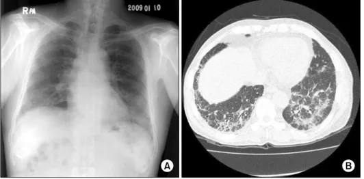
(B) A chest computed tomog- raphy obtained on the day of admission demonstrates multifocal diffuse, patchy ground glass opacity and interlobular septal thickening in both

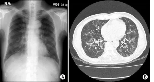
A chest computed tomography scan with a lung window set- ting shows multiple ill-defined ground glass opacities and multiple nodules with ground glass opacity (arrowheads) in the