Pneumatosis cystoides intestinalis (PCI) was first de- scribed in 1730 as an uncommon condition that is char- acterized by multiple air-containing cysts in the submu- cosa or subserosa of the gastrointestinal wall (1). This condition is classified into primary (idiopathic) and sec- ondary types. The latter arises in the presence of under- lying causes, most commonly gastrointestinal disorders (2). In rare cases, collagen vascular disease such as pro- gressive systemic sclerosis, systemic lupus erythemato- sus (SLE) and dermatomyositis can cause PCI (3-6). In Korea, there has been only one report of PCI associated with collagen vascular disease, in which the patient was diagnosed with progressive systemic sclerosis (6).
Unlike PCI associated with gastrointestinal disorders or neoplasms, PCI occurring in association with collagen vascular disease is treated conservatively, even in the presence of a pneumoperitoneum (4-6). Computed to-
mography (CT) is useful for diagnosing PCI and can ex- clude predisposing intraabdominal conditions, which can assist in the treatment plan (7).
We report two cases of PCI involving the colon in two patients, one with antinuclear antibody (ANA) negative SLE and the other with dermatomyositis.
Case Report
Case 1
An 18-year-old girl was admitted complaining of an abdominal pain, fever, nausea and vomiting. She had a 6-year history of progressive arthritis. SLE had been di- agnosed 5 years ago and she was being treated with steroid medication. The serologic test was negative for the lupus preparation and antinuclear antibodies. The plain abdominal radiographs showed a honeycomb ap- pearance along the right colon due to extensive gases within the colonic wall (Fig. 1A). A CT scan with a lung window setting (window width: 2,000 HU, window lev- el: -500 HU) revealed the mottled appearance of gas collections in the walls of the ascending and transverse colons (Fig. 1B). After 7 days without treatment, the in- tramural gases had disappeared spontaneously. There
J Korean Radiol Soc 2007;56:361-364
─ 361 ─
Pneumatosis Cystoides Intestinalis in Patients with Antinuclear Antibody Negative Systemic Lupus Erythematosus and Dermatomyositis: Report of Two Cases1
Soo-Yeon Kim, M.D., On Koo Cho, M.D., Byunghee Koh, M.D., Yongsoo Kim, M.D.2, Soon-Young Song, M.D.
1Department of Diagnostic Radiology, College of Medicine, Hanyang University
Received June 24, 2006 ; Accepted November 17, 2006
Address reprint requests to : On Koo Cho, M.D., Department of Diagnostic Radiology, Hanyang University Seoul Hospital, 17, Haengdang-dong, Seongdong-gu, Seoul, 133-792, South Korea.
Tel. 82-2-2290-9164 Fax. 82-2-2293-2111 E-mail: [email protected]
Pneumatosis cystoides intestinalis (PCI) occurring in association with collagen vas- cular disease is an unusual combination that presents with intramural gas in the gas- trointestinal tract. We report two cases of PCI, one with antinuclear antibody (ANA) negative SLE and the other with dermatomyositis, with a review of the relevant litera- ture.
Index words :Colon, CT
Colon, perforation
Computed tomography (CT) Pneumoperitoneum
was no recurrence during the follow-up period.
Case 2
A 42-year-old man with dermatomyositis visited the rheumatology clinic for regular check-up. Dermato- myositis was diagnosed 18 months earlier. At that time, the chest CT revealed interstitial lung disease (Fig. 2A) and the patient was prescribed low-dose steroid. The pa- tient had no significant symptoms. The plain abdominal radiographs revealed honeycomb appearance due to ex- tensive gases within the walls of the ascending, trans- verse and proximal descending colons (Fig. 2B). A CT scan with a lung window setting (window width: 2,000 HU, window level: -500 HU) showed the multiple curvilinear appearance of intramural gas in the colon with a pneumoperitoneum (Fig. 2C). The patient was managed conservatively with high dose corticosteroid pulse therapy. After 5 days, the plain radiographs showed an improvement in the PCI.
After three months, the patient was again admitted to our hospital because of abdominal pain. The abdomen CT again revealed intramural gases in the transverse colon with a pneumoperitoneum. The patient was treat- ed with antibiotics and high-flow oxygen therapy. After one month, he was completely asymptomatic and was discharged with the radiographs showing the resolution of the PCI and pneumoperitoneum.
Discussion
Pneumatosis cystoides intestinalis (PCI) is a rare con- dition that manifests as the cystic accumulation of gas in the bowel wall. Two subtypes, primary (idiopathic) and secondary, of PCI have been described (1). A review by Heng et al. (2) showed that secondary PCI occurs with several distinctive clinical settings: 1) in premature in- fants with necrotizing enterocolitis; 2) in adults with ob- structive pulmonary disease; 3) in adults and infants with a mechanical bowel obstruction, infection, drug therapy (particularly steroids), and collagen vascular dis- ease; and 4) as an incidental finding on endoscopy.
In most cases, the pathogenesis of PCI is unknown.
Two hypotheses, mechanical and bacterial, have been suggested (1). According to the mechanical hypotheses, intestinal intramural air might develop from a retroperi- toneal dissection extending down from the mediastinum.
It occurs as a result of an alveolar rupture with air dis- secting interstitially along the bronchopulmonary bun- dles to the mediastinum and then retroperitoneally along the vascular supply of the viscera. This dissection of gas can accounts for the retroperitoneal gas and the pneu- moperitoneum when the cysts have ruptured. PCI may also result from the escape of intraluminal gas secondary to the loss of mucosal integrity (8). According to the bac-
Soo-Yeon Kim, et al : Pneumatosis Cystoides Intestinalis in Patients with Antinuclear Antibody Negative Systemic Lupus Erythematosus and Dermatomyositis
─ 362 ─
A B
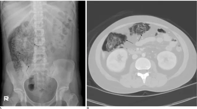
Fig. 1. Case 1: A 18-year old girl with SLE.
A. Plain abdominal radiograph shows the curvilinear collection of intramural gases (arrows) in the right upper abdomen.
B. CT scan with lung window setting (window width: 2,000 HU, window level: -500 HU) shows multiple gaseous cysts (arrows) within the ascending and transverse colons.
terial hypothesis, increased fermentation by gas-forming bacteria and the subsequent production of a large vol- ume of gas predispose an individual to developing PCI (9). In rare occasions, collagen vascular disease can cause PCI. In 1966, Hughes et al. (10) reported the first case in a patient with collagen vascular disease. Since then, oth- er cases have been described, which have mostly been associated with progressive systemic sclerosis (3, 6).
Other collagen vascular diseases that predispose an indi- vidual to PCI such as rheumatoid arthritis (3), dermato- myositis (5), and SLE (3, 4) have been reported. In Korea, there has been only one report of PCI associated with collagen vascular disease, in which the patient was diag-
nosed with progressive systemic sclerosis (6).
Several hypotheses for the pathogenesis of PCI associat- ed with progressive systemic sclerosis has been suggest- ed. These include mucosal stretching and fissuring due to a chronic distention and an elevated intraluminal pres- sure, or from small focal areas of ischemia and ulceration due to vascular compression in the submucosa by fibrous connective tissue deposition (3). In contrast to the exten- sive fibrosis observed in progressive systemic sclerosis, vasculitis is the mechanism of PCI in cases of SLE . PCI can occur due to focal areas of mucosal ischemia or ulcer- ation as a result of a small vessel occlusion. Lupus vasculi- tis results in ischemia that affects the mucosa more than
J Korean Radiol Soc 2007;56:361-364
─ 363 ─
A B
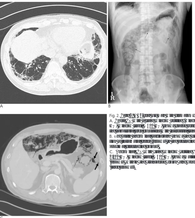
Fig. 2. Case 2: A 42-year old man with dermatomyositis.
A. Chest CT with a lung window setting (window width: 2,000 HU, window level: -500 HU) shows coarse reticular densities in the subpleural area consistent with interstitial lung disease.
B. The plain abdominal radiograph shows the curvilinear collec- tion of intramural gases (arrows) in the ascending, transverse and proximal descending colons.
C. Abdominal CT with lung window setting (window width:
2,000 HU, window level: -500 HU) shows multiple gaseous cysts (arrows) within the transverse colon and pneumoperitoneum (large arrows).
C
the submucosa (4). There have been only a few reports on PCI occurring in conjunction with dermatomyositis, par- ticularly in adults. The mechanism of PCI in cases of der- matomyositis may involve an intramuscular thrombosis because there is inflammation of the intramuscular arteri- oles with a thrombus and infarction (5).
The radiological features of PCI vary. The aim of imaging techniques is to differentiate between the pri- mary type, which generally has a favorable prognosis, and the secondary type, the course of which is deter- mined by the underlying disease. In the primary type, PCI is usually located in the submucosal layer of the left hemicolon or its mesentery, and the intramural gas col- lections are a spherical, well-defined cystic appearance.
In the secondary type, PCI is usually limited to the sub- serosal layer of the stomach, small intestine and right colon, and intramural gas may appear as curvilinear lu- cencies that circumscribe a loop of the bowel when ob- served en face and parallel to a long segment when seen lengthwise (4, 7). It is widely accepted that abdominal radiographs and CT can demonstrate the intraluminal gas. However, a recent report suggested that CT shows the cysts quite clearly and also allowed the localization of the affected bowel segments, thereby making CT a more sensitive and discriminating diagnostic tool.
Diverticulosis, gas-containing abscess, and gas inclu- sions or gas layer resulting from coprostasis are potential sources of error related to the CT diagnosis of PCI (7).
After excluding the treatable cause or an occult malig- nancy, the treatment for PCI in association with colla- gen vascular disease is largely conservative. Oxygen therapy leads to a relative decrease in the partial pres- sure of the non-oxygen gases in the blood, which in turn
diffuse out of the cysts leading to resorption. Antibiotics and elemental diets can provide temporary relief. PCI may be complicated by pneumoperitoneum. In contrast to the pneumoperitoneum of other origins, a PCI-related pneumoperitoneum does not usually cause signs of dif- fuse peritonitis. A pneumoperitoneum can be managed non-operatively if it occurs without peritonitis. The CT demonstration of cysts and the patient’s history can help to avoid the need for an unnecessary exploratory laparo- tomy in patients with PCI (4-7).
References
1. Feldman M, Scharschmidt BF, Sleisenger MH, Sleisenger &
Fordtran’s Gastrointestinal and liver disease: pathophysiology/diagno- sis/management. 6th ed. Philadelphia: W. B. Saunders, 1998: 1979- 1980
2. Heng Y, Schuffler MD, Haggitt RC, Rohrmann CA. Pneumatosis intestinalis: a review. Am J Gastroenterol 1995;90:1747-1758 3. Morrison WJ, Siegelman SS. Pneumatosis intestinalis in associa-
tion with connective tissue disease. South Med J 1976;69:1536-1539 4. Kleinman P, Meyers MA, Abbott G, Kazam E. Necrotizing entero- colitis with pneumatosis intestinalis in systemic lupus erythemato- sus and polyarteritis. Radiology 1976;121:595-598
5. Morris-Stiff GJ, Williams RJ. Pneumatosis cystoides intestinalis in a patient with dermatomyositis. J R Soc Med 1999;92:366-367 6. Kwun NK, Min JK, Park KS, Jun EJ, Cho CS, Pyun JY, et al. A case
of pneumatosis cystoides intestinalis in a patient with systemic sclerosis. J Korean Rheum Assoc 1998;5:281-286
7. Scheidler J, Stabler A, Kleber G, Neidhardt D. Computed tomogra- phy in pneumatosis intestinalis: differential diagnosis and thera- peutic consequences. Abdom Imaging 1995;20:523-528
8. Keyting WS, McCarver RR, Kovarik JL, Daywitt AL. Pneumatosis intestinalis: a new concept. Radiology 1961;76:733-741
9. Yale CE, Balish E, Wu JP. The bacterial etiology of pneumatosis cystoides intestinalis. Arch Surg 1974;109:89-94
10. Hughes DT, Gordon KC, Swann JC, Bolt GL. Pneumatosis cys- toides intestinalis. Gut 1966;7:553-557
Soo-Yeon Kim, et al : Pneumatosis Cystoides Intestinalis in Patients with Antinuclear Antibody Negative Systemic Lupus Erythematosus and Dermatomyositis
─ 364 ─
대한영상의학회지 2007;56:361-364
항핵항체 음성 전신성홍반성루푸스 환자와 피부근염 환자에서의 장벽낭상기종: 증례 보고1
1한양대학교 의과대학 서울병원 진단방사선과
2한양대학교 의과대학 구리병원 진단방사선과
김수연・조온구・고병희・김용수2・송순영
교원성 질환과 연관된 장벽낭상기종은 위장관의 장벽 내에 공기를 보이는 드문 소견이다. 저자들은 항핵항체 음 성 전신성홍반성루푸스 환자와 피부근염 환자에서 발생한 장벽낭상기종 및 복강기종을 경험하였기에 문헌고찰과 함께 보고하고자 한다.