• 검색 결과가 없습니다.

이 논문은 2014 년도 충북대학교병원 임상연구비에 의하여 연구되었음 .

N/A
N/A
Protected

Academic year: 2021

Share "이 논문은 2014 년도 충북대학교병원 임상연구비에 의하여 연구되었음 ."

Copied!
5
0
0

로드 중.... (전체 텍스트 보기)

전체 글

(1)

접수일 : 2014428, 게재승인일 : 20141015 일 책임저자 : 방희제 , 충북 청주시 서원구 1 순환로 776

󰂕 361-711, 충북대학교병원 재활의학과

Tel: 043-269-6227, Fax: 043-269-6228 E-mail: [email protected]

이 논문은 2014 년도 충북대학교병원 임상연구비에 의하여 연구되었음 .

신경섬유종증이 동반된 골형성부전증 환자에서 초음파를 이용한 수근관 증후군 진단

󰠏 증례 보고 󰠏

충북대학교 의과대학 재활의학교실

김효종ㆍ방희제

Diagnostic Ultrasonography for Carpal Tunnel Syndrome of the Patient with Osteosgenesis Imperfecta and Neurofi- bromatosis

󰠏 A Case Report 󰠏

Hyo Jong Kim, M.D. and Heui Je Bang, M.D.

Department of Rehabilitation Medicine, Chungbuk National University College of Medicine, Cheongju, Korea

Carpal tunnel syndrome occurs with pressure on the me- dian nerve passing through the carpal tunnel, and is diag- nosed by typical symptoms, signs and electrodiagnostic examination. A twenty-one year old woman suffered from osteogenesis imperfecta and neurofibromatosis was re- ferred for severe both hands tingling sensation. On physical examination, both carpal tunnel syndrome was suspected.

We test electrodiagnostic examination, but we could not find any abnormalities. We could diagnose the carpal tunnel syndrome with diagnostic ultrasonography by measuring the cross-sectional area of the median nerve at the pisiform bone level. In this case, both carpal tunnel syndrome were diagnosed by ultrasonography, and the symptoms were im- proved after surgical treatment. (Clinical Pain 2014;13:94-98) Key Words: Carpal tunnel syndrome, Osteogenesis imperfecta,

Ultrasonography

서 론

전통적인 수근관 증후군은 전형적인 임상 증상 및 징후 여부와 전기진단 검사 등을 통하여 진단하지만, 최근에는

초음파 사용이 가치 있는 진단도구로서 인정받고 있다.

1

최 근 많은 연구들은 초음파상에서 정중신경의 단면적(cross- sectional area)이 수근관 내에서 정중신경이 눌리는 정도를 반영한다고 보고하였다.

1

수근관 증후군의 진단에 있어서 전기진단 검사와 초음파 상 정중신경의 단면적(cross-sectional area)간에 상관관계 가 있다는 사례들은 많이 보고되고 있으며,

1-3

임상에서도 수근관 증후군의 진단을 위하여 환자의 증상, 이학적 검사 및 전기진단 검사 이외에도 초음파를 이용하는 경우가 점 점 늘고 있다.

이에 저자들은 외상 등의 뚜렷한 과거력 없이 수근관 증 후군의 증상과 징후를 보이며, 신경섬유종증 및 골형성부 전증을 앓고 있는 21세 여자에게서 전기진단 검사는 정상 이었지만 초음파 검사로 수근관 증후군을 진단하고 수술적 치료 후 증상이 호전된 사례를 경험하였기에 간단한 문헌 고찰과 함께 보고하는 바이다.

증 례

21세 여자 환자가 양측 수부 통증 및 저린 증상으로 의뢰 되었다. 환자는 1남 1녀의 장녀로 영유아 시기에 제 2형 골 형성부전증 및 제 1형 신경섬유종증으로 본원에서 입원 및 통원치료를 받아왔으며, 골형성부전증으로 인한 잦은 골절 로 수차례 수술 등의 치료를 받았다. 환자의 아버지는 신경 섬유종증, 어머니는 골형성부전증, 환자의 남동생은 신경 섬유종증을 진단 받았다.

재활의학과로 의뢰 당시 환자는 우측 제 4족지의 단지증 으로 정형외과에서 수술 후 입원중인 상태로, 특별한 외상 없이 내원 약 1년 전부터 주로 양측 제 1수지에서 제 3수지 까지 시각적 상사척도 3점의 통증 및 저린 증상을 보였다.

내원 7∼8개월 전부터는 잠에서 깰 정도로 증상이 악화되

어 양측 수근관증후군이 의심되어 전기진단 검사를 위하여

의뢰되었다. 환자의 신장은 118 cm로 매우 작았으며, 양측

하지에 병적골절로 수차례 수술 받은 흉터가 있었다. 환자

는 Hems 등

4

에 의한 수근관 증후군 점수 체계(scoring sys-

(2)

Table 2. Results of Nerve Conduction Study

Right Left

CMAP DML (ms) M Amp (mV) NCV (m/s) DML (ms) M Amp (mV) NCV (m/s)

Median 2.70 8.8 63.0 2.65 8.6 63.0

Ulnar 2.35 11.1 66.7 2.75 10.4 66.7

SNAP DSL (ms) S Amp (μV) DSL (ms) S Amp (μV)

Median 2.25 59.8 2.20 58.2

Ulnar 2.15 53.3 2.10 40.2

Median-digit IV 2.20 41.5 2.25 35.0

Ulnar-digit IV 2.15 38.6 2.20 27.4

DSL: Distal sensory latency, S Amp: Sensory amplitude, DML: Distal motor latency, M Amp: Motor amplitude, NCV: Nerve con- duction velocity, Median-digit IV: Nerve conduction study in median nerve on 4th finger recording, Ulanr-digit IV: Nerve conduction study in ulnar nerve on 4th finger recording. Motor and sensory amplitudes are measured onset-to-peak. The median motor response was recorded over the thenar eminence, after stimulating the median nerve 8 cm proximally in the wrist. The ulnar motor response was recorded over the hypothenar eminence, after stimulating the ulnar nerve 8 cm proximally in the wrist. Mixed nerve responses were obtained by stimulating the median and ulnar nerves in the wrist and recording at the finger, using a distance of 14 cm.

Table 1. Scoring System for Carpal Tunnel Syndrome by Hems et al.

4

Case Age

Below 60 years (2 points) 2

Above 60 years (0 points) 0

Symptoms

Night pain (2 points) 2

Paraesthesia median nerve distribution (2 points) 2 Relief pain by shaking (2 points) 0 Relief pain by splint (1 points) 0

Clumsiness (1 points) 1

Signs

Tinel’s sign (2 points) 2

Phalen’s test (1 points) 1

Alteration in sensation in distribution of

median nerve (2 points) 0

Wasting Thenar eminence (2 points) 0

Total (17 points) 10

tem for CTS)에서 10점(60세 이하, 야간 동통, 정중신경 지 배영역의 이상 감각, 미세 수부 기능 저하, 양성 Tinel’s sign 및 Phalen’s test)으로 수근관 증후군이 의심되었으나, 양측 상지의 근력을 포함한 신경학적 검사에서는 특이 소 견이 없었다(Table 1).

전기진단 검사에서 근전도 기기는 Medelec Synergy sys- tem (Oxford Instruments, Oxford, UK)을 사용하였다. 신경 전도 검사상 정중신경 및 척골신경의 운동 신경과 감각 신 경의 신경전도 속도, 잠시와 진폭 등은 정상범주였고, 침근

전도 검사상 특이소견은 관찰되지 않았다(Table 2).

전기진단 검사 결과 상 상지의 운동 및 감각 신경전도 검사에서 정상치를 보인 것에 비하여 환자의 증상과 이학 적 검사 소견이 수근관 증후군으로 강하게 의심되어 초음 파 검사를 추가적으로 시행하였다. 초음파 검사 기기는 iU22 (Philips

, Bothell, USA)를 사용하였고, 앉은 상태에 서 상완을 완전 회외 상태로 손바닥이 위를 향하게 한 자세 에서 손목은 중립으로 유지하고 손가락은 약간 구부린 상 태로 검사하였다. 5∼17 MHz 소식자를 두상골(pisiform bone)에서 직각으로 놓고 정중신경의 단면적(cross- sectional area)을 측정하였다. 정중신경의 단면적을 측 정하기 위하여 저음영의 정중신경 주변에 고음영을 갖 는 신경초의 내측 경계선을 기계적으로 따라가며 직접 추적 측정한 연속추적방법(continuous trace method)을 사용하였다(Fig. 1). 두상골 부위에서 측정한 양측 정중 신경의 단면적은 우측 및 좌측에서 각각 0.114 cm

2

, 0.112 cm

2

였고, Duncan 등

5

에 의한 진단기준에 따라 양측 수근관 증후군으로 진단할 수 있었다.

환자는 초음파 검사를 통해 양측 수근관 증후군 진단 후 우측 횡손목인대 유리술을 받았고, 그로부터 1개월 후 좌측 횡손목인대 유리술을 받았다. 수술 후 환자의 증상은 Hems 등

4

의 점수체계상 2점으로 호전되었으며, 수술 4개월째 추 적관찰에서 시행한 초음파 검사 상 양측 정중신경의 단면 적은 우측 및 좌측에서 모두 0.084 cm

2

였다(Fig. 2).

고 찰

수근관 증후군은 손목관절의 굽힘근지지띠 부위에서 정

(3)

Table 3. Results of Needle Electromyography

Muscle

Right Left

SA

MU Rec

SA

MU Rec

Fib PSW Fib PSW

Deltoid 0 0 NMU C 0 0 NMU C

BB 0 0 NMU C 0 0 NMU C

TB 0 0 NMU C 0 0 NMU C

ECR 0 0 NMU C 0 0 NMU C

FCR 0 0 NMU C 0 0 NMU C

PT 0 0 NMU C 0 0 NMU C

APB 0 0 NMU C 0 0 NMU C

FDI 0 0 NMU C 0 0 NMU C

Paraspinalis 0 0 0 0

BB: Biceps brachii, TB: Triceps brachii, ECR: Extensor carpi radialis, FCR: Flexor carpi radialis, PT: Pronator teres, APB: Abductor pollicis brevis, FDI: First dorsal interossei, SA: Spontaneous activity, Fib: Fibrillation potential, PSW: Positive sharp wave, MU: Motor unit, Rec: Recruitment pattern, NMU: Normal motor unit, C: Complete.

Fig. 1. Cross sectional area of median nerve at the level of pisiform bone was obtained from continuous tracing method by ultrasonography. (A) Right side (cross sectional area: 0.114 cm

2

), (B) Left side (cross sectional area: 0.112 cm

2

).

중신경이 압박됨에 따라 정중신경이 지배하는 영역에 근력 약화, 이상감각, 저림 증상 등의 증상을 보이며, 약물, 주사 및 보조기 등의 비수술적 치료 또는 수술 등의 치료로 호전 될 수 있다. Hems 등

4

의 연구에서는 수근관 증후군이 의심 되는 환자에서 진단을 위하여 증상과 징후에 대한 점수체 계를 제시하였는데(Table 1), 총점 17점 중 7점 이상이면 전기진단 검사 없이도 수술적 치료를 고려할 수 있다고 하 였다. 증례의 경우 Hems 등

4

의 점수체계상 10점으로 수근 관 증후군이 의심되었으나, 정확한 진단을 위해 전기진단 검사를 시행하였다. 전기진단 검사 상 특이 소견이 관찰되 지 않아서 보다 정확한 진단을 위하여 초음파 검사를 추가

적으로 시행하였고, 이를 통하여 수근관 증후군을 확진할 수 있었던 증례이다.

신경섬유종증은 임상적으로 광범위한 표현형을 보이는

모반증으로서, 신경 및 피부 조직이 분포한 다양한 장기를

침범하는 질환이다. 제 1형 신경섬유종증은 신경압박을 특

징으로 하며, 이로 인하여 말초신경병증이 유발될 수 있다

는 연구가 보고되어 있다.

6

이미 1981년 총상 신경섬유종

(plexiform neurofibroma) 환자에서 수근관 증후군이 발생

한 증례가 보고되었고,

7

Freitas 등

8

은 청소년기에 제 1형 신

경섬유종증으로 진단받은 56세 환자에서 수근관 증후군이

발생하였다고 보고하였다.

(4)

Fig. 2. Cross sectional area of median nerve at pisiform bone measured by ultrasonography after carpal tunnel release operation.

(A) Right side (cross sectional area: 0.084 cm

2

), (B) Left side (cross sectional area: 0.084 cm

2

).

증례의 경우 비교적 젊은 나이에 양측 정중신경이 압박 된 것으로 보아 제 1형 신경섬유종증의 영향에 의하여 신경 손상에 다소 취약하였던 것일 수도 있지만, 전기진단 검사 에서는 특이소견을 관찰할 수 없었다.

수근관 증후군의 진단을 위하여 임상증상, 이학적 소견, 그리고 전기진단 검사를 주로 사용하게 되는데, 증례와 같 이 임상증상 및 이학적 소견으로는 수근관 증후군이 강하게 의심되지만 전기진단 검사에서 특이소견이 발견되지 않을 때는 수술적 치료 여부를 결정하기가 쉽지 않다. Duncan 등

5

은 초음파상에서 두상골 부위의 정중신경 단면적이 0.09 cm

2

이상이면 수근관 증후군으로 진단할 수 있다고 하였고, Park 등

9

은 한국 성인에서 두상골 부위의 정중신경 단면적 의 임계값을 8.5 mm

2

로 제시하였다. Park 등

10

은 정상 한국 성인에서 나이에 따른 수근관내 정중신경의 초음파 소견을 연구하였는데, 40세 이하의 정상 한국 성인에서 두상골 부 위의 정중신경 단면적은 6.39±0.26 mm

2

로 보고하였다. 증 례의 경우 두상골에서 측정한 양측 정중신경의 단면적이 각각 0.114 cm

2

, 0.112 cm

2

이었으며, 단면적이 0.09 cm

2

이 상으로서 수근관 증후군으로 진단할 수 있었다. 초음파 검 사는 비침습적, 무통 및 실시간 검사가 가능하다는 장점을 가진 검사이므로, Lee 등

11

은 진단초음파를 수근관 증후군 의 초기 진단 방법으로 사용할 수 있다고 제안하였다.

골형성부전증은 골밀도 저하, 골격 이상, 저신장 등을 특 징으로 하는 유전질환으로서, 주로 제 1형 콜라겐 합성에 영향을 주는 돌연변이에 의해 발생한다.

12

증례의 경우 골 형성부전증으로 인해 신장이 같은 연령에 비하여 매우 작 았는데, 신장이 전기진단 검사에서의 신경전달속도나 잠시 에 미치는 영향에 대해서는 아직 명확하게 밝혀져 있지 않 다. Rivner 등

13

에 따르면 신장과 신경전도속도와의 관계에

대한 연구에서 신장이 클수록 신경전도속도와 잠시가 느려 지는 경향이 있다고 보고하였다. 이와는 반대로 Kato 등

14

은 신장과 신경전달속도와는 관계가 없다고 주장하였으며, Soundmand 등

15

은 신경의 종류에 따라 신장에 의한 영향이 달라지는데, 특히 정중신경의 경우 신경전달속도는 신장과 는 관계가 없다고 하는 등 일치된 소견을 보이고 있지 않다.

Rivner 등

13

의 연구에 의하면 신장이 클수록 신경전도속도 가 느려진다고 하였는데, 증례의 경우 신장이 118 cm로서 정상 신장의 성인에 비하여 신경전도속도가 매우 빨라서 수근관 증후군이 있음에도 불구하고 신경전도속도가 정상 범위 소견을 보인 것으로 가정할 수 있다. 하지만, 연령대비 평균보다 신장이 작은 사람들을 대상으로 신경전도검사의 정상치를 조사한 대규모 연구는 현재까지 없으므로, 증례 와 같이 말초신경 압박에 의한 증상들이 있음에도 불구하 고 전기진단 검사상 이상소견이 나타나지 않고 병적으로 작은 신장에 의하여 차폐(masking)되는 효과가 있는지 여 부에 대한 추후 연구가 필요할 것으로 생각된다. 신장과 관 계없이 수근관 증후군의 증상 및 이학적 검사 소견이 명확 한 경우에도 정상 전기진단 검사 소견을 보일 수 있다는 점을 감안한다면, 이러한 경우 초음파 검사가 유용한 대안 이 될 수 있다.

16

또한 Wong 등

17

은 수근관 증후군이 의심되 는 환자에게 우선적으로 초음파 검사를 통하여 수근관 증 후군을 진단하고, 만약 초음파 검사에서 이상을 보이지 않 는 경우 근전도 검사를 시행할 것을 제안하고 있다.

수근관 증후군의 증상과 징후를 명확하게 보이는 신경섬

유종증과 골형성부전증환자에서 전기진단 검사상 특이소

견이 관찰되지 않았으나, 초음파 검사를 통하여 수근관 증

후군을 진단한 증례에 대하여 문헌고찰과 함께 보고한다.

(5)

참 고 문 헌

1. Seror P. Sonography and electrodiagnosis in carpal tunnel syndrome diagnosis, an analysis of the literature. Eur J Radiol 2008; 67: 146-152

2. Klauser AS, Halpern EJ, De Zordo T, Feuchtner GM, Arora R, Gruber J, et al. Carpal tunnel syndrome assess- ment with US: value of additionalcross-sectional area meas- urements of the median nerve in patients versus healthy volunteers. Radiology 2009; 250: 171-177

3. Buchberger W, Judmaier W, Birbamer G, Lener M, Schmidauer C. Carpal tunnel syndrome: diagnosis with high-resolution sonography. AJR 1992; 159: 793-798 4. Hems TE, Miller R, Massraf A, Green J. Assessment of a

diagnostic questionnaire and protocol for management of carpal tunnel syndrome. J Hand Surg Eur Vol 2009; 34:

665-670

5. Duncan I, Sullivan P, Lomas F. Sonography in the diag- nosis of carpal tunnel syndrome. AJR 1999; 173: 681-684 6. Drouet A, Wolkenstein P, Lefaucheur JP, Pinson S,

Combemale P, Gherardi RK, et al. Neurofibromatosis 1-as- sociated neuropathies: a reappraisal. Brain 2004; 127:

1993-2009

7. Monballiu G. Plexiform neurofibroma of the median nerve.

Chirurgia Plastica 1981; 6: 141-145

8. Freitas D, Aido R, Sousa M, Costa L, Oliveira V, Cardoso P. Carpal tunnel syndrome due to a plexiform neurofibroma of the median nerve in a neurofibromatosis type 1 patient:

clinical approach. BMJ Case Rep 2013; 13: 1

9. Park GY, Bae JH, Lee SY, Oh JS, Lim JG, Son DG.

Ultrasonographic findings of mild and very mild carpal tun- nel syndrome. J Korean Acad Rehab Med 2008; 32: 67-72 10. Park JY , Park SR, Lee SH, Choi KH. The ultrasonographic findings of the median nerve in the carpal tunnel according to age and sex of normal Korean adults. J Korean Acad Rehab Med 2008; 32: 564-569

11. Lee D, van Holsbeeck MT, Janevski PK, Ganos DL, Ditmars DM, Darian VB. Diagnosis of carpal tunnel syndrome. Ultrasound versus electromyography. Radiol Clin North Am 1999; 37: 859-872

12. Biggin A, Munns CF. Osteogenesis Imperfecta: Diagnosis and Treatment. Curr Osteoporos Rep 2014; 12: 279-288 13. Rivner MH, Swift TR, Crout BO, Rhodes KP. Toward

more rational nerve conduction interpretations: the effect of height. Muscle Nerve 1990; 13: 232-239

14. Kato M. The conduction velocity of the ulnar nerve and the spinal reflex time measured by means of the H wave in average adults and athletes. Tohoku J Exp Med 1960; 25:

74-85

15. Soudmand R, Ward LC, Swift TR. Effect of height on nerve conduction velocity. Neurology 1982; 32: 407-410 16. Koyuncuoglu HR, Kutluhan S, Yesildag A, Oyar O, Guler

K, Ozden A. The value of ultrasonographic measurement in carpal tunnel syndrome in patients with negative electro- diagnostic tests. Eur J Radiol 2005; 56: 365-369

17. Wong SM, Griffith JF, Hui AC, Lo SK, Fu M, Wong KS.

Carpal tunnel syndrome:diagnostic usefulness of sono-

graphy. Radiology 2004; 232: 93-99

수치

Table  1.  Scoring  System  for  Carpal  Tunnel  Syndrome  by  Hems  et  al. 4
Table  3.  Results  of  Needle  Electromyography Muscle Right LeftSA MU Rec SA MU Rec Fib PSW Fib PSW
Fig.  2.  Cross  sectional  area  of  median  nerve  at  pisiform  bone  measured  by  ultrasonography  after  carpal  tunnel  release  operation

참조

관련 문서

Selective fascicular involvement of median nerve in carpal tunnel syndrome (CTS) has been reported, such as predominant involvement of volar-radially located motor and

Key Words : Carpal tunnel syndrome, Hand configuration, Hand dimensions, Median nerve, Electrodi- agnositc evaluation.. Address reprint requests to Young

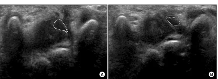
Increased signal with proximal swelling and distal flattening of the median nerve were seen in all patients of carpal tunnel syndrome.. Among the eight

Hence, we report an unusual case and surgical outcome of thrombosed persistent median artery with bifid median nerve causing acute carpal tunnel syndrome.. Informed consent

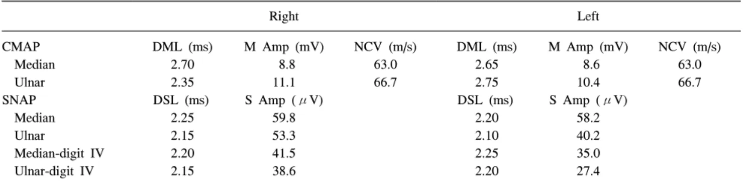
amp.: amplitude, CMAP: compound muscle action potential, CSA: cross-sectional area, CTS: carpal tunnel syndrome, CV: conduction velocity, DM: dia- betes mellitus, lat.: latency,

Box plot showing the median nerve CSAs measured using 3D ultrasonography at the carpal tunnel inlet (left) and the maximal swell- ing point (right) in patients with CTS who

Methods : 20 patients suffering from carpal tunnel syndrome were randomly divided into two groups: 10 subjects treated both with electrical therapy and median

─ CSAa = cross sectional area (CSA) of median nerve proximal to tunnel inlet, CSAb = CSA at pisiform bone level, CSAb-a = area difference between CSAb and CSAa, % area = percentage