서 론
역위(inversion, inv)는 균형재배열을 나타내는 염색체의 구 조적 이상 중 하나로 대부분 정상 표현형을 나타내게 된다. 그러 나 생식 세포의 감수분열 단계에서 역위 고리(inversion loop)를 만들면 염색체의 결실 또는 중복을 보이는 재조합염색체(recom- binant chromosome, rec)가 형성되어 자녀에서 비정상 표현 형을 나타낼 수 있다[1]. 4번 염색체의 구조적 이상에 동반된 4p 결실은 Wolf-Hirschhorn 증후군(WHS)을 초래할 수 있다[2].
저자들은 아버지의 inv(4)로 기인한 4p 결실 및 4q 중복을 갖 는 재조합염색체를 관찰하여 문헌고찰과 함께 보고하는 바이다.
증 례
1. 임상상양쪽 폐의 가슴막삼출을 주소로 입원한 미숙아에서 말초혈액 염색체 검사가 의뢰되었다. 환아는 정상 표현형을 보이는 부부의 두 번째 임신으로 사중검사와 초음파 등 산전관찰상 이상 소견 없이 임신이 유지되었다. 재태연령 33주 6일에 태아 가슴막삼 출과 양수과다증 및 태아절박가사를 주소로 전원되어 응급제왕 절개술로 태어난 여아였다. 출생 시 키 41 cm, 몸무게 1,530 g, 두위 29.5 cm로 모두 10-50 백분위수에 해당하였다. 출생 시 심박수가 100회 미만으로 감소되고, 청색증 및 호흡곤란을 보였 으나, 흉막천자 및 양압환기 시행 후 호전되었다. 신체검사상 오 른쪽 귓바퀴 앞 피부돌출(preauricular skin tags) 외에 다른 비 정상 소견은 관찰되지 않았다.
두부 자기공명영상에서 뇌실내출혈 및 뇌실주위 백질연화증 (periventricular leukomalacia)이 관찰되었고, 뇌파검사는 정 상이었다. 복부초음파검사에서 0.3 cm의 작은 신낭종이 보였다.
심장초음파상 양측성 상대정맥 기형이 존재하였고, 2차공 심방
89 Korean J Lab Med 2010;30:89-92
DOI 10.3343/kjlm.2010.30.1.89
89 89 89 Received :August 27, 2009 Manuscript No :KJLM09-110 Revision received :November 12, 2009
Accepted :November 20, 2009 Corresponding author :Soo Jin Yoo, M.D.
Department of Laboratory Medicine, Sanggye Paik Hospital, Inje University, 761-1 Sanggye 7-dong, Nowon-gu, Seoul 139-707, Korea
Tel : +82-2-950-1242, Fax : +82-2-950-1244 E-mail : [email protected]
*본 논문은 2008년도 인제대학교 학술연구조성비 보조에 의한 것임.
Pericentric inversion of chromosome 4 can give rise to 2 alternate recombinant (rec) chromosomes- by duplication or deletion of 4p. The deletion of distal 4p manifests as Wolf-Hirschhorn syndrome (WHS). Here, we report the molecular cytogenetic findings and clinical manifestations observed in an infant with 46,XX,rec(4)dup(4q)inv(4)(p16q31.3)pat. The infant was delivered by Cesarean sec- tion at the 33rd week of gestation because pleural effusion and polyhydramnios were detected on ultrasonography. At birth, the infant showed no malformation or dysfunction, except for a preauricu- lar skin tag. Array comparative genomic hybridization analysis of neonatal peripheral blood sam- ples showed a gain of 38 Mb on 4q31.3-qter and a loss of 3 Mb on 4p16.3, and these results were consistent with WHS. At the last follow-up at 8 months of age (corrected age, 6 months), the infant had not achieved complete head control. (Korean J Lab Med 2010;30:89-92)
Key Words : Inversion, Recombinant, Wolf-Hirschhorn syndrome
�Case Report∙Diagnostic Genetics�
Recombinant Chromosome 4 with Partial 4p Deletion and 4q Duplication Inherited from Paternal Pericentric Inversion
Se Jin Mun, M.D.
1, Eun Hae Cho, M.D.
2, Myoung-Jae Chey, M.D.
3, Gyu-Hong Shim, M.D.
3, Bo-Moon Shin, M.D.
1, Rae-Kyung Lee, M.T.
1, Ji-Kyung Ko, M.D.
4, and Soo Jin Yoo, M.D.
1Departments of Laboratory Medicine1, Pediatrics3, and Obstetrics and Gynecology4, Sanggye Paik Hospital, Inje University College of Medicine, Seoul; Greencross Reference Laboratory2, Yongin, Korea
90 Se Jin Mun, Eun Hae Cho, Myoung-Jae Chey, et al.
중격결손과 동맥관 개존증은 추적 검사에서 소실되었다. 추적관 찰상 생후 8개월, 분만예정일을 기준으로 한 교정연령 6개월에 목 가누기가 완전하지 않았다.
2. 가족력
2년 전 환아 부모의 첫 번째 임신 시 비정상 삼중 검사 소견, 양수 내 AFP 증가(200 mg/mL 이상, 9.91 over MoM) 및 ace- tylcholinesterase 양성, 초음파소견 등이 신경관 결손에 합당 하여 15주에 인공유산하였다. 당시 다른 기관에서 실시한 양수 염색체 검사에서 4번 염색체 단완의 구조적 이상이 있음이 보고
되었고(Fig. 1B), 추가로 실시한 부모의 말초혈액 염색체 검사에 서 어머니는 정상 핵형이었으나, 아버지에서 46,XY,inv(4)(p16- q31.3) 핵형이 보고되었다(Fig. 1A).
3. 유전학적 검사
환아의 말초혈액을 phytohaemagglutinin를 첨가한 RPMI 배지에서 3일간 배양하였고, 제작된 슬라이드를 G-분염법을 통 해 염색하였다. 첫번째 임신 시 관찰된 핵형과 동일한 핵형이 관 찰되었고, 이는 아버지의 inv(4)에서 기인한, 4p 결실과 4q 중복 을 동반한 재조합 4번 염색체로 추정되어 46,XX,rec(4)dup(4q)- inv(4)(p16q31.3)pat로 핵형을 보고하였다(Fig. 1C). 그러나, 결 실 부위가 작아 정확한 진단을 위해 추가 검사가 요구되었다.
환아의 말초혈액 DNA에 대해 각 염색체 아종말체(subtelom- ere)에 대한 소식자를 포함하는 SALSA MLPA kit P069 human telomere-4 (MRC-Holland, Amsterdam, The Netherlands) 로 검사하여 4p 말단의 소실 및 4q 말단의 증폭을 확인하였다 (Fig. 2A) [3]. 또한, SALSA MLPA kit P096 mental retarda- tion-2 (MRC-Holland) 키트 중 4p 말단 2 Mb 내의FLJ20265, GAK,FGFRL1,SPON2, CTBP1, SLBP,TACC3, FGFR3, LETM1,WHSC1,WHSC2유전자에 대한 소식자로 검사를 실 시한 결과 이들 모두 소실되어 WHS 염색체 소실 부위에 합당 함을 확인하였다(Fig. 2B).
WHS는 결실의 크기에 따라 표현형이 달라지므로, 정확한 절 단점을 알기 위해 array comparative genomic hybridization (aCGH)를 시행하였다. 1 Mb 간격의 해상도를 보이는 MacAr- rayTMKaryo 4500 (Macrogen Inc., Seoul, Korea)칩을 사용 하였고, 정상인의 DNA와 비교하여 정상화된 log2 비율이 0.25 초과일 경우 증폭으로, -0.25 미만일 경우 소실로 판단하였다.
Fig. 1. GTG-banded partial karyotypes of chromosome 4 from 3 members of a family. The chromosomes on the right hand side of each of the 3 sets (indicated by arrows) are the abnormal chro- mosomes. (A) Chromosomes of the father showed balanced peri- centric inversion, inv(4)(p16q31.3). (B) Amniotic fluid obtained dur- ing the first pregnancy, which ended in abortion (C) peripheral blood of the second offspring. (B) and (C) showed rec(4)dup(4q)- inv(4)(p16q31.3)pat.
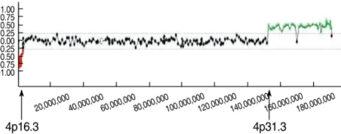
Fig. 3. Result of the array comparative genomic hybridization (aCGH) analysis for the neonatal peripheral blood. The y-axis rep- resents the log2 of the intensity ratios of patient/control DNA. The x-axis represents the distance from the 4p telomere of the BAC clones to the site. The arrows indicate the location of the clones flanking the breakpoints. Array CGH showed a loss of 3 Mb on in 4p16.3 and a gain of 38 Mb in 4q31.3-qter.
A B C
Fig. 2. Results of multiplex ligation-dependent probe amplification (MLPA) obtained using the neonatal peripheral blood samples were analyzed using the Genemarker version 1.6 software. (A) SALSA MLPA kit P069 human telomere-4 showed 2 dots outside the reference range (0.75-1.3) indicating the gain in 4q and the loss in 4p. (B) SALSA MLPA kit P096 mental retardation-2 showed the loss in all 16 probes for 4p, including the Wolf-Hirschhorn syn- drome-related genes.
Peak ratio
2
1
0
100 200 300 400 500
Size (bps) A
Peak ratio
2
1
0
100 200 300 400 500
A
Peak ratio
2
1
0
100 200 300 400 500
Size (bps) B
Peak ratio
2
1
0
1.00 0.75 0.50 0.250.00 -0.25 -0.50 -0.75 -1.00
20,000,00040,000,00060,000,00080,000,000100,000,000120,000,000140,000,000160,000,000180,000,000
4p16.3 4p31.3
실험 결과, 4p16.3에서 2,995 Kb의 소실이 있었으며, 4q31.3- qter에서는 38,253 Kb의 증폭이 있었다(Fig. 3).
환아의 핵형분석, MLPA, aCGH를 종합한 결과, ISCN 2009 명명법에 준해 46,XX,rec(4)dup(4q)inv(4)(p16.3q31.3)pat.arr 4p16.3(76,938-3,072,186)x1, 4q31.3(152,752,722-191,006,683) x3.mlpa 4psubtel(P069)x1,4qsubtel(P069)x3,4p16.3(P096) x1로 표기할 수 있었다[4].
고 찰
WHS의 표현형을 결정하는 중요한 부위는 4p 말단(4p16.3)으 로 대부분 자연발생적으로 4p 부분결실이 일어나며, 10-15%는 상호전좌 등 균형재배열을 가진 보인자인 부모로부터 유전된다 [5, 6]. ‘그리스 병사 투구’모양으로 불리는 얼굴 모양이 공통적 특징으로, 튀어나온 미간과 넓고 뾰족한 코, 높은 이마선, 두눈 먼거리증, 짧은 인중, 작은 턱, 그 외에 소두증, 장두증, 낮은 귀, 귓바퀴 앞 피부돌출 등을 동반한다[2, 6, 7]. 그 외의 표현형은 결 실의 위치에 따라 다양하여, 결실 부위가 말단 3.5 Mb 미만으로 작은 경우는 90% 정도에서 신생아기 근육긴장 저하, 성장지연이 관찰되고, 이후 경련, 발달 지연 등이 보이며, 약 70%에서 가벼 운 정신 지체가 나타난다. 결실 부위가 큰 경우 정신 지체, 심기형, 신장, 골격계 등의 기형이 증가한다[6, 7]. WHS의 원인이 되는 critical region (WHSCR)은 말단 2 Mb 내에 존재하며, WHSC1, WHSC2,LETM1,FGFR3등의 유전자가 있고, 특히LETM1 소실은 경련과 관련된다고 알려졌다[8, 9].
본 환아에서는 귓바퀴 앞 피부돌출 외에 다른 전형적인 증상 이 관찰되지 않았다. 그러나, 본 환아는 미숙아로 태어나 특징적 얼굴 형태를 발견하지 못했을 가능성도 배제할 수 없고, 현재 교 정연령 6개월까지 추적되었기 때문에 추후 다른 소견이 발생할 가능성을 배제할 수 없다. 다른 보고에서도, 신생아기에는 특징적 인 증상을 보이지 않다가 성장하면서 발달 지연 및 지능 저하로 WHS로 진단받은 예를 찾아볼 수 있다[2, 10]. WHS에서 관찰되 는 경련은 9-10개월 사이에 가장 많이 시작되며, 성장 및 발달 지연도 출생 후 1년 후부터 두드러지는 것으로 알려져 있다[11].
환아의 양측성 상대정맥은 일반적으로 심기형을 가진 신생아 의 2.8-11% 정도에서 비특이적으로 발생하며[12], WHS에서 심 기형을 동반하는 것은 대개 5 Mb 이상의 결실이다[6]. 따라서 환아의 심기형은 WHS과 직접적 관련성이 적을 것으로 추정되 었다.
환아의 경우 4q 중복도 관찰되었는데, 기존 보고에서 작은 4q 중복에서는 얼굴 기형, 성장 지연, 정신지체, 사지기형, 신장 기
형 등이 관찰되고[13, 14], 크기가 큰 경우 정신지체, 발달지연, 심기형, 경련 등이 동반되었다[15, 16]. 그러나 환아에서는 이런 표현형을 관찰할 수 없었고, 심기형과 4q 중복과의 관련성도 판 단하기 어려웠다.
inv(4)로 인한 재조합 염색체를 보이는 가계에서 80%는 4p 중복과 4q 결실을 보여 이러한 형태의 재조합을‘recombinant 4 syndrome’이라고 명명하기도 하였다. 본 가계와 같이 inv(4) 로 인한 4p 결실과 4q 중복의 재조합의 발생이 적은 이유 중 하 나는 4q 중복을 가진 태아의 자궁 내 생존율이 더 낮기 때문인 것으로 추정하고 있다[17]. 하지만 본 증례에서는 특별한 이상 소견 없이 임신 3분기까지 유지되었고, 생후 임상 증상도 심하 지 않았던 것을 보면 4q 중복을 동반한 rec(4)의 임상상에 대한 더 많은 증례의 축적이 필요할 것으로 생각된다.
본 가계의 경우, 첫 번째 수태산물에 대해서는 분자세포유전 학적 방법으로 4p 결실을 확인하지는 못하였으나, 해상도가 550 bands 정도인 핵형분석상 환아와 동일한 소견을 보였으므로, 환아와 같이 WHS에 합당한 결실을 가졌을 것으로 예측할 수 있 다. 그러나 태아는 15주에 인공유산되었고, 의무기록에도 당시 소견이 자세히 남아있지 않아 WHS에 합당한 표현형을 확인하 기 어려웠다. 신경관결손은 4번 염색체의 결실 및 중복에서 보 고된 바 없어, 많은 비유전적 원인이나 염색체 검사의 해상도에 서는 확인할 수 없는 다른 유전학적 원인에 의해 발생했을 가능 성을 배제할 수 없다.
본 환아는 산전 사중 검사 및 초음파에서 정상 소견을 보여 산 전 염색체 검사를 실시하지 않았다. 그러나 이전 임신력 및 아버 지의 역위 핵형을 고려할 때, 불균형 4번 염색체의 재발 및 WHS 의 가능성이 존재하는 경우였다. 이런 경우, 세포유전 검사실에 서는 형광제자리부합법(Fluorescence in situ hybridization), MLPA, aCGH 중 접근 가능한 분자세포유전학적 방법 하나를 시행하여 WHS 관련 부위의 미세 결실 여부를 확인하고 이에 합당한 유전상담과 생후 적절한 의학적 조치를 취하는 것이 도 움이 되겠다.
요 약
4번 염색체의 완간역위는 4p 중복 또는 4p 결실을 보이는 2 가지 형태의 재조합염색체를 초래할 수 있으며, 4p 원위부의 부 분 결실은 Wolf-Hirschhorn syndrome (WHS)의 원인이 된 다. 저자들은 46,XX,rec(4)dup(4q)inv(4)(p16q31.3)pat의 핵형 을 가지는 환아의 분자세포유전학적 소견과 임상상을 보고하는 바이다. 본 환아는 33주에 태아 가슴막삼출 및 양수과다증이 관
Rec (4) with del 4p and dup 4q 91
찰되어 제왕절개술로 출생하였다. 환아는 출생 당시 귓바퀴 앞 피부돌출 외에는 심각한 기형이나 기능장애가 관찰되지 않았다.
말초혈액의 array comparative genomic hybridization 분석 을 통해 4q31.3-qter의 38 Mb의 획득과 WHSCR을 포함하는 4p16.3의 3 Mb의 소실을 확인하였으며, 4p의 소실은 WHS에 합당한 소견이었다. 환아는 생후 8개월, 분만예정일을 기준으로 한 교정연령 6개월까지의 추적 관찰에서 목가누기가 완전하지 않았다.
REFERENCES
1. Gardner RJ and Sutherland GR, eds. Chromosome abnormalities and genetic counseling. 3rd ed. New York: Oxford University Press, 2004; 142-62.
2. Rauch A, Schellmoser S, Kraus C, Dorr HG, Trautmann U, Altherr MR, et al. First known microdeletion within the Wolf-Hirschhorn syndrome critical region refines genotype-phenotype correlation.
Am J Med Genet 2001;99:338-42.
3. Cho EH, Park BYN, Cho JH, Kang YS. Comparing two diagnostic laboratory tests for several microdeletions causing mental retarda- tion syndromes: multiplex ligation-dependent amplification vs flu- orescent in situ hybridization. Korean J Lab Med 2009;29:71-6. (조 은해, 박보야나, 조정희, 강유선. 정신지체의 원인인 여러 미세결실증후 군의선별검사로이용되는 Multiplex Ligation-Dependent Probe Am- plification로형광동소교잡법의비교평가. 대한진단검사의학회지 2009;
29:71-6.)
4. Shaffer LG, Slovak ML, et al. eds. An international system for human cytogenetic nomenclature (ISCN) 2009. Basel: Karger 2009.
5. Engbers H, van der Smagt JJ, van’t Slot R, Vermeesch JR, Hochsten- bach R, Poot M. Wolf-Hirschhorn syndrome facial dysmorphic fea- tures in a patient with a terminal 4p16.3 deletion telomeric to the WHSCR and WHSCR 2 regions. Eur J Hum Genet 2009;17:129-32.
6. Zollino M, Murdolo M, Marangi G, Pecile V, Galasso C, Mazzanti L, et al. On the nosology and pathogenesis of Wolf-Hirschhorn syn- drome: genotype-phenotype correlation analysis of 80 patients and literature review. Am J Med Genet C Semin Med Genet 2008;148C:
257-69.
7. Zollino M, Di Stefano C, Zampino G, Mastroiacovo P, Wright TJ, Sorge G, et al. Genotype-phenotype correlations and clinical diag- nostic criteria in Wolf-Hirschhorn syndrome. Am J Med Genet 2000;
94:254-61.
8. Schlickum S, Moghekar A, Simpson JC, Steglich C, O’Brien RJ, Win- terpacht A, et al. LETM1, a gene deleted in Wolf-Hirschhorn syn- drome, encodes an evolutionarily conserved mitochondrial protein.
Genomics 2004;83:254-61.
9. Bergemann AD, Cole F, Hirschhorn K. The etiology of Wolf-Hirs- chhorn syndrome. Trends Genet 2005;21:188-95.
10. Van Buggenhout G, Melotte C, Dutta B, Froyen G, Van Hummelen P, Marynen P, et al. Mild Wolf-Hirschhorn syndrome: micro-array CGH analysis of atypical 4p16.3 deletions enables refinement of the genotype-phenotype map. J Med Genet 2004;41:691-8.
11. Kagitani-Shimono K, Imai K, Otani K, Kamio N, Okinaga T, Toribe Y, et al. Epilepsy in Wolf-Hirschhorn syndrome (4p-). Epilepsia 2005;
46:150-5.
12. Huhta JC, Smallhorn JF, Macartney FJ, Anderson RH, de Leval M.
Cross-sectional echocardiographic diagnosis of systemic venous return. Br Heart J 1982;48:388-403.
13. Carrascosa RM, Garcia MO, Vidal CA, Cabezas TM, Gonzalvez PJ.
Partial duplication of chromosome 4q (q31, q35): auriculo-acro-renal syndrome. An Pediatr (Barc) 2008;68:361-4.
14. Cui YX, Wang YH, Hao LJ, Hou L, Li W, Huang YF. Partial trisomy 4q: a case report. Chin Med J (Engl) 2006;119:1136-9.
15. Halal F, Vekemans M, Chitayat D. Interstitial tandem direct dupli- cation of the long arm of chromosome 4 (q23-q27) and possible assign- ment of the structural gene encoding human aspartylglucosamini- dase to this segment. Am J Med Genet 1991;39:418-21.
16. Jeziorowska A, Ciesla W, Houck GE Jr, Yao XL, Harris MS, Truszc- zak B, et al. Cytogenetic and molecular identification of a de novo direct duplication of the long arm of chromosome 4(q21.3-->q31.3).
Am J Med Genet 1993;46:83-7.
17. Garcia-Heras J and Martin J. A rec(4) dup 4p inherited from a mater- nal inv(4)(p15q35): case report and review. Am J Med Genet 2002;
109:226-30.
92 Se Jin Mun, Eun Hae Cho, Myoung-Jae Chey, et al.