479
목 적 :흰쥐의 가골 조직에서 연령에 따른 구성성분비율을 관찰하고, 노화관련 단백질들의 발현 양상을 면역 조직 화학
염색법으로 확인하여, 이들 단백질들이 골 조직에서의 역할과 골 유합 과정에 미치는 영향에 대하여 알아보고자 하였다.
대상 및 방법 :18마리의 흰 쥐(8주: 7, 32주: 6, 70주: 5)를 사용하였으며, 대퇴골에 골절을 유발한 후 2주째의 가골을 채취하여 증식성 연골세포, 비후성 연골세포 및 간엽성 조직 부위군으로 구분하여 관찰하였고, 항 p16, p21, c-fos, c-jun 항체를 이용한 면역 조직 화학 염색법을 통하여 부위별 발현을 관찰하였다.
결 과 : 각 연령군별 노화 관련 단백질(p16, p21) 및 조기 발현 단백질(c-fos, c-jun)의 발현은 의미있는 차이를 보이지 않 았고, 가골의 부위별 발현은 p16의 경우 증식성 연골; 54.93%, 간엽조직; 46.48%, 비후성 연골; 10.85% 순으로, c-fos의 경우 노화 관련 단백질에서처럼 증식성 연골(73.32%), 간엽 조직(51.84%), 비후성 연골(9.64%)의 순으로 발현하였다.
결 론 :흰 쥐의 가골조직에서 노화관련 단백질들이 연골세포의 분화와 골절후 골화과정에 관여하고 있음을 보여 주었고,
골조직에서의 노화 유도에 대한 기능은 분명하지 않은 것으로 생각되었다.
색인 단어 : 노화관련 단백질, 가골, 면역조직화학 염색법, 노화, p16, p21, c-fos, c-jun
479
쥐의 골절 후 가골 표본에서 본 노화관련 단백질의 발현
송상호**∙박영의*∙최영희*∙심창구∙조성우
인천기독병원 정형외과학교실, 한림대학교 의과대학 병리학교실*, 송상호 정형외과**
서 론
최근 고령 인구의 증가에 따라 퇴행성 관절질환과 골다공증과 같은 골 질환들에 대한 연구가 많이 진행되고 있고, 이들 질환 들은 노화에 따라 노화와 관련된 단백질이 관여하는 것으로 알 려져 있다.
섬유아세포의 연구에서 세포의 분열과 증식이 활발하게 나타 나는 젊은 연령에서는 c-fos, c-jun의 발현이 높았고, 노령에서 는 세포의 활성의 감소와 더불어 p16, p21 등의 발현이 증가하 여 이들 단백질이 노화와 관계하는 것으로 알려져 있다2). c-fos 와 c-jun은 조기 발현 유전자(immediate early response gene)로써 조골 세포의 증식 및 분화에 관여하고7,16,21), 연골세 포와 파골 세포의 분화에도 관여하는 것으로 알려져 있다. p16, p21은 CDK (cyclin dependent kinase) inhibitor로서 세포 주 기를 정지시키고1,6,29), 노화를 유도하는 기능을 갖고 있으며2), 연골 세포의 말단 분화에도 관여하는 것으로 알려져 있다10,11,19).
본 저자들은 면역 조직 화학적 염색법을 이용하여 8주, 32주, 70주의 흰쥐 대퇴골을 골절시킨 후 2주 후의 가골에서 c-fos, c- jun, p16, p21의 연령에 따른 발현을 관찰하였고, 이 단백질들 의 골 조직에서 기능과, 골 유합에 대한 영향에 대하여도 알아 보고자 하였다.
대상 및 방법
1. 실험동물 및 골절유발실험동물은 8주, 32주, 70주령의 수컷 Sprague-Dawley (SD)계 흰쥐 18마리(8주, 7; 32주, 6; 70주, 5)를 사용하였다.
에테르로 전신마취를 한 후 피부절개를 하여 반경 1 mm인 드 릴로 3곳을 구멍낸 후 외반력을 가하여 우측 대퇴골 간부에 골 절 시켰고, 골절에 대한 고정은 시행하지 않았다. Bolander4)에 의하면, 골절 후 조직학적으로 네 단계의 복구과정이 존재하는 데, 골조직과 연골조직, 섬유화조직의 모든 구조물을 관찰할 수 있는 2주를 택하여, 골절 후 제 14일에 에테르로 전신 마취하여 가골부위를 박리한 후 근위 및 원위 골편을 붙인 채로 가골을 분리해냈다. 조직은 먼저 실온에서 4% 파라포름알데하이드 용 액에 8시간 고정한 후 15% EDTA용액에 4일간 탈 석회화하고 파라핀 블록을 제작하여 H&E 염색을 하였다(Fig. 1).
2. 실험방법
1) 면역조직화학적 염색법
탈파라핀과 함수과정을 거친 슬라이드에 과산화수소수(Ultra- vision Mouse Tissue Detection System, Anti-Mouse, HRP/
AEC; Lab vision, UMTDS)를 실온에서 30분간 가하여 내인 성 과산화 효소 활성을 억제하였다. 증류수로 씻은 후 펩신 (pepsin)을 37℃에서 30분간 반응하였다. Ultra block 479
479 통신저자 : 송 상 호
경기도 부천시 원미구 중동 1035-3 송상호 정형외과
TEL: 032-323-5114∙FAX: 032-327-2333 E-mail: [email protected]
(UMTDS)을 실온에서 5분간 반응한 후 Rodent block (UMTDS)을 실온에서 120분간 반응하였다. 그 후 항 p16 (sc- 1661, Santa Cruz, 1:30), p21 (sc-6246, Santa Cruz, 1:50), c-fos (sc-52-G, Santa Cruz, 1:10) 및 c-jun (sc-822, Santa Cruz, 1:10)항체를 4℃에서 밤새 반응하였다. 그 후 실온에서 PBS로 30분간 씻은 후 Biotinylated anti-Mouse secondary antibody (UMTDS)를 실온에서 60분간 반응하였다. Strep- toavidin peroxidase (UMTDS)를 실온에서 30분간 반응하고 1% Triton x-100 용액으로 30초간 반응한 후 DAB로 발색하 여 Mayer's Hematoxylin으로 대조 염색한 후 봉입하였다.
2) 가골 부위의 현미경적 관찰
연골내 골화과정에 의해 생긴 가골 부위를 증식성 연골세포, 비후성 연골세포 및 간엽성 조직 부위군의 세 군으로 구분하여
관찰하였다(Fig. 1). 가골 중 가장 골화가 진행된 부분을 택하 여 관찰하였으며, 400배의 배율에서 중첩시킨 eyepiece grid를 통해 grid내의 100개의 세포중, p16, p21은 핵에 염색된 것을 양성으로, c-fos와 c-jun은 핵이나 세포질에서 염색된 것을 양 성으로 판독하여, Harris와 Weinberg12)의 방법에 따라 Point- counting technique으로 세어, 총 세포수 중 양성세포수의 백분 율을 구하였다.
3) 통계학적 분석
통계학적 검색은 PC-SAS version 6.04 (SAS Institute Inc., Cary, NC, USA)로 student t-test, Mantel-Haenszel chi square test, Kruskal-Wallis test 및 Spearman's correlation test를 시행하여 95%의 유의수준으로 검정하였다.
결 과
1. p16과 p21의 발현p16의 발현은 비후성 연골세포에서는 32주령, 증식성 연골세 포와 간엽성 조직 부위에서는 70주령에서 발현이 높았고(Table 1), p21의 발현은 비후성 연골세포에서는 70주령, 증식성 연골 세포와 간엽성 조직 부위에서는 32주령에서 발현이 높았다 (Table 2).
부위별 관찰에서 p16과 p21 모두 증식성 연골세포에서 높은 발현을 나타내었다(Table 5, Fig. 2).
2. c-jun과 c-fos 발현
c-jun의 발현에서는 가골의 세 부위 모두 8주령에서 발현이 높았으며(Table 3), c-fos의 발현에서는 비후성 연골세포에서 Fig. 1.Light microscopic finding of the fracture callus from 32
week-old rat femur (Hematoxylin-Eosin stain). A: The fracture cal- lus is morphologically divided into a proliferating zone, a hyper- trophic zone and a mesenchymal zone (×100). B: Higher magni- fication of the rectangular frame (×400).
A B
A B
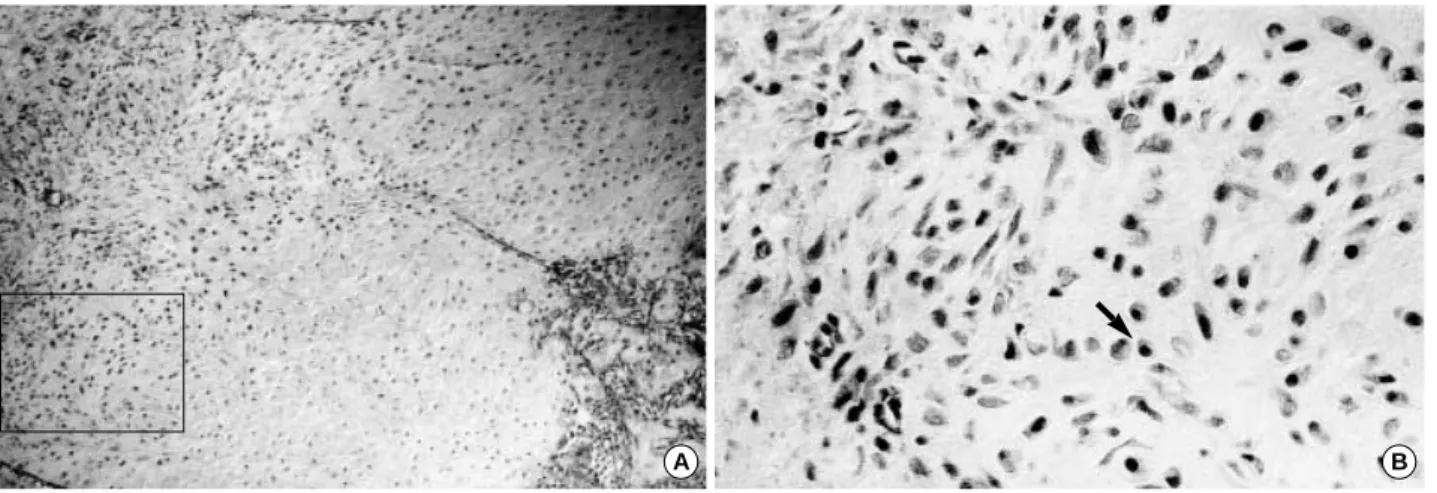
Fig. 2. Light microscopic finding of the fracture callus from a 70 and an 8 week-old rat femur, respectively (Immunohistochemical stain A, B;×400). Immunohistochemical staining for p16 (A) and p21 (B) showed the higher cytoplasmic and nuclear positive reactivity in the proliferating zone (arrow).
는 70주령, 증식성 연골세포에서는 8주령, 간엽성 조직 부위에 서는 32주령에서 발현이 높았다(Table 4). 각 부위별로 발현에 서는 c-jun과 c-fos 모두 증식성 연골세포에서 발현이 높았다 (Table 5, Fig. 3, 4).
고 찰
노화 세포의 원인에 대하여는 아직 밝혀져 있지 않지만 텔로 미어(telomere)가 짧아져서 복제가 중지되거나28), 이미 유전학
Age Hypertrophic chondrocyte
Proliferative chondrocyte
Mesenchymal layer 8 wk 10.53±11.99 54.43±14.41 47.98±12.64 32 wk 11.43±14.83* 51.92±16.80 43.69±3.72 70 wk 10.37±15.18 60.69±8.34� 49.32±13.28� Table 1.Positive ratio of p16, according to the age, in the cal- lus of the fracture site (value=mean±SD)
*, The positive ratio of p16 in the hypertrophic chondrocyte zone was higher than in any other age group (p<0.05); �, The positive ratio of p16 in the proliferative chondrocyte zone and in the mesenchymal layer was higher than in the younger age groups (p<0.05).
Age Hypertrophic chondrocyte
Proliferative chondrocyte
Mesenchymal layer 8 wk 9.12±10.53 33.07±25.82 7.05±5.29 32 wk 6.14±5.90 41.24±27.55� 15.53±14.04� 70 wk 11.89±11.02* 27.23±12.96 8.39±3.97 Table 2.Positive ratio of p21, according to age, in the callus of the fracture site (value=mean±SD)
*, The positive ratio of p21 in the zone of the hypertrophic chondro- cytes was higher than in any other age group (p<0.05); �, The positive ratio of p21 in the zone of the proliferative chondrocytes and in the mesenchymal layer was higher than in any other age group (p<0.05).
Age Hypertrophic chondrocyte
Proliferative chondrocyte
Mesenchymal layer 8 wk* 8.66±4.96 52.85±13.00 57.23±9.34 32 wk 7.92±13.43 50.83±20.88 49.06±14.04 70 wk 7.55±12.25 44.93±14.18 42.37±23.82 Table 3.Positive ratio of c-jun, according to the age, in the cal- lus of the fracture site (value=mean±SD)
*, The positive ratios of c-jun in the zone of hypertrophic chondrocytes, proliferative chondrocytes and in the mesenchymal layer were higher than in any other age group (p<0.05).
Age Hypertrophic chondrocyte
Proliferative chondrocyte*
Mesenchymal layer 8 wk 6.43±8.96 75.75±9.56� 46.03±15.78 32 wk 9.11±16.94 71.98±13.14 58.28±16.23� 70 wk 15.96±23.35* 73.23±8.90 49.54±14.69 Table 4.Positive ratio of c-fos, according to the age, in the cal- lus of the fracture site (value=mean±SD)
*, The positive ratio of c-fos in the zone of the hypertrophic chondro- cytes was higher than in any other age group (p<0.05); �, The positive ratio of c-jun in the zone of the proliferative chondrocytes was higher than in any other age group (p<0.05); �, The positive ratio of c-jun in the zone of the mesenchymal layer was higher than in any other age group (p<0.05).
A B
Fig. 3. Light microscopic finding of the fracture callus from an 8 week-old rat femur (Immunohistochemical staining for c-jun). A: Immuno- histochemical staining for c-Jun showing higher cytoplasmic and nuclear positive reactivity in the proliferating zone (×100). B: Higher magnification of the rectangular frame (×400). Black arrow indicates positive nuclear reactivity in the proliferating chondrocytes.
Age Hypertrophic chondrocyte
Proliferative chondrocyte
Mesenchymal layer p16 10.85±13.50 54.93±14.39 46.48±12.94 p21 8.68±9.00 34.75±23.66 10.67±9.97 c-jun 8.12±10.35 49.68±16.67 49.26±17.64 c-fos 9.64±16.02 73.32±11.00 51.84±16.01 Table 5.Positive ratio of p16, p21, c-jun and c-fos, according to the callus zone (value=mean±SD).
*, The positive ratios of p16, p21, c-fos and c-jun in the zone of prolifer- ative chondrocyte was higher than the others (p<0.05).
적으로 예정되어 있는 과정23)으로 설명하는 등 몇 개의 가설이 존재한다8). 노화 세포는 세포가 일정 기간동안 배가한 후 DNA 와 세포 분열이 정지되어 세포주기의 합성전기(G1 phase)에 정지하여 있고30), 어떤 분열 촉진제에도 반응하지 않으나 신진 대사나 생활력은 유지되는 세포로 정의한다13). 세포주기를 조절 하는데 있어서 CDK라고 하는 단백질이 있는데 이는 cyclin의 활성화, subunit의 인산화 혹은 탈인산화, CDK inhibitor의 조 절에 의하여 효소의 기능이 조절된다14). 세포가 노화하면 p16과 p21이 대표적으로 증가하는데, p16은 CDK inhibitor로서 CDK4와 CDK6를 억제하며25), p21은 cyclin/CDK complex에 붙어서 kinase의 기능을 억제시켜 세포 주기를 정지시킨다고 하 였다1,6,29).
사람의 섬유모세포 배양하여 복제 노화를 시킨 결과 노화 초 기에는 p21의 mRNA와 단백질이 증가하였다가 감소한 후 p16 의 mRNA와 단백질이 40배 가량 증가 하였고, 증가한 p16 단 백질은 CDK4 및 CDK6와 결합하여 억제하는 것을 발견하여, 노화 과정은 여러 단계로 구성되어 노화 초기에는 p21이 노화를 유도하고, 노화의 후기에는 p16이 세포노화를 유도하리라 생각 하였다2). 또한 p21은 여러 종류의 세포에서 말단 분화에 있어 중요한 기능을 담당하는 것으로 알려져 있다10,11,19).
골 조직에서 이들 노화 관련 단백질의 발현에 대한 연구는 많 지 않은데, Stewart 등27)은 연골세포에서 p21의 발현을 관찰하 여 성장판의 증식성 및 비후성 연골세포, 골단 골화 중심에 위 치하는 연골세포, 관절의 연골세포 등에서 높게 발현하여 p21이 연골세포의 세포 주기 조절에 관여하고, 비후성 연골세포로의 분화에 관여한다고 하였다. 또한 Bellido 등3)은 p21이 조골세포 의 분화를 촉진하고 apoptosis에 저항하는 기능을 갖게 하는데 있어 중요한 중계 역할을 한다고 하였다. 이와 같이 p16은 노화 후기에 세포 노화를 유도하고, p21은 노화 초기에 노화를 유도 하며, 연골 세포의 말단 분화에 관여하고 노화세포가 apoptosis
에 저항하게 하는 기능을 갖는 것으로 보인다.
본 실험에서는 p16과 p21은 증식성 연골세포와 간엽성 조직 에서 높게 발현하여 연골세포의 분화와 골절후의 골화과정에 관 여하고 있음을 보여 주었다. 이들 단백질들의 발현에 있어 연령 에 따른 발현은 의미 있는 차이를 보이지 않아, 이는 골절이라 는 조직의 자극에 대하여 세포가 새로운 세포주기를 갖기 때문 인 것으로 생각되어, 골조직에서 이들 단백질의 노화 유도 기능 은 분명하지 않은 것으로 보인다. 또한 본 실험에서 골절부의 고정을 시행하지 않아, 각 대조군별 가골의 크기가 일정치 않을 수 있어서 실험 결과에 영향이 있을 수 있었으리라 생각되어, 향후 연구에서는 Bonnarens 등5)이 제시한 바와 같이 표준화된 내고정 및 폐쇄적 골절 유발 방법을 사용하는 것이 실험상의 오 류를 줄일 수 있는 방법으로 생각되었다20).
c-jun과 c-fos 단백질은 세포주기의 휴지기(G0 phase) 세포 들이 어떤 자극에 대한 조기 발현 유전자(early reponse gene) 로서 헤테로다이머(heterodimer)를 형성하여 DNA 전사조절 단백질인 AP1 (activation protein factor 1)과 반응하므로서 세포의 분화 조절에 관여 하는 것으로 알려져 있다24). 골 조직 에 대한 연구는 transgenic mice와 chimeric mice에서 c-fos가 과발현할 때 각각 골육종과 연골 육종의 발생을 보고하였고31,32), c-fos의 발현이 없는 쥐에서 골화석증의 발생을 보고하면서32), 이들 단백질들이 조골세포와 연골세포의 specific transforma- tion에 영향을 미치는 것으로 생각하게 되었다. 이들 단백질은 골조직의 자극에 대한 조기 발현 유전자로서 자극이 없는 골조 직에서는 발현하지 않고26), 골절 등 외부 자극에 대하여 자극 직후에 높게 발현하였다가 서서히 감소하는 양상을 보인다15). 세포 배양 연구에서 조골 세포 발생의 증식기와 발생 말기 apoptosis가 나타나는 시기에 증가 양상을 나타내어 조골 세포 의 증식과 분화에 관여 하는 것을 보여 주었고18), 연골 세포에 서 조골 세포로 직접 분화하기도 하여 연골 세포의 분화에도 관
A B
Fig. 4. Light microscopic finding of the fracture callus from an 8 week-old rat femur (Immunohistochemical staining for c-fos). A: Immuno- histochemical staining for c-fos showing higher cytoplasmic and nuclear positive reactivity in the proliferating zone (×100). B: Higher magnification of rectangular frame (×400). Black arrow indicates positive nuclear reactivity in the proliferating chondrocytes.
여한다9). Oyama 등22)은 쥐의 대퇴골의 성장판과 골조직에서 이들 단백질의 관찰에서 성장판에서는 증식성 연골 조직과 상부 비후성 연골 조직에서 발현이 높았고, 해면골 조직에서는 조골 세포의 세포질에서 발현이 높은 것을 발견하여, c-fos가 조골 세포의 활성에 관여하고 정상 연골세포의 분화와 증식에 있어 필수적인 요소라고 하였다. Kuroki 등17)은 c-fos가 조골세포에 서 과발현하면 조골세포의 콜라겐 합성을 억제시킬 뿐 아니라 파골세포에도 영향을 미쳐 파골세포의 성숙을 촉진시키고, 골흡 수를 자극하여 파골세포의 분화와 골 재형성에 있어서도 중요한 역할을 한다고 하였다. c-fos는 조골세포 뿐 아니라 연골세포와 파골세포의 분화 및 골의 재형성에도 밀접하게 관여하는 것을 알 수 있다. 본 실험에서는 c-jun와 c-fos가 증식성 연골세포 및 간엽성 조직 순으로 높게 발현하여 이들이 연골세포의 증식과 분화에 관여하는 것을 보여주었다. 그리고 이들 단백질은 연령 에 관계없이 골절 후 2주째에 비슷한 발현을 나타내었는데, 이 는 이들 단백질의 발현이 연령보다는 골절이라는 자극과 관계가 있는 것을 보여 주는 것이라 하겠다. 또한 이들 단백질의 발현 이 가골의 각 부위에 따라 다른 발현을 보였는데, 이는 이들 단 백질이 가골의 형성 조절에 관여하고 있는 것으로 생각된다.
결 론
p16, p21, c-fos, c-jun 등의 노화관련 단백질의 발현을 골 조직에서 확인하였고, 이를 면역 조직 화학 염색법을 통하여 직 접 육안으로 확인하여, 이들 단백질이 연골세포의 분화와 골절 후 골화 과정에 관여하고 있음을 보여 주었다.
참고문헌
1. Afshari CA, Nichols MA, Xiong Y and Mudryj M:A role for a p21- E2F interaction during senescence arrest of normal human fibroblasts.
Cell Growth and Differentiation, 7: 979-988, 1996.
2. Alcorta DA, Xiong Y, Phelps D, Hannon G, Beach D and Barrett C:Involvement of the cyclin-dependent kinase inhibitor p16(INK4a) in replicative senescence of normal human fibroblasts. Proc Natl Acad Sci USA, 93: 13742-13747, 1996.
3. Bellido T, O’’Brien CA, Roberson PK and Manlagas SC:Transcrip- tional activation of the p21WAF1,CIP1,SDI1gene by interleukin-6 type cytokines.
J Biol Chemi, 273: 21137-21144, 1998.
4. Bolander ME:Regulation of fracture repair by growth factors. Poc soc Exp Biol Med, 200: 165-170, 1992.
5. Bonnarens and Thomas AE: Production of a standard closed fracture in laboratory animal bone. J orthop Res, 2: 97-101, 1984.
6. Brugarolas J, Chandrasekaran C, Gordon JI, Beach D, Jacks T
and Hannon GJ:Radiation-induced cell cycle arrest compromised by p21 deficiency. Nature, 377: 522-557, 1995.
7. Clohisy JC, Scott DK, Brakenhoff KD, Quinn CO and Partridge NC:Parathyroid hormone induces c-fos and c-jun messenger RNA in rat osteoblastic cells. Mol Endocrinol, 6: 1834-1842, 1992.
8. Cristofalo VJ and Pignolo RJ:Replicative senescence of human fibrob- last-like cells in culture. Physiol Rev, 73: 617-638, 1993.
9. Closs EI, Murray AB, Schmidt J, Schon A, Erfle V and Strauss PG:
c-fos Expression precedes osteogenic differentiation of cartilage cells in vitro. J Cell Biol. 111: 1313-1323, 1990.
10. Evers BM, Tien CK, Jing L and Thompson EA:Cell cycle protein suppression and p21 induction in differentiation Caco-2 cells. Am J Phys- iol, 271: G722-G727, 1996.
11. Halevy O, Novitch BG, Spicer DB, et al:Correlation of terminal cell cycle arrest of skeletal muscle with induction of p21 by MyoD. Science, 267:
1018-1021, 1995.
12. Harris WH and Weinberg ZH:Microscopic method of measuring increases in cortical bone volume and mass. Calcif Tissue Res, 8: 190-197, 1972.
13. Hayflic L: The cell biology of human aging. N Engl J Med, 295: 1302- 1308, 1976.
14. Hunter T and Pines J:Cyclins and Cancer II: cyclin D and CDK inhi- bitors come of age. Cell, 79: 573-582, 1994.
15. Inaoka T, Lean JM, Bessho T, et al:Sequential analysis of gene expres- sion after an osteogenic stimulus: c-fos expression is induced in osteocytes.
Biochem Biophys Res Commun, 217: 264-270, 1995.
16. Jackson ME, Shalhoub V, Lian JB, Stein GS and Marks SC Jr:
Aberrant gene expression in cultured mammalian bone cells demonstrates an osteoblast defect in osteopetrosis. J Cell Biochem, 55: 366-372, 1994.
17. Kuroki Y, Shiozawa S, Sugimoto T, et al:Constitutive c-fos expres- sion in osteoblastic MC3T3-E1 cells stimulates osteoclast maturation and osteoclastic bone resorption. Clin Exp Immunol, 95: 536-539, 1994.
18. McCabe LR, Kockx M, Lian J, Stein J and Stein G:Selective Expres- sion of Fos- and jun-related genes during osteoblast proliferation and dif- ferentiation. Experimental Cell Res, 218: 255-262, 1995.
19. Missero C, Calautti E, Eckner R, et al:Involvement of the cell cycle inhibitor Cipl/Wafl and the EIA-associated p300 protein in terminal dif- ferentiation. Proc Natl Acad Sci USA, 92: 5451-5455, 1995.
20. Nunamarker DM:Experimental models of fracture repair. Clin Orthop Rel Res, 355: 56-65, 1998.
21. Okamoto A, Demetrick DJ, Spillare EA, et al:Mutations and altered expression of p16INK4in human cancer. Proc Natl Acad Sci USA, 91:
11045-11049, 1994.
22. Oyama M, Chiba J, Kato Y, et al:Distribution and expression of mRNAs for the proto-oncogenes c-fos and c-jun in bone cells in vivo. Histol Histo- pathol, 13: 671-678, 1998.
23. Pereira-Smith OM and Smith JR:Genetic analysis of indefinite divi- sion in human cells: identification of four complementation groups. Proc Natl Acad Sci USA, 85: 6042-6046, 1988.
24. Riabowol K, Schiff J and Gilman MZ:Transcription factor AP-1 activity is required for initiation of DNA synthesis and is lost during cel- lular aging. Proc Natl Acad Sci USA, 89: 157-161, 1992.
25. Sherr CJ and Roberts JM:Inhibitors of mammalian G1 cyclin-depen- dent kinases. Genes and Dev, 9: 1149-1163, 1995.
26. Ohta S, Yamamuro T, Lee K, et al:Fracture healing induces expres- sion of the Fos protein in osteoblastic differentiation. Febs, 284: 42-45, 1991.
27. Stewart MC, Farnum CE and MacLeod JN:Expression of p21CIP1/WAF1 in chondrocytes. Calcif Tissue Int, 61: 199-204, 1997.
28. Wright WE and Shay JW: Telomere positional effects and the regulation
of cellular senescence. Trends Genet, 8: 193-197, 1992.
29. Xiong Y, Hannon GJ, Hui Z, Casso D, Kobayashi R and Beach D:
p21 is a universal inhibitor of cyclin kinases. Nature, 336: 701-704, 1993.
30. Yanishevsky R, Mendelsohn ML, Mayall BH and Cristofalo VJ:
Proliferative capacity and DNA content of aging human diploid cells in culture: a cytophotometric and autoradiographic analysis. J Cell Physiol, 84: 165-170, 1974.
31. Wang ZQ, Grigoriadis AE and Wagner EF:Stable murine chondro- genic cell ines derived from c-fos-induced cartilage tumors. J Bone Miner Res, 8: 839-847, 1993.
32. Wang ZQ, Ovitt C, Grigoriadis AE, Mohle-Steinlein U, Ruther U and Wagner EF:Bone and hematopoietic defects in mice lacking c-fos.
Nature, 360: 741-745, 1992.
Purpose :To evaluate the expression of senescence-related proteins according to the aging process and to determine the role of senescence-related proteins in the bone tissue and their effects on the process of bone union.
Materials and Methods :18 Sprague-Dawley rats (8 weeks old: 7, 32 weeks old: 6, and 70 weeks old: 5) were used in the experiment. A unilateral closed femur fracture was made, and the fracture callus was obtained 2 weeks after the fracture. The ossification process was observed in prolifera- tive chondrocytes, the hypertrophic chondrocytes, and in the mesenchymal layer individually by immunohistochemistry, using p16, p21, c-fos and c-jun antibodies.
Results : There was no significant differences in the manifestation of p-16, p-21, c-fos, c-jun gene according to the age. The positive ratio of p-16 was maximal in proliferative chondrocytes (54.93%) and decreased in the mesenchymal layer (46.48%), and in hypertrophic chondrocytes (10.85%), in order. The positive ratio of c-fos was maximal in proliferative chondrocytes (73.32%) and decreased in the mesenchymal layer (51.84%), and in hypertrophic chondrocytes (9.64%), in order.
Conclusion :We believe that senescent genes in the bone tissue participate in the differentiation of osteochondral cells and in the process of fracture callus ossification.
Key Words : Senescence-related protein, Callus, Immunohistochemistry, Aging, p16, p21, c-fos, c-jun
Expression of Senescence-related Proteins in Rat Fracture Callus
Sang Ho Song, M.D.**, Young Euy Park, M.D.*, Young Hee Choi, M.D.*, Chang Gu Shim, M.D., and Seong Woo Cho, M.D.
Department of Orthopaedic Surgery, Inchon Christian Hospital, Inchon; Department of Pathology, College of Medicine, Hallym University*, Chunchon; Song’s Orthopedic Surgery, Puchon**, Korea
Abstract
Address reprint requests to Sang Ho Song, M.D.
Song’s Orthopedic Surgery
1035-3 Jung-dong, Wonmi-gu, Puchon 420-020, Korea Tel : +82.32-323-5114, Fax : +82.32-327-2333 E-mail: [email protected]
. .