Anti-Inflammatory Effect of Licochalcone E, a Constituent of Licorice, on Lipopolysaccharide-Induced Inflammatory Responses in Murine Macrophages
Geun-Mook Park1, Jong-Gab Jun2 and Jin-Kyung Kim1*
1Department of Biomedical Science, College of Natural Science, Catholic University of Daegu, Gyeoungsan-Si 700-712, Korea
2Department of Chemistry and Institute of Natural Medicine, Hallym University, Chuncheon 200-702, Korea Received March 3, 2011 /Accepted May 16, 2011
Licochalcone, a major phenolic constituent of the licorice species Glycyrrhiza inflata, a constituent of licorice, exhibits various biological properties, including chemopreventive-, antibacterial-, and anti- spasmodic activities. Recently, Licochalcone E (LicE) was isolated from the roots ofGlycyrrhiza inflate, however its biological functions have not been fully examined. In the present study, we investigated the ability of LicE to regulate inflammation reactions in macrophages. Ourin vitro experiments using murine macrophages, RAW264.7 cells, showed that LicE suppressed not only nitric oxide (NO) and prostaglandin E2 generation, but also the expression of inducible NO synthase and cyclooxygenase-2 induced by lipopolysaccharide (LPS). Similarly, LicE inhibited the release of proinflammatory cyto- kines induced by LPS in RAW264.7 cells, including tumor necrosis factor-α and interleukin-6. The underlying mechanism of LicE on anti-inflammatory action correlated with down-regulation of the nu- clear factor-κB. Our data collectively indicate that LicE inhibited the production of several in- flammatory mediators and might be used in the treatment of various inflammatory diseases.
Key words : Inflammation, licochalcone E, NF-κB, macrophage, inflammatory mediators
*Corresponding author
*Tel:+82-53-850-3774, Fax:+82-53-850-3774
*E-mail : [email protected]
Introduction
Inflammation is part of complex biological responses to harmful stimuli, such as pathogens, damaged cells, or irritants. An inflammatory reaction is self-limiting and in- volves the down-regulation of proinflammatory protein ex- pression and increased expression of anti-inflammatory pro- teins that facilitate the initial immune cell recruitment proc- ess [5,22,23,29]. During this response, inflammatory cells are exposed to apoptosis-inducing stimuli and subsequently un- dergo phagocytosis. Prolonged inflammation contributes to the pathogenesis of many inflammatory diseases [11,31].
The macrophage plays a critical role in the inflammatory response and serves as an essential interface between innate and adaptive immunity. Following activation, macrophages modulate the expression of accessory molecules, such as CD14 and Toll-like receptor (TLR) 4 [27,35]. Stimulation of TLR4 by lipopolysaccharide (LPS) in the presence of LPS-binding protein, CD14, and the MD2 protein triggers the recruitment of the cytoplasmic adaptor protein MyD88 and subsequently culminates in the activation of down- stream signaling pathways, such as the transcription factor
nuclear factor-κB (NF-κB) pathway [10]. This pathway in- duces the expression of various inflammatory mediators, in- cluding nitric oxide (NO), prostaglandins (PGs), and the in- flammatory cytokines [3,12,14,28].
NF-κB, the key molecule in inflammatory responses, is a generic term for a dimeric transcription factor that is formed by the dimerization of proteins in the Rel family [8].
NF-κB activity is exerted through its ability to regulate the expression of genes that encode inflammatory cytokines, ad- hesion molecules, chemokines, growth factors, and inducible enzymes, such as cyclooxygenase (COX)-2 and inducible NO synthase (iNOS) [2].
PGs are produced by a variety of cell types, including the activated macrophage [19]. The rate-limiting enzyme in the synthesis of PGs is COX. Two main isoforms of COX have been described: COX-1 and COX-2. COX-1 is expressed constitutively in most tissues and is responsible for the ho- meostatic production of PGs. In contrast, COX-2 is induced by several stimuli, including growth factors, mitogens, cyto- kines, and tumor promoters. Its uncontrolled activity is thought to play an important role in the pathogenesis of many chronic inflammatory diseases [1,19]. In addition to PGs, NO is synthesized from l-arginine by NO synthases.
Overproduction of NO by iNOS has been involved in the pathology of several inflammatory disorders, including sep-
tic shock, tissue damage following inflammation [16,20,21].
Based on these observations, it has been hypothesized that the suppression of NO and PG production in macrophages could be key mechanisms to utilize in anti-inflammatory drug development.
Currently, a strong interest exists in developing new an- ti-inflammatory drugs from various natural products used in traditional medicine. Licorice has been used for more than four millennia as a flavoring agent in foods, beverages, and tobacco, as well as an alternative medicine to treat in- dividuals with gastric or duodenal ulcers, sore throat, bron- chitis, cough, arthritis, adrenal insufficiency, and allergies [34]. Among the constituents of licorice, licochalcone E (LicE) was isolated recently, however, its biological functions have yet to be fully tested [33]. Therefore, the anti-inflammatory effects of LicE were investigated using LPS-induced in- flammatory response in the present study.
Materials and Methods Chemicals and reagents
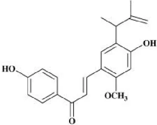
LPS derived from Escherichia coli and dimethylsulfoxide (DMSO) was obtained from Sigma (St Louis, Mo, USA). The Dulbecco’s modified Eagle’s medium (DMEM), fetal bovine serum (FBS), penicillin, and streptomycin used in this study were obtained from Hyclone (Logan, Utah, USA). Synthetic LicE (Fig. 1) was provided by Dr. Jong-Gab Jun [18]; it was dissolved in DMSO and added directly to the culture media.
The final concentrations of DMSO never exceeded 0.1%, which did not affect the assay systems. The antibodies (Abs) used in this study were: anti-iNOS rabbit polyclonal, an- ti-COX-2 mAb, anti-IκB mAb, anti-phospho-JNK (pT183, pY185) rabbit polyclonal, anti-ERK1/2 rabbit polyclonal, an- ti-phospho-p38 (pT180, pY182) rabbit polyclonal, anti-p38 rabbit polyclonal (Cell Signaling Technology, Danvers, MA), and anti-β-actin mAb (Sigma).
Cell culture and cell viability assay
RAW264.7 murine macrophages were obtained from the Korean Cell Bank (Seoul, Korea) and cultured in DMEM con- taining 10% FBS, 100 U/ml penicillin, and 100 μg/ml strep- tomycin at 37oC in 5% CO2. The effects of LicE on cell via- bility were tested using the CellTiter 96® AQueous One Solution Assay of cell proliferation (Promega, Madison, WI), which uses colorimetry to count the number of viable cells.
RAW264.7 cells were plated at a density of 2×10⁴ cells in
Fig. 1. Chemical structure of LicE.
a 96-well flat-bottom plate, and LicE were added to each plate at indicated concentrations. After a 24 hr incubation period, the number of viable cells was counted according to the manufacturer's instructions. This assay is based on the reduction of a tetrazolium compound, MTS, to formazan, which has an optimum absorption at 490 nm. Thus, the quantity of the product in the cell culture is indicated by the optical density of formazan at 490 nm, which is directly proportional to the number of living cells.
Measurement of nitrite and PGE2
The amount of nitrite and PGE2produced by the mouse macrophages was measured in the RAW264.7 cell culture supernatant. RAW264.7 cells were plated at a density of 2.5×10⁵ cells in a 48-well cell culture plate with 500 μl of culture medium and incubated for 12 hr. They were then treated with indicated concentrations of LicE plus LPS (100 ng/ml) and incubated for another 18 hr. The amount of ni- trite produced was measured using the Griess reagent sys- tem (Promega). The amount of PGE2 produced was meas- ured using an enzyme-linked immunosorbent assay (ELISA) kit (R & D, Minneapolis, MI,) according to the manu- facturer’s instructions.
Cytokine assays
The amount of TNF-α and IL-6 in the cell culture super- natant was measured using an ELISA kit (eBioscience, San Diego, CA). RAW264.7 cells were plated in a 48-well cell culture plate at a density of 2.5×10⁵cells and incubated with indicated concentrations of LicE in 100 ng/ml LPS for 24 hr. The culture supernatant was collected and assayed ac- cording to the manufacturer’s instructions to determine the
0 10 20 30
*** ***
***
***
NO(mM)
vehicle
LPS + LicE (mM)
0 2.5 5 10 20
1000
0 2000
PGE2(pg/ml)
vehicle
LPS + LicE (mM)
0 2.5 5 10 20
***
*** ***
***
A B
0 10 20 30
*** ***
***
***
NO(mM)
vehicle
LPS + LicE (mM)
0 2.5 5 10 20
1000
0 2000
PGE2(pg/ml)
vehicle
LPS + LicE (mM)
0 2.5 5 10 20
***
*** ***
0 ***
10 20 30
*** ***
***
***
NO(mM)
vehicle
LPS + LicE (mM)
0 2.5 5 10 20
0 10 20 30
*** ***
***
***
*** ***
***
***
NO(mM)
vehicle
LPS + LicE (mM)
0 2.5 5 10 20
vehicle
LPS + LicE (mM)
0 2.5 5 10 20
0 2.5 5 10 20
1000
0 2000
PGE2(pg/ml)
vehicle
LPS + LicE (mM)
0 2.5 5 10 20
***
*** ***
***
1000
0 2000
PGE2(pg/ml)
vehicle
LPS + LicE (mM)
0 2.5 5 10 20
vehicle
LPS + LicE (mM)
0 2.5 5 10 20
0 2.5 5 10 20
***
*** ***
***
A B
Fig. 3. Effects of LicE on LPS-induced NO and PGE2 release. RAW264.7 cells were treated with 0-20 μM of LicE in the presence of 100 ng/ml of LPS or with LPS alone for 18 hr, and NO (A) and PGE2 (B) release were determined. The results are reported as mean±SE of four independent experiments in triplicate. Statistical significance is based on the difference when compared with LPS-stimulated cells (***p<0.001).
amount of TNF-α and IL-6 that had been released from the cell.
Immunoblotting analysis
Whole cell extracts (30 μg protein/lane) were separated on 10% SDS-polyacrylamide gels. The separated proteins were electrophoretically transferred onto nitrocellulose membranes. Immunoreactive bands were detected by in- cubating the samples with horseradish peroxidase (HRP)- conjugated secondary antibodies and WesternBreeze®
Chromogenic Kits (Invitrogen, Carlsbad, CA).
NF-κB activity
RAW264.7 cells were placed in a 24-well cell culture plate at a density of 5×10⁵cells in 0.5 ml of culture medium and incubated for 12 hr. The cells were then exposed to 0-20 μM LicE and LPS (100 ng/ml) for 4 hr. The transcriptional activ- ities of NF-κB were measured using TransAM (Active Motif, Carlsbad, CA), according to the manufacturer’s instructions.
Statistical analysis
The data are depicted as the means±SE. The values were evaluated by one-way analysis of variance (ANOVA) with Bonferroni multiple comparison post tests using the GraphPad Prism 4.0 software (GraphPad Software Inc., San Diego, CA). Differences withp<0.05 were considered to be statistically significant.
Results
LicE inhibits the releases of NO and PGE2
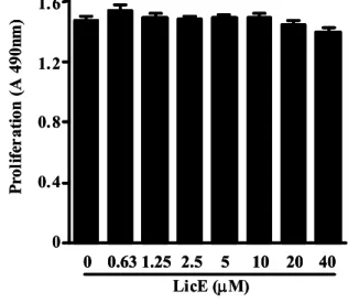
To evaluate LicE-induced anti-inflammatory effects, we used an in vitro model with the murine RAW264.7 macro- phage cell line. Since LicE showed no cytotoxicity with con- centrations up to 20 μM in RAW264.7 macrophages (Fig. 2), we used up to 20 μM LicE for the rest of the experiments.
We first sought to determine the effect of LicE on the
0 0.4 0.8 1.2 1.6
0 0.631.25 2.5 5 10 20 40 LicE (mM)
Proliferation (A 490nm)
0 0.4 0.8 1.2 1.6
0 0.631.25 2.5 5 10 20 40 0 0.631.25 2.5 5 10 20 40
LicE (mM)
Proliferation (A 490nm)
Fig. 2. Effects of LicE on murine macrophage viability.
RAW264.7 cells were treated with indicated concen- trations of LicE for 24 hr, and proliferation was de- termined as described in Materials and Methods.
0 10 20 30 40
***
TNF-a(ng/ml)
***
**
vehicle
LPS + LicE (mM)
0 2.5 5 10 20
*** 0
1 2 3 4 5 6
***
IL-6(ng/ml) ***
vehicle
LPS + LicE (mM)
0 2.5 5 10 20
***
A B
0 10 20 30 40
***
TNF-a(ng/ml)
***
**
vehicle
LPS + LicE (mM)
0 2.5 5 10 20
0 10 20 30 40
***
TNF-a(ng/ml)
***
**
vehicle
LPS + LicE (mM)
0 2.5 5 10 20
vehicle
LPS + LicE (mM)
0 2.5 5 10 20
0 2.5 5 10 20
*** 0
1 2 3 4 5 6
***
IL-6(ng/ml) ***
vehicle
LPS + LicE (mM)
0 2.5 5 10 20
***
0 1 2 3 4 5 6
***
IL-6(ng/ml) ***
vehicle
LPS + LicE (mM)
0 2.5 5 10 20
0 1 2 3 4 5 6
***
IL-6(ng/ml) ***
vehicle
LPS + LicE (mM)
0 2.5 5 10 20
vehicle
LPS + LicE (mM)
0 2.5 5 10 20
0 2.5 5 10 20
***
A B
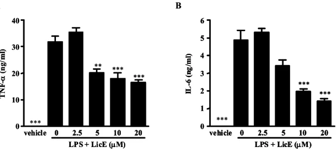
Fig. 5. Effects of LicE on LPS-induced inflammatory cytokine production in murine macrophages. RAW264.7 cells were treated with 0-20 μM of LicE in the presence of 100 ng/ml LPS or with LPS alone for 24 hr. The cell culture media were then collected, and the amount of TNF-α (A) and IL-6 (B) released was measured as described in Materials and Methods. The results are reported as mean±SE of three independent experiments in triplicate. Statistical significance is based on the difference when compared with LPS-stimulated cells (**p<0.01, ***p<0.01).
LPS-stimulated release of the inflammatory mediators, NO and PGE2, by RAW264.7 cells. As shown in Fig. 3A, LicE inhibited LPS-induced NO and PGE2 production in a dose-dependent manner. The nitrite concentrations in LPS-stimulated cells and in those exposed to 20 μM LicE were 26.3±0.9 μM and 13.5±0.5 μM, respectively. The in- hibitory effects of LicE on PGE2production in LPS-exposed cells were similar to their effects on NO production (Fig.
3B). Consistent with the findings related to NO and PGE2
production, immunoblotting analysis showed that LPS-re- lated iNOS and COX-2 induction in RAW264.7 cells was also reduced by LicE treatment (Fig. 4). This indicates that the LicE-induced reduction in iNOS and COX-2 expression was responsible for the inhibition of NO and PGE2
production.
LicE inhibits the release of proinflammatory cyto- kines
We next examined if LicE reduced the release of proin- flammatory cytokines in LPS-stimulated RAW264.7 cells.
The release of TNF-α and IL-6 was significantly impaired by LicE co-treatment of LPS-exposed cells (Fig. 5). These re- sults indicate that LicE suppressed various inflammatory mediators including NO and PGE2, as well as proin- flammatory cytokines.
iNOS
COX-2
b-actin
vehicle
LPS + LicE (mM)
0 2.5 5 10 20
iNOS
COX-2
b-actin
vehicle
LPS + LicE (mM)
0 2.5 5 10 20
0 2.5 5 10 20
Fig. 4. Effects of LicE on LPS-induced iNOS and COX-2 expression. RAW264.7 cells treated the same as in Fig.
2. Thirty μg of protein obtained from each cell lysate was resolved on 10% SDS-PAGE for iNOS and COX-2 determination. β-actin expression is shown as a loading control.
LicE-reduced inflammatory mediator release is regulated by NF-κB
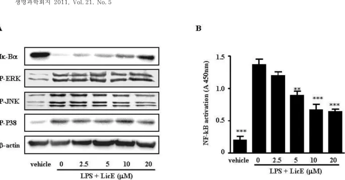
We further investigated the mechanism by which LicE in- hibited the production of inflammatory mediators. NF-κB is a major transcription factor involved in the release of pro- teins that mediate the inflammatory response, and the degra- dation and phosphorylation of inhibitor of κB (Iκ-B) are nec- essary to release NF-κB from the cytoplasmic NF-κB/Iκ-Bα complex and allow its subsequent translocation to the cell nucleus. We evaluated the effect of LicE on NF-κB activation to determine if it is mediated by Iκ-B degradation. We found
Fig. 6. Effect of LicE on LPS-induced Iκ-B degradation and DNA-binding of NF-κB in LPS-stimulated murine macrophages. (A) RAW264.7 cells were plated in 100-mm dishes. After 12 hr of seeding, cells were treated with different doses of LicE for 1 hr, followed by stimulation with 100 ng/ml of LPS for 20 min. Whole cell extracts were immunoblotted with the indicated Abs. β-actin was used as a control. (B) RAW264.7 cells were treated with various concentrations of LicE in the presence of 100 ng/ml LPS or with LPS alone for 4 hr. After nuclear protein extraction, 2 μg of nuclear protein were assayed for the ability to bind to the immobilized NF-κB consensus site as described in Materials and Methods. The results are reported as mean±SE of three independent experiments in triplicate. Statistical significance is based on the difference when compared with LPS-stimulated cells (**p<0.01, ***p<0.001).
that LPS-induced Iκ-B degradation was inhibited after 20 mi- nutes of exposure to LicE (Fig. 6A).
LPS activates not only NF-κB, but also MAPK family members such as ERK, p38, and JNK, and it plays a crucial role in mediating the induction of proinflammatory cytokines. We sought to determine if LicE modulated the MAPK signal transduction pathway. As shown in Fig. 5A, the activation of ERK, JNK, and p38 via LPS stimulation in RAW264.7 cells was not altered by LicE.
To directly observe the effect of LicE on the nuclear trans- location and DNA binding of NF-κB, we performed a tran- scription factor ELISA assay. We found that LPS-induced DNA-binding of NF-κB was significantly inhibited in LPS-stimulated RAW264.7 cells exposed to LicE in a dose-dependent manner (Fig. 6B).
Discussion
Numerous studies have focused on herbal remedies and botanicals since they offer much promise in health benefits and disease treatments without excessive side effects and cytotoxicity [17]. A wide variety of plant-derived products have shown an anti-inflammatory effect, but only a few have
been examined to determine the molecular mechanism of this inhibitory action. It has been reported that licorice is one of the most common plants prescribed by practitioners of traditional alternative medicine [12,16,21]. Indeed, our previous study demonstrated that ethanol extracts of licorice exert suppressive effects on LPS-induced inflammatory re- actions [20]. Moreover, glabridin, a major component of the hydrophobic fraction of licorice, has preventive and ther- apeutic effects on DSS-induced colonic inflammation [21].
The focus of this report is on the anti-inflammatory potency of LicE, the characteristic phenolic constituent of licorice [3,27].
In this study, we investigated the anti-inflammatory prop- erties of LicE and dissected its possible molecular mecha- nism of action. The results presented herein showed that LicE significantly decreased NO and PGE2 production in LPS-stimulated murine macrophages. The inhibition of NO and PGE2was due to the inhibition of iNOS and COX-2 ex- pression, respectively, at protein levels as shown by immunoblot. Moreover, we demonstrated that LicE sup- pressed iNOS and COX-2 expression as well as the release of proinflammatory cytokines, possibly via the inhibition of LPS-induced NF-κB activation. Several recent studies re-
ported that reduced NF-κB activation by LicE induces apop- tosis in endothelial cells [7] and inhibits the symptoms of chronic contact dermatitis in mice [9]. Although the exact mechanisms by which LicE inhibits NF-κB activation are not known, one possible explanation is that reduction of NO, a reactive oxygen species, by LicE may be involved in NF-κB inactivation because licochalcones are known to be reactive oxygen scavengers [16].
Accumulated evidence indicates that dysregulation of cy- tokines, such as TNF-α and IL-6, plays an essential role in many inflammatory conditions like septic shock, hemor- rhagic shock, rheumatoid arthritis, and atherosclerosis [13,25]. Inhibition of inflammatory cytokine and mediator production or function serves as a key mechanism in the control of inflammation, and agents that suppress the ex- pression of these inflammation-associated genes have ther- apeutic potential in the treatment of inflammatory diseases.
Therefore, natural phytochemicals have attracted clinical in- terest as potential therapeutic agents for the treatment of inflammation. In addition to proinflammatory cytokines, COX-2 is an essential molecule to regulate inflammatory responses. The PG production in LPS-treated macrophages is primarily due to transcriptional activation of the COX-2 gene [24]. Thus, potential COX-2 inhibitors have been con- sidered as anti-inflammatory agents.
Expression of iNOS, COX-2, TNF-α, and IL-6 requires the activation of NF-κB, which is an important proximal mecha- nism for the overproduction of the inflammatory mediators in macrophages in response to LPS and cytokines [26].
Meanwhile, NF-κB plays a critical role in apoptosis, various autoimmune diseases and inflammation [2,15]. Recently, many investigations demonstrated that many anti-in- flammatory drugs including dexamethasone can inhibit the production of IL-1β, TNF-α, and NO by suppression of NF- κB activation [32]. Due to its ubiquitous role in the patho- genesis of inflammatory gene expression, NF-κB is a current target for treating various inflammation diseases [30]. Since multiple studies have demonstrated the importance of these inflammation factors in the inflammation process, the an- ti-inflammatory action of several drugs at the cellular level could be explained by their effects on these factors. Hence we studied these key inflammation cytokines and mediators in LPS-stimulated RAW 264.7 cells to elucidate the anti-in- flammatory effects of LicE.
In conclusion, our observations suggest that LicE strongly inhibits the production of inflammation-associated media-
tors by suppressing NF-κB activation, and therefore, LicE may be a potential candidate for treating various in- flammatory diseases.
Acknowledgement
This research was supported by Basic Science Research Program through the National Research Foundation of Korea (NRF) funded by the Ministry of Education, Science and Technology (2010-0004728) and Priority Research Centers Program through the National Research Foundation of Korea (NRF) funded by the Ministry of Education, Science and Technology (2010-0029642).
References
1. Aktan, F. 2004. iNOS-mediated nitric oxide production and its regulation. Life Sci. 75, 639-653.
2. Baker, R. G., M. S. Hayden, and S. Ghosh. 2011. NF-κB, in- flammation, and metabolic disease. Cell Metab. 13, 11-22.
3. Barfod, L., K. Kemp, M. Hansen, and A. Kharazmi. 2002.
Chalcones from chinese liquorice inhibit proliferation of T cells and production of cytokines.Int. Immunopharmacol.2, 545-555.
4. Bielory, L. 2004. Complementary and alternative inter- ventions in asthma, allergy, and immunology.Ann. Allergy Asthma Immunol. 93, S45-54.
5. Blackwell, T. S. and J. W. Christman. 1997. The role of nu- clear factor-kappa B in cytokine gene regulation. Am. J.
Respir. Cell Mol. Biol. 7, 3-9.
6. Carbonell-Barrachina, A. A., P. Aracil, E. Garcia, F. Burlo, and F. Martinez-Sanchez. 2003. Source of arsenic in licorice confectionery products. J. Agric. Food Chem.51, 1749-1752.
7. Chang, H. J., G. Yoon, J. S. Park, M. H. Kim, M. K. Baek, N. H. Kim, B. A. Shin, B. W. Ahn, S. H. Cheon, and Y.
D. Jung. 2007. Induction of apoptosis by the licochalcone E in endothelial cells via modulation of NF-kappaB and Bcl-2 Family. Biol. Pharm. Bull. 30, 2290-2293.
8. Cho, M. K., S. H. Suh, and S. G. Kim. 2002. JunB/AP-1 and NF-kappa B-mediated induction of nitric oxide synthase by bovine type I collagen in serum-stimulated murine macrophages. Nitric Oxide6, 319-332.
9. Cho, Y. C., S. H. Lee, G. Yoon, H. S. Kim, J. Y. Na, H. J.
Choi, C. W. Cho, S. H. Cheon, and B. Y. Kang. 2010.
Licochalcone E reduces chronic allergic contact dermatitis and inhibits IL-12p40 production through down-regulation of NF-kappa B. Int. Immunopharmacol. 10, 1119-26.
10. Fu, Y. T., C. Hsieh, J. Guo, J. Kunicki, M. Y. Lee, Z.
Darzynkiewicz, and J. M. Wu. 2004. Licochalcone-A, a novel flavonoid isolated from licorice root (Glycyrrhiza glabra), causes G2 and late-G1 arrests in androgen-independent PC-3 prostate cancer cells. Biochem. Biophys. Res. Commun.
322, 263-270.
11. Fukai, T., A. Marumo, K. Kaitou, T. Kanda, S. Terada, and T. Nomura. 2002. Anti-Helicobacter pylori flavonoids from licorice extract. Life Sci. 71, 1449-1463.
12. Fukai, T., K. Satoh, T. Nomura, and H. Sakagami. 2003.
Preliminary evaluation of antinephritis and radical scaveng- ing activities of glabridin fromGlycyrrhiza inflate.Fitoterapia 74, 624-629.
13. Ghezzi, P. and A. Cerami. 2005. Tumor necrosis factor as a pharmacological target. Mol. Biotechnol. 31, 239-244.
14. Guha, M. and N. Mackman. 2001. LPS induction of gene expression in human monocytes. Cell Signal. 13, 85-94.
15. Gyrd-Hansen, M. and P. Meier. 2010. IAPs: from caspase inhibitors to modulators of NF-kappaB, inflammation and cancer. Nat. Rev. Cancer 10, 561-74.
16. Haraguchi, H., H. Ishikawa, K. Mizutani, Y. Tamura, and T. Kinoshita. 1998. Antioxidative and superoxide scaveng- ing activities of retrochalcones inGlycyrrhiza inflata. Bioorg.
Med. Chem. 6, 339-347.
17. Jachak, S. M. and A. Saklani. 2007. Challenges and oppor- tunities in drug discovery from plant. Curr. Sci. 92, 1251-1257.
18. Jeon, J. H., M. R. Kim, E. M. Kwon, N. R. Lee, and J. G.
Jun. 2011. Highly efficient synthesis of licochalcone E through water-accelerated [3,3]-sigmatropic rearrangement of allyl aryl ether. Bull. Korean Chem. Soc. 32, 1059-1062.
19. Kamimura, D., K. Ishihara, and T. Hirano. 2003. IL-6 signal transduction and its physiological roles: the signal orches- tration model. Rev. Physiol. Biochem. Pharmacol. 149, 1-38.
20. Kim, J. K., S. M. Oh, H. S. Kwon, Y. S. Oh, S. S. Lim, and H. K. Shin. 2006. Anti-inflammatory effect of roasted licorice extracts on lipopolysaccharide-induced inflammatory re- sponses in murine macrophages. Biochem. Biophys. Res.
Commun. 345, 1215-1223.
21. Kwon, H. S., S. M. Oh, and J. K. Kim. 2008. Glabridin, a functional compound of liquorice, attenuates colonic in- flammation in mice with dextran sulphate sodium-induced colitis. Clin. Exp. Immunol. 151, 165-173.
22. Lang, R. M., M. Hammer, and J. Mages. 2006. DUSP meet immunology: dual specificity MAPK phosphatases in con- trol of the inflammatory response. J. Immunol. 177, 7497-7504.
23. Lawrence, T. D., A. Willoughby, and D. W. Gilroy. 2002.
Anti-inflammatory lipid mediators and insights into the res- olution of inflammation. Nat. Rev. Immunol.2, 787-795.
24. Lo, C. J., M. Fu, F. R. Lo, and H. G. Cryer. 2000.
Cyclooxygenase 2 (COX-2) gene activation is regulated by cyclic adenosine monophosphate. Shock13, 41-45.
25. Montecucco, F. and F. Mach. 2009. Common inflammatory mediators orchestrate pathophysiological processes in rheu- matoid arthritis and atherosclerosis.Rheumatology48, 11-22.
26. Müller, J. M., H. W. Ziegler-Heitbrock, and P. A. Baeuerle.
1993. Nuclear factor kappa B, a mediator of lip- opolysaccharide effects. Immunobiology 187, 233-56.
27. Nielsen, S. F., T. Boesen, M. Larsen, K. Schonning, and H.
Kromann. 2004. Antibacterial chalcones–bioisosteric re- placement of the 4′-hydroxy group.Bioorg. Med. Chem.12, 3047-3054.
28. Ohsawa, M., T. Koyama, N. Nara, and S. Hirosawa. 2003.
Induction of tissue factor expression in human monocytic cells by protease inhibitors through activating activator pro- tein-1 (AP-1) with phosphorylation of Jun-N-terminal kinase and p38. Throm. Res. 112, 313-320.
29. O'Shea, J. J. and P. J. Murray. 2008. Cytokine signaling mod- ules in inflammatory responses. Immunity 28, 477-487.
30. Renard, P. and M. Raes. 1999. The proinflammatory tran- scription factor NFkappaB: a potential target for novel ther- apeutical strategies. Cell Biol. Toxicol. 15, 341-3444.
31. Tominaga, K., S. Saito, M. Matsuura, and M. Nakano. 1999.
Lipopolysaccharide tolerance in murine peritoneal macro- phages induces downregulation of the lipopolysaccharide signal transduction pathway through mitogen-activated protein kinase and nuclear factor-kappaB cascades, but not lipopolysaccharide-incorporation steps. Biochim. Biophys.
Acta. 1450, 130-144.
32. Yamamoto, Y. and R. B. Gaynor. 2001. Role of the NF-kappaB pathway in the pathogenesis of human disease states. Curr. Mol. Med. 1, 287-296.
33. Yoon, G., Y. D. Jung, and S. H. Cheon. 2005. Cytotoxic allyl retrochalcone from the roots of Glycyrrhiza inflata. Chem.
Pharm. Bull. 53, 694-695.
34. Zhou, S., H. L. Koh, Y. Gao, Z. Y. Gong, and E. J. Lee. 2004.
Herbal bioactivation: the good, the bad and the ugly.Life Sci. 74, 935-968.
35. Ziegler, H. L., H. S. Hansen, D. Staerk, S. B. Christensen, H. Hagerstrand, and J. W. Jaroszewski. 2004. The anti- parasitic compound licochalcone A is a potent echinocyto- genic agent that modifies the erythrocyte membrane in the concentration range where antiplasmodial activity is observed. Antimicrob. Agents. Chemother. 48, 4067-4071.
초록:Licochalcone E의 항염증 효과와 그 기전에 대한 연구 박근묵1․전종갑2․김진경1*
(1대구가톨릭대학교 의생명과학과, 2한림대학교 화학과)
Licochalcone은 감초의 주요 생리활성 물질로 항균작용, 항암작용 등의 다양한 효과가 있는 것으로 알려져 있 다. 최근 감초로부터 licochalcone E가 분리, 동정 되었을 뿐만 아니라, 효과적인 licochalcone E의 합성을 위해 다양한 합성법이 개발되고 있다. 반면, licochalcone E의 생리활성 연구는 매우 미비한 상태이다. 본 연구는 li- cochclcone E의 항염증 활성과 그 기전의 일단을 밝히는 것을 목적으로 진행 되었다. 생쥐의 대식세포주인 RAW264.7 세포에 lipopolysaccharide (LPS)를 처리 염증반응을 유도하고, licochalcone E를 처리한 한 결과, li- cochalcone E는 LPS 처리에 의한 nitric oxide (NO), prostaglandin E2 (PGE2) 및 염증성 사이토카인의 분비를
현저히 억제시키는 것을 관찰 할 수 있었으며, NO와 PGE2 생합성효소인 iNOS와 COX-2 단백질의 발현 또한
억제시킴을 확인할 수 있었다. 이러한 licochalcone E의 항염증 활성의 기전을 밝히기 위해 염증반응에 핵심적인 역할을 하는 전사인자인 nuclear factor-κB (NF-κB)의 활성을 관찰한 결과, licochalcone E의 처리가 NF-κB의 DNA결합을 억제하는 것을 확인 하였다. 이러한 연구결과로 볼 때 licochalcone E가 NF-κB의 활성을 억제하여,
염증반응의 매개물질인 NO, PGE2, 염증성 사이토카인 등의 생성을 억제함으로 항염증활성을 나타내는 것으로
판단된다.