40(3) : 233 237 (2009)
233
생강나무 추출물의 알레르기성 염증반응 억제 효과
김상현1·손준호2·이승호*
1경북대학교의과대학, 2대구경북한방산업진흥원, 영남대학교약학대학
Inhibitory Effects of Water Extract of Lindera obtusiloba on the Mast Cell-Mediated Allergic Inflammation
Sang-Hyun Kim
1, Jun-Ho Son
2and Seung-Ho Lee
*1Department of Pharmacology, School of Medicine, Kyungpook National University, Daegu 700-422, Korea
2Daegu.Gyeongbuk Institute for Oriental medicine Industry, Kyongsan, Korea College of Pharmacy, Yeungnam University,Kyongsan 712-749, korea
Abstract −Lindera obtusiloba has been used for centuries as a traditional medicine in Korea and recently known to have an anti-fibrotic effect. In this report, we investigated the effect of hot water extract from L. obtusiloba (WELB) on the mast cell- mediated allergic inflammation and studied its possible mechanisms of action. WELB inhibited phorbol-12-myristate 13-acetate and calcium ionophore A23187 (PMACI)-induced histamine release in HMC-1 human mast cells. WELB reduced PMACI- induced gene expression and secretion of proinflammatory cytokines such as tumor necrosis factor-α, interleukin (IL)-1β, IL- 6, and IL-8. The inhibitory effect of WELB on the expression of proinflammatory cytokines was c-jun N-terminal kinase and nuclear factor-κB dependent. These results indicate that WELB may be beneficial in the treatment of mast cell-mediated allergic inflammation.
Key words −Lindera obtusiloba, histamine, proinflammatory cytokine, mast cells, allergic inflammation.
비만세포는피부, 호흡기, 림프관주위, 혈관주위, 위장
관의점막, 뇌등전신의장기에분포하고있으며, 천식이
나알레르기성비염과같은알레르기반응을매개하는중 요한세포이다.1)비만세포로부터히스타민의유리는알레르
기반응의병리적진행과정에서필수적인단계인데, 비만세
포표면에존재하는면역글로블린 E (IgE)의수용체인 FcεRI
에항원이결합하여유발되는비만세포활성화에의해히 스타민이유리된다. 비만세포가활성화되면비만세포는탈
과립되고또한아라키돈산대사물질과염증반응을유발하 는다양한사이토카인이분비된다.2, 3)비만세포에서분비되
는다양한염증유발물질중히스타민은즉시형과민성반 응을유발하는가장강력한생리활성물질로알려져있다.4)
비만세포의탈과립반응은 IgE 수용체를통한자극이외
에도 칼슘 ionophore, codeine, 합성부신피질자극호르몬,
compound 48/80과같은약리학적복합물에의한자극등이
있다. Compound 48/80은비만세포내의칼슘수준을증가
시켜아나필락시반응을일으키는데가장많이사용되고있 으며, 이러한비만세포의탈과립을유도하는자극에의해
세포내과립에저장되어있는히스타민등의화학적매개 물질이유리되고, 그결과말초혈관에대한투과성항진과
확장작용, 점막표면에대한선세포의분비항진작용, 기관
지평활근에대한수축작용등을일으켜알레르기반응이 발현된다.5)비만세포의활성화후유발되는탈과립과정의
신호전달경로에대해서는지금까지많은연구가진행되었 는데특히 tyrosine kinase의인산화와칼슘의세포내유입
이중요하다.6,7)또한비만세포에서의히스타민의유리에는 cAMP가중요한역할한다고알려져있다.8)
다양한자극에의한비만세포의활성화는히스타민, 세로
토닌, 류코트리엔과같은생리활성물질이외에도 tumor necrosis factor (TNF)-α, interleukin (IL)-1β, IL-6, IL-8과
같은사이토카인을분비하고, 이러한염증유발성물질에의
해알레르기성염증질환이유발된다.9,10)전사인자인 mitogen- activated protein kinase (MAPK)와 NF-κB는세포활성, 염
*교신저자(E-mail):[email protected] (Tel):053-810-4654
증과같은다양한세포내반응을조절하는인자로서특히
TNF-α, IL-1β, IL-6 및 IL-8와같은염증유발사이토카인의
발현을조절하고세포외부로부터의자극을세포내부로전 달하는역할을주로담당한다고알려져있다.10-12)
생강나무는녹나무과의다년생관목으로약명은黃梅木 이라하며속명으로는 楸木, 楸樟, 山薑, 香麗木, 檀香
梅, 산동백나무등으로불린다. 우리나라전역에분포하는
낙엽관목으로꽃은황색이며잎보다먼저핀다. 관상용, 공
업용, 약용으로쓰이고, 관상수및열매로기름을짜며향
료로사용된다. 민간에서는열매, 잎, 가지등을해열, 강심
제, 학질, 건위제 등으로 사용한다. 성분으로는 가지에 sitosterol, stigmasterol, campesterol 등이, 가지와잎에는방
향유가 0.4-0.6% 함유되어있고주성분은 l-borneol이다. 잎
은탄소수 16-33개의파라핀이함유되어있고종자유중에
는 carpric acid, lauric acid, myristic acid, linderic acid 등
이함유되어있다.13)
본연구에서는천연물에서비만세포에의해유발되는알 레르기성염증반응에효과가있는생강나무를시료로하 여히스타민유리와염증유발성사이토카인억제에대한효 과및작용기전을실험하여유의성있는결과를얻었기에 보고하고자한다.
재료 및 방법
시약및세포배양 −Phorbol-12-myristate 13-acetate (PMA), calcium ionophore A23187 (PMACI)은 Sigma사에서구입하
였다. 항 TNF-α와 IL-6 항체는 R&D사제품을사용하였다.
인체비만세포주인 HMC-1 세포는 10% FBS가첨가된 Life Technologies사의 Iscove's media에서배양하였다.
시료의제조 − 실험에사용된생강나무는 2009년 4월대
구지역에서채집한것으로충남대학교배기환교수에게감 정받아사용하였으며 (SH09018), 표본은영남대학교약학
대학에보관하고있다. 생강나무줄기 1 kg을잘게썰어 2
L의증류수를가하고 100oC에서 4시간추출후용매를 유
거하고동결건조하여 115 g의추출물을얻었다.
히스타민정량 − 세포배양액에있는히스타민은형광분석 법으로정량하였다.12)즉에펜돌프튜브에시료 500µl를취
하여 0.1 M HCl 450µl와 60% 과염소산용액 50µl를혼
합한후원심분리(400 g, 20분)하였다. 그상등액 800µl를
취해 5 M NaOH 용액 500µl, 증류수 3 ml, n-butanol 10 ml, NaCl 1.2 g을혼합한시험관에넣고진탕후원심분
리(500 g, 10분)하였다. n-butanol층 8 ml를 취해 0.1 M HCl 3 ml, n-heptane 10 ml를 가하여 진탕 후 원심분리 (500 g, 10분)하였다. 여기서얻어진수층 2 ml에 1 M NaOH 400µl, 1% o-phthaldialdehyde 용액 100µl를가하여혼합
하고 2분 동안방치한 다음 emission 438 nm, excitation
353 nm에서형광강도를측정하였다.
RT-PCR
을이용한유전자 발현분석 −RNA 분리 kit를사용해 세포로부터 total RNA를 분리하고, 분리된 total RNA의흡광도를측정하여정량한후 1µg의 RNA로 cDNA
를합성한후이 cDNA를주형으로다양한 primer (Table I)를사용해 PCR로증폭하였다. 이때대조군으로β-actin에
대한 PCR을함께 실시하며 얻어진 DNA 생성물은 2%
agarose gel에서전기영동후 Ethidium bromide로염색하여 UV하에서관찰하였다.
세포활성물질분비측정
(Enzyme-linked immnosorbent assay, ELISA)
−각세포활성물질에대한단클론항체를PBS (pH 7.4)로희석하여 96 well plate에 100µl씩각각코
팅하고, 0.05% tween이 함유된 PBS로 씻어낸 다음 1%
BSA, 5 % sucrose, 0.05% NaN3를함유한 PBS로 1시간동
안 blocking 하였다. 여러번씻어낸다음 원심분리하여
얻은혈청과표준세포활성물질을첨가한후 37oC에서 2시
간동안 방치하였다. Biotin이 결합된 2차항체 첨가후, avidin peroxidase를첨가하고기질인 ABTS 용액을첨가하
고, 발색반응은 ELISA reader를사용하여 405 nm에서측
정하였다.
Table I. Primers and PCR conditions for gene expression
Gene Primers Annealing
temperature (oC) Amplified size(bp) TNF-α s 5'-cct acc aga cca agg tca ac-3'
as 5'-agg ggg taa taa agg gat tg-3' 57 279
IL-1β s 5'-aaa cag atg aag tgc tcc tt-3'
as 5'-tgg aga aca cca ctt gtt gc-3' 57 391
IL-6 s 5'-aaa gag gca ctg gca gaa aa-3'
as 5'-atc tga ggt gcc cat gct ac-3' 59 412
IL-8 s 5'-aca gca gag cac aca agc tt-3'
as 5'ctg gca acc cta caa cag ac-3' 59 247
β-actin s 5'-gga ctt cga gca aga gat gg-3'
as 5'-agc act gtg ttg gcg tac ag-3' 54 234
Western blot
분석 − 세포를 2 mM PMSF와 10µg/ml leupeptin를 함유한 추출완충액(1% triton X-100, 0.5%sodium deoxycholate, 0.1% SDS를함유한 PBS용액)으로용
해시킨후 syndication해서 DNA를조각낸다. 단백량은 BSA
를표준으로이용해서 Bio-Rad protein assay kit를이용하
여측정하였다. 세포총단백 10-50µg을 0.1% SDS를함유
한 8-12% (w/v) polyacrylamide gel에서전기영동한후 gel
에존재하는단백을 electroblotting 방법으로 nitrocellulose (NC) filter에옮기고난후, 비특이적인결합을차단하기위
하여 NC filter를 5% 탈지분유를함유한 tris-buffered saline- tween (TBS-tween) 용액에넣고실온에서 1시간동안반응
시켰다. Filter를타겟단백질에대한항체를함유한 TBS- tween 용액에넣고실온에서 1시간동안방치한후 HRP로
표지된 2차항체로표지하였다. Enhanced chemiluminescence (ECL)을이용하여 band들을가시화하였다.
통계학적분석 − 실험결과는 mean±SEM으로표시하였고 ANOVA와 Duncan's multiple range tests에의해유의성을
검정하여 P<0.05인결과를얻었을때유의성이있는것으
로하였다.
결과 및 고찰
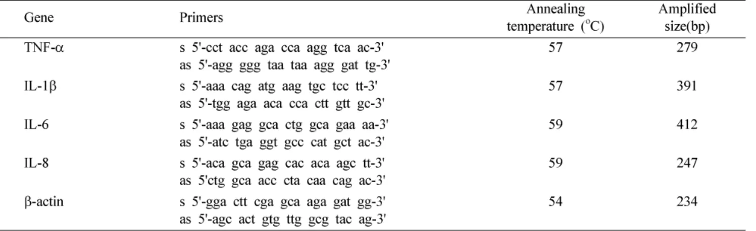
비만세포로부터히스타민의유리에 미치는
WELB
의 효과 − 비만세포에 PMACI를투여하면비만세포가활성화되
면서세포내의주함유물질이며알레르기유발물질인히스 타민이유리된다. 인체비만세포주인 HMC-1 세포에미치
는 WELB의효과를검토하기위하여 PMACI를투여하기
10분전에 WELB를처리하였다. Fig. 1에서와같이 WELB
는비만세포에서 PMACI에의한히스타민의유리를농도
의존적으로억제하였다. WELB의비만세포에대한독성여
부를 확인하기 위해 MTT 실험을 한 결과 WELB 농도
1 mg/ml까지세포독성이나타나지않았다. (data not shown)
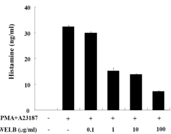
염증유발 사이토카인유전자의 발현 및 분비에 미치는
WELB
의효과 −TNF-α, IL-1β, IL-6 및 IL-8은잘알려진염증유발물질로서 PMACI의자극에의해비만세포에서발
현된다. 비만세포유래의염증유발에대한 WELB의효과를
검토하기위하여 HMC-1 세포를 PMACI로자극하기 1시간
전에 WELB를처리하였다. Fig. 2에서와같이 WELB는비
만세포에서 PMACI에의한 TNF-α, IL-1β, IL-6 및 IL-8의
발현을농도의존적으로억제하였다. 또한 Fig. 3에서와같
이 WELB는 PMACI로자극된비만세포에서 TNF-α와 IL- 6의분비를농도의존적으로억제하였다.
WELB
의염증유발 사이토카인발현억제기전 − 전사인자인 mitogen-activated protein kinase (MAPK)와 NF-κB는
세포활성, 염증과같은다양한 세포내반응을조절하는인
자로서특히 TNF-α, IL-1β, IL-6 및 IL-8와같은염증유발
사이토카인의발현을조절한다고알려져있다. MAPK는 extracellular signal-regulated kinase (ERK), c-jun N- terminal kinase (JNK), 및 p38 MAPK로구성되어있으며,
이들은세포외부로부터의자극을세포내부로전달하는역 할을 주로담당한다. 염증유발사이토카인발현에대한 WELB의억제효과에대한작용기전을 검토하기위하여 HMC-1 세포를 PMACI로자극하기 1시간전에 WELB를
처리하고 MAPK와 NF-κB의활성에대한 WELB의효과를
측정하였다. Fig. 4에서와 같이 WELB는비만세포에서 PMACI에의한 NF-κB의활성을억제하였다. 또한 WELB
는 ERK 및 p38 MAPK에대한영향없이 JNK의인산화를
특이적으로억제하였다.
활성화된비만세포는히스타민과같은생리활성물질및 염증유발성사이토카인분비를통해알레르기성염증질환 을유발한다. 또한염증유발성사이토카인은 MAPK와 NF-
κB와같은전사인자를통해분비된다. 따라서비만세포에
의해분비되는히스타민및염증유발성사이토카인분비 를억제하는물질을발굴하고작용기전을밝히는일은알 레르기성염증질환치료에서중요한분야이다. 본연구에서
는 WELB가히스타민유리를억제하고, NF-κB와 JNK의
활성을억제하여염증유발사이토카인의발현을억제함을 시사하고있다.
결 론
비만세포에의해매개되는알레르기성염증반응에대한
WELB의효과실험 결과 WELB는인체비만세포주에서
Fig. 1. Effect of the water extract of L. obtusiloba (WELB) on PMACI-induced histamine release from HMC-1 cells. The cells (2×10⁶ cells/ml) were preincubated with WELB at 37oC for 10 min prior to incubation with PMA (20 nM) and A23187 (1µM). Each data represents the mean±SEM of three independent experiments. *Statistically significant at P<0.05.
Fig. 2. Effect of the water extract of L. obtusiloba (WELB) on the gene expression of proinflammatory cytokines in HMC-1 cells.
HMC-1 (2×10⁶ cells/ml) was pretreated with WELB for 1 h and stimulated by PMA (20 nM) and A23187 (1µM) for 4 h. Gene expression of TNF-α, IL-1β, IL-6, and IL-8 was quantified by RT-PCR. Products were electrophoresed on a 1.5% agarose gel, visualized by staining with ethidium bromide and certificated using a Kodak DC 290 digital camera. Each data represents the mean±SEM of three independent experiments. *Statistically significant at P<0.05.
Fig. 3. Effect of the water extract of L. obtusiloba (WELB) on the secretion of proinflammatory cytokines in HMC-1 cells. HMC- 1 (2×10⁶ cells/ml) was pretreated with WELB for 1 h and stimulated by PMA (20 nM) and A23187 (1µM) for 12 h. The secretion of TNF-α and IL-6 was measured by ELISA. Each data represents the mean±SEM of three independent experiments. *Statistically significant at P<0.05.
PMACI에의해유도된히스타민의유리를억제하였다. 또
한 WELB는전사인자인 NF-κB와 JNK의활성화를억제하
여다양한염증유발성사이토카인의발현과분비를억제하 였다.
이러한결과로미루어볼때생강나무추출물인 WELB
는비만세포매개알레르기성염증질환의예방과치료에 사용될수있음을시사하고있다.
인용문헌
1. Metcalfe, D. D., Kaliner, M. and Donlon, M. A. (1981) The mast cell. Crit. Rev. Immunol. 3: 23.
2. Miyajima, I., Dombrowicz, D., Martin, T. R., Ravetch, J. V., Kinet, J. P. and Galli, S. J. (1997) Systemic anaphylaxis in the mouse can be mediated largely through IgG1 and Fc gam- maRIII. Assessment of the cardiopulmonary changes, mast
cell degranulation, and death associated with active or IgE- or IgG1-dependent passive anaphylaxis. J. Clin. Invest.99: 901.
3. Church, M. K. and Levi-Schaffer, F. (1997) The human mast cell. J. Allergy Clin. Immunol. 99: 155.
4. Petersen, L. J., Mosbech, H. and Skov, P. S. (1996) Allergen- induced histamine release in intact human skin in vivo assessed by skin microdialysis technique: characterization of factors influencing histamine releasability. J. Allergy Clin.
Immunol.97: 672.
5. Ennis, M., Pearce, F. L. and Weston, P. M. (1980) Some stud- ies on the release of histamine from mast cells stimulated with polylysine. Br. J. Pharmacol. 70: 329.
6. Alfonso, A., Cabado, A. G., Vieytes, M. R. and Botana, L. M.
(2000) Functional compartments in rat mast cells for cAMP and calcium on histamine release. Cell Signal.12: 343.
7. Beaven, M. A., Rogers, J., Moore, J. P., Hesketh, T. R., Smith, G. A. and Metcalfe, J. C. (1984) The mechanism of the cal- cium signal and correlation with histamine release in 2H3 cells. J. Biol. Chem. 259: 7129.
8. Botana, L. M. and MacGlashan, D. W. (1994) Differential effects of cAMP-elevating drugs on stimulus-induced cyto- solic calcium changes in human basophils. J. Leukoc. Biol.
55: 798.
9. Galli, S. J., Gordon, J. R. and Wershil, B. K. (1991) Cytokine production by mast cells and basophils. Curr. Opin. Immunol.
3: 865.
10. Galli, S. J., Kalesnikoff, J., Grimbaldeston, M. A., Piliponsky, A. M., Williams, C. M. and Tsai, M. (2005) Mast cells as
“tunable” effector and immunoregulatory cells: recent advances. Annu. Rev. Immunol. 23: 749.
11. Azzolina, A., Bongiovanni, A. and Lampiasi, N. (2003) Sub- stance P induces TNF-alpha and IL-6 production through NF kappa B in peritoneal mast cells. Biochim. Biophys. Acta.
1643: 75.
12. Kim, S. H., Jun, C. D., Suk, K., Choi, B. J., Lim, H., Park, S., Lee, S. H., Shin, H. Y., Kim, D. K. and Shin, T. Y. (2006) Gallic acid inhibits histamine release and pro-inflammatory cytokine production in mast cells. Toxicol. Sci. 91: 123.
13. 중약대사전상해과학기술출판사 1913.
(2009년 8월 20일 접수) Fig. 4. Effect of the water extract of L. obtusiloba (WELB) on
the activation of MAPKs and NF-κB in HMC-1 cells. HMC-1 (2×10⁶cells/ml) was pretreated with WELB for 1 h and stimulated by PMA (20 nM) and A23187 (1µM) for 1 h.
Phosphorylation of MAPK and activation of NF-κB were analyzed by Western blot. C-NF-κB, cytoplasmic NF-κB; N- NF-κB, nucleus NF-κB.