INTRODUCTION
Previous studies indicated that proinflammatory cytokines such as interleukin (IL)-1, IL-6, tumor necrosis factor (TNF)- and their receptors are increased in patients with both decom- pensated heart failure (New York Heart Association (NYHA) functional class III and IV as well as compensated (NYHA functional class II) (1-3). Furthermore, proinflammatory cyto- kines have been attributed as contributors to the syndrome of congestive heart failure and the underlying cardiomyopathic process of adverse left ventricular remodeling and thus, evolv- ing into progressive left ventricular dysfunction (4-6).
The origin of proinflammatory cytokines in patients with heart failure remains unclear, hence the implication of such proinflammatory cytokines in the pathogenesis of heart fail- ure is still controversial, which was reflected by the failure of multicenter clinical trials that used “targeted” approaches to neutralize TNF- in patients with moderate to advanced heart failure (7).
Some authors suggested endotoxin may induce cytokine activation via intestinal bacterial translocation (8). However, this hypothesis failed to explain elevated cytokines in patients with compensated heart failure (9). Other studies proposed myocardium itself produce cytokines due to elevated left ven- tricular diastolic wall stress (10). Tissue hypoxia and free radi-
cal production was also hypothesized to cause NF- B medi- ated cytokine production especially from skeletal muscles (11).
In order to determine the cellular source of circulating proinflammatory cytokines and their soluble receptors, we measured the proinflammatory cytokine concentrations in systemic artery, infra-renal inferior vena cava, and coronary sinus and their concentrations were compared with one anoth- er to determine the origin of its production. Furthermore, we correlated the proinflammatory cytokine levels with the clinical variables and hemodynamic parameters assessed both at admission and at recovery status.
MATERIALS AND METHODS Study Subjects
We consecutively enrolled 30 patients who were admitted to Ajou University Hospital for congestive heart failure due to idiopathic dilated cardiomyopathy with various function- al classes. At admission, we assessed NYHA functional classes, performed routine clinical evaluations and measured echocar- diographic parameters. All patients were treated with stan- dard medical therapy depending upon the severity of heart failure. After patients achieved hemodynamic stabilization,
Hyuk-Jae Chang, Jaehoon Chung, Byoung-Joo Choi, Tae-Young Choi, So-Yeon Choi, Myeong-Ho Yoon, Gyo-Seung Hwang, Joon-Han Shin, Seung-Jea Tahk, Byung-il William Choi
Department of Cardiology, Ajou University School of Medicine, Suwon, Korea
Address for correspondence Byung-il William Choi, M.D.
Department of Cardiology, Ajou University School of Medicine, San 5 Wonchon-dong, Paldal-gu, Suwon 442-721, Korea
Tel : +82.31-219-5712, Fax : +82.31-219-5708 E-mail : [email protected]
791
The Origin of Proinflammatory Cytokines in Patients with Idiopathic Dilated Cardiomyopathy
Proinflammatory cytokines and their receptors are increased in the peripheral blood of patients with heart failure. We measured cytokines and their receptors in sys- temic artery (SA), coronary sinus (CS) and infra-renal inferior vena cava (IVC), in order to investigate their origin and influential factors. Thirty patients with idiopathic dilated cardiomyopathy were performed echocardiography at admission, and right heart catheterization after stabilization. Blood was drawn from 3 sites for measure- ment of tumor necrosis factor- (TNF- ), interleukin-6 (IL-6) and soluble tumor necrosis factor- receptor (sTNFR) I, II. TNF- at CS (3.25±0.34 pg/mL) was higher than those of SA (1.81±0.39 pg/mL) and IVC (1.88±0.38 pg/mL, p<0.05).
IL-6 at CS (18.3±3.8 pg/mL) was higher than that of SA (5.8±1.2 pg/mL, p<0.01).
The levels of sTNFR I, II showed increasing tendency in sequence of SA, IVC and CS. TNF- and sTNFR I, II from all sites were proportional to worsening of func- tional classes at admission (p<0.05). E/Eaby Doppler study at admission, which reflects left ventricular end-diastolic pressure (LVEDP) was positively correlated with TNF- from SA (R=0.71, p<0.01), CS (R=0.52, p<0.05) and IVC (R=0.46, p<0.05). Thus, elevated LVEDP during decompensation might cause cytokine release from myocardium in patients with idiopathic dilated cardiomyopathy.
Key Words : Heart Failure, Congestive; Cytokines; Myocardial Diseases
Received : 9 July 2003 Accepted : 21 August 2003
cardiac catheterization was performed for blood sampling at femoral artery as a systemic artery, infra-renal inferior vena cava representative of cytokine production from skeletal mus- cle and coronary sinus from myocardium, and measurement of hemodynamic variables including pulmonary capillary wedge pressure, mean pulmonary arterial pressure, mean arterial pressure and cardiac index.
Patients with the following criteria were excluded; (1) any identifiable cause of dilated cardiomyopathy such as history of heavy alcohol abuse, severe hypertension, primary valvu- lar disease or history of ischemic heart disease, (2) elevated cardiac enzyme or EKG change compatible to myocarditis, (3) history of infectious disease within recent 4 weeks or ele- vated acute phase reactants (e.g. ESR, C-Reactive protein), (4) contraindication for cardiac catheterization
We also selected 11 age- and gender-matched control sub- jects (aged 38 to 60, mean 49 yr, male: 7) who visited Ajou University Hospital for electrophysiologic study. All the control subjects had normal coronary angiogram.
All participants gave written informed consents before entering the study and the study protocol was reviewed by the ethical committee of Ajou University Hospital.
Blood Sampling
After proper medical therapy, right heart catheterization and coronary angiography were performed. Under fluoro- scopic guidance, 7Fr NIH catheter was positioned in the coronary sinus via subclavian vein. Correct catheter position was confirmed with contrast injection and oxygen satura- tion. Pulmonary capillary wedge pressure, mean pulmonary arterial pressure, mean arterial pressure and cardiac index were measured during blood sampling.
Cytokine Assay
Blood samples were immediately centrifuged with 5,000
rpm for 15 min at 4℃.Then, serum was separated into aliquots and stored at -70℃for a period not exceeding 6 months. Cir- culating levels of cytokines and their receptors were measured using commercially available ELISA kits (Quantikine, R &
D systems, Minneapolis, MN, U.S.A.). The intra-assay coef- ficients of variation were as follows; TNF- (6.0%), sTNFR I (4.7%), sTNFR II (2.4%) and IL-6 (3.5%).
Statistical Analysis
Statistical analysis was performed with SPSS 8.0 statisti- cal software (SPSS, Inc. Illinois, U.S.A.). All data are pre- sented as mean±SEM. Student’s t-test was used for differ- ences in continuous variables and 2test was used for cate- gorical variables. The differences in cytokine levels by NYHA functional classes were verified using ANOVA. Correlations of cytokine levels with demographic characteristics were measured by Pearson’s analysis. p<0.05 was considered sta- tistically significant.
RESULTS Characteristics of Study Subjects
Among 30 patients enrolled, 21 were male and 9 were female, with age ranging from 32 to 67 yr (51.3±9.3 yr).
Mean left ventricular ejection fraction was 27.7±9.5%. Mean duration of symptoms was 18.5±1.8 days before admis- sion (Table 1). Seven patients were classified as NYHA func- tional class I, 8 as II, 6 as III, and 9 as IV at admission (Table 1). At the time of cardiac catheterization after appropriate medical therapy, all patients with heart failure were com- pensated and their functional classes had improved (Table 2).
Circulating Levels of Cytokines according to Site of Origin
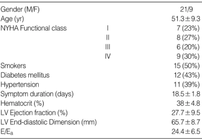
Mean values of all the cytokines were at least fivefold high- er in the patient group than those in the control group from all the three sampling sites (Fig. 1). We could not measure TNF- from systemic artery in 9 patients and from inferior
Data are expressed mean±SD. MAP, mean arterial pressure; MPAP, mean pulmonary arterial pressure; NYHA, New York Heart Association;
PCWP, pulmonary capillary wedge pressure.
Duration from enrollment (days) 4.8±0.5
NYHA Functional class I 11 (37%)
II 19 (63%)
PCWP (mmHg) 13.6±1.8
MAP (mmHg) 92.6±11.2
MPAP (mmHg) 23.4±4.6
Cardiac Index (L/min/m2) 2.2±0.1 Table 2.Patient characteristics at cardiac catheterization
Data are expressed mean±SD. LV, left ventricle; NYHA, New York Heart Association.
Gender (M/F) 21/9
Age (yr) 51.3±9.3
NYHA Functional class I 7 (23%)
II 8 (27%)
III 6 (20%)
IV 9 (30%)
Smokers 15 (50%)
Diabetes mellitus 12 (43%)
Hypertension 11 (39%)
Symptom duration (days) 18.5±1.8
Hematocrit (%) 38±4.8
LV Ejection fraction (%) 27.7±9.5
LV End-diastolic Dimension (mm) 65.7±8.7
E/Ea 24.4±6.5
Table 1.Patient characteristics at enrollment
vena cava in 8 patients, due to limited minimal detection level. In contrast, TNF- at coronary sinus was measurable in all patients.
TNF- of 3.25±0.34 pg/mL at coronary sinus was high- er than that at inferior vena cava (1.88±0.38 pg/mL, p<0.05) and systemic artery (1.81±0.39 pg/mL) (Fig. 1). The levels of sTNFR I and II had a tendency to increase in order of systemic artery, inferior vena cava and coronary sinus with- out statistical significance, i.e., sTNFR I in systemic artery, 2,088±212 pg/mL; inferior vena cava, 2,125±216 pg/mL;
coronary sinus, 2,368±220 pg/mL (p=NS) and sTNFR II in systemic artery, 2,880±306 pg/mL; inferior vena cava, 2,945±301 pg/mL; coronary sinus, 3,242±316 pg/mL (p=NS) (Fig. 1). However, the levels of IL-6 increased sig- nificantly in the same order (systemic artery, 5.78±1.23 pg/mL; inferior vena cava, 12.1±2.7 pg/mL; coronary sinus, 18.3±3.84 pg/mL, p<0.01) (Fig. 1). Significant differences were detected in the levels of IL-6 between coronary sinus and systemic artery (p<0.01), but not between coronary sinus
and inferior vena cava (Fig. 1).
Significant correlations were noted between TNF- from coronary sinus, and IL-6 from coronary sinus (R=0.44, p<0.05) and inferior vena cava (R=0.51, p<0.01).
Circulating Levels of Cytokines and Clinical, Hemo- dynamic Variables
TNF- , sTNFR I and II from inferior vena cava increased in relation to worsening NYHA functional classes at admis- sion (R=0.59, 0.64 and 0.52, respectively; p<0.01). The con- centrations of IL-6 showed a tendency to increase in accor- dance with worsening functional status without statistical significance (R=0.32, p=0.06).
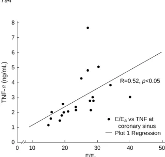
Early diastolic mitral inflow versus mitral annular velocity ratio (E/Ea) at admission by Doppler study, which reflects left ventricular end diastolic pressure, was positively corre- lated with TNF- from systemic artery (R=0.71, p<0.01), coronary sinus (R=0.52, p<0.05) and inferior vena cava (R=
TNF (pg/mL)
5
4
3
2
1
0 Systemic artery IVC Coronary artery
sTNFR1 (pg/mL)
4,000 3,500
3,000
2,500 2,000
1,500
1,000 500
0
Peripheral artery IVC Coronary sinus
*
sTNFR2 (pg/mL)
5,000 4,500 4,000 3,500 3,000 2,500 2,000 1,500 1,000 500 0
Peripheral artery IVC Coronary sinus
IL-6 (pg/mL)
30
25
20
15
10
5
0
Peripheral artery IVC Coronary sinus
�
control patient
control patient control patient
control patient
Fig. 1.Levels of proinflammatory cytokines at different sites. Data are expressed mean±SEM. *: p<0.05 compared with TNF- from IVC,�: p<0.01 compared with IL-6 from systemic artery, IVC: inferior vena cava.
0.46, p<0.05) (Fig. 2). In contrast, pulmonary capillary wedge pressure measured during right catheterization did not show any significant correlation with cytokines.
DISCUSSION
Since Levine et al. first reported that, among patients with congestive heart failure, circulating levels of TNF- are most markedly increased in those with the most advanced disease and cachexia (13), many other studies have observed that cytokines are sufficient to mimic some aspects of the so-called heart failure phenotype, including progressive left ventricu- lar dysfunction, pulmonary edema, left ventricular remodel- ing, fetal gene expression and cardiomyopathy (4-6). Thus, the “cytokine hypothesis” holds that heart failure progresses, at least in part, as a result of the toxic effects exerted by endoge- nous cytokine cascades on the heart and the peripheral cir- culation (14). However, the site of cytokine production in patients with congestive heart failure is still poorly understood.
Anker et al. proposed that elevated plasma cytokines in heart failure resulted from a cascade of events consisting of mesen- teric venous congestion, bowel wall edema, and intestinal bacterial translocation (8). But elevations in plasma cytokines have been observed in patients with functional class II and even in many patients without peripheral edema (9). Other studies proposed elevated left ventricular diastolic wall stress stimulates myocardium to produce cyto-kines (10). Tissue hypoxia and free radical production due to impaired vasodila- tor reserve was also hypothesized to cause NF- B mediated cytokine production especially from skeletal muscles.
Deliargyris et al. suggested that elevated IL-6 in patients with heart failure is the result of extracardiac production (15).
In contrast, Petretta et al. found no significant differences in
the levels of cytokines among coronary sinus, ascending aorta, inferior vena cava and hepatic vein (16). However, there is substantial limitation in their results because their subjects had various causes of heart failure including ischemic heart disease. Our study may be more relevant, for all the cases in the study were diagnosed as idiopathic dilated cardiomyo- pathy with various functional classes, and left ventricular end- diastolic pressure was measured twice; first, at admission, indirectly by Doppler study and after proper medical therapy, second, at recovery status, by cardiac catheterization.
The TNF- molecule (a 157-amino acid polypeptide) exists bioactive as a membrane-bound and secreted molecule (17).
The activated macrophage is the main source of TNF- , although other cells releasing TNF- include lymphocytes, fibroblasts, neutrophils, smooth muscle cells and mast cells (18). Furthermore, adult mammalian myocardial cells can produce TNF- with extracellular stimuli such as endotox- in, hypoxia or increased mechanical stress (10).
Our data with the highest concentration of TNF- at coro- nary sinus seem to support the concept of the myocardial cyto- kine production. Torre-Amione et al. demonstrated myocar- dial expression of TNF- in failing human hearts (19). Myocar- dial stretch was a sufficient stimulus for the induction of TNF- and mRNA biosynthesis in feline myocardium (10).
Nagueh et al. investigated E/Eaby Doppler study provides the best index for the prediction of pulmonary capillary wedge pressure, irrespective of the diastolic filling pattern (12). Significant correlation between TNF- and baseline E/Eawas observed in the present study (Fig. 2). However, after compensation with appropriate medical therapy, no significant correlation was noted between TNF- and pul- monary capillary wedge pressure gained at cardiac catheteri- zation. Consequently, we deduced stretched myocardium may be the stimulus of increased level of TNF- .
TNF- also orchestrates the inflammatory cascade through regulation of transduction for proinflammatory cytokines such as IL-1 and IL-6, and nitric oxide synthase (20), which was reflected as a significant correlation between TNF- and IL-6 in the present study.
TNF- acts at the cellular level via both type I (p55) and type II (p75) receptors and recently, it has been suggested that both of them are present in the human myocardium.
The extracellular domain fragments of both TNF receptors shed from cell surfaces can be detected as soluble forms in the blood and urine (21). At physiologic concentrations, sTN- FRs may act as ‘slow-release reservoir’ of bioactive TNF- , but at higher concentrations, as in patients with severe heart failure, they could inhibit the pathological increase of TNF- activity (19).
Saraste et al. reported sTNFR II identified a subgroup of heart failure patients with increased cardiomyocyte apopto- sis (22). Finally, the increased levels of sTNFR II were sig- nificantly correlated with poor short-term prognosis of heart failure patients (23).
TNF-(ng/mL)
8 7 6 5 4 3 2 1 0
0 10 20 30 40 50
E/Ea
Fig. 2.Correlation between TNF- at coronary sinus and E/Eaby Doppler study. TNF- at coronary sinus was positively correlated with E/Eaby Doppler study (R=0.52, p<0.05).
R=0.52, p<0.05
E/Eavs TNF at coronary sinus Plot 1 Regression
Although our study failed to demonstrate any significant difference in the levels of sTNFR I and II from the different sites of sampling, the absence of a transmyocardial gradient of cytokines does not rule out myocardial production of cytokines due to the diffusion barrier between interstitial space and coronary venous effluent and relatively high molec- ular weight compared with TNF- and IL-6 (24).
Serum IL-6 was known as the most powerful independent predictor of heart failure episodes and mortality or need for heart transplantation during heart failure management (25).
Tsutamoto et al. implicated IL-6 is produced mainly in the periphery in heart failure patients (26), but their study population is different from ours in background; 60% of subject patients had history of myocardiac infarction. In the setting of ischemic heart failure, both myocardial necrosis and successful reperfusion, and peripheral vascular tissue may affect to increase myocardial IL-6 production (26, 27).
The mechanism for elaboration of IL-6 in idiopathic dilated cardiomyopathy has not been elucidated. TNF- may be sufficient to induce IL-6 gene and protein expression in a variety of cell types, suggesting that there may be a ‘cytokine cascade’ in the setting of heart failure (28). We also identi- fied a significant positive correlation between the levels of TNF- and IL-6, which is similar to other study (29). On the basis of our results, we suggested that IL-6 might be produced mainly in the myocardium. But it remains con- troversial to determine whether the local myocardial pro- duction of IL-6 is secondary to increased TNF- , or directly related to the myocardial tissue or some combination of both.
In our study, we conclude that myocardium might be the major source of proinflammatory cytokines in idiopathic dilated cardiomyopathy. It might be relevant in determin- ing the origin for all the subjects had non-ischemic heart failure with various functional status.
Accordingly, we presumed that increased myocardial wall stress might lead to sustained expression of stretch-derived genes and increased oxidative stress as a result of subendo- cardial hypoperfusion, with resultant activation of families of genes which might be translated in subsequent produc- tion of cytokines.
REFERENCES
1. Paulus WJ. How are cytokines activated in heart failure? Eur J Heart Fail 1999; 1: 309-12.
2. Munger MA, Johnson B, Amber IJ, Callahan KS, Gilbert EM. Cir- culating concentrations of proinflammatory cytokines in mild or moderate heart failure secondary to ischemic or idiopathic dilated cardiomyopathy. Am J Cardiol 1996; 77: 723-7.
3. Kubota T, Miyagishima M, Alvarez RJ , Kormos R, Rosenblum WD, Demetris AJ, Semigran MJ, Dec GW, Holubkov R, McTiernan CF, Mann DL, Feldman AM, McNamara DM. Expression of proinflam- matory cytokines in the failing human heart: Comparison of recent-
onset and end-stage congestive heart failure. J Heart Lung Trans- plant 2000; 19: 819-24.
4. Bozkurt B, Kribbs S, Clubb FJ Jr , Michael LH, Didenko VV, Horns- by PJ, Seta Y, Oral H, Spinale FG, Mann DL. Pathophysiologically relevant concentrations of tumor necrosis factor- promote pro- gressive left ventricular dysfunction and remodeling in rats. Circu- lation 1998; 97: 1382-91.
5. Thaik CM, Calderone A, Takahashi N, Colucci WS. Interleukin-1 ‚ modulates the growth and phenotype of neonatal rat cardiac myocytes.
J Clin Invest 1995; 96: 1093-9.
6. Kubota T, McTiernan CF, Frye CS, Slawson SE, Lemster BH, Koret- sky AP, Demetris AJ, Feldman AM. Dilated cardiomyopathy in trans- genic mice with cardiac specific overexpression of tumor necrosis factor- . Circ Res 1997; 81: 627-35.
7. Feldman AM, Kadokami T, Higuichi Y, Ramani R, McTiernan CF.
The role of anticytokine therapy in heart failure: recent lessons from preclinical and clinical trials. Med Clin North Am 2003; 87: 419-40.
8. Anker SD, Egerer KR, Volk H-D, Kox WJ, Poole-Wilson PA, Coats AJS. Elevated soluble CD 14 receptors and altered cytokines in chronic heart failure. Am J Cardiol 1997; 79: 1426-30.
9. Torre-Amione G, Kapadia S, Benedict CR, Oral H, Young JB, Mann DL. Proinflammatory cytokine levels in patients with depressed left ventricular ejection fraction: a report from the Studies on Left Ven- tricular Dysfunction (SOLVD). J Am Coll Cardiol 1996; 27: 1201-6.
10. Kapadia SR, Oral H, Lee J, Nakano M, Taffet GE, Mann DL. Hemo- dynamic regulation of tumor necrosis factor- gene and protein expression in adult feline myocardium. Circ Res 1997; 81: 187-95.
11. Adams V, Jiang H, Yu J, Mobius-Winkler S, Fiehn E, Linke A, Weigl C, Schuler G, Hambrecht R. Apoptosis in skeletal myocytes of patients with chronic heart failure is associated with exercise intolerance. J Am Coll Cardiol 1999; 33: 959-65.
12. Nagueh SF, Middleton KJ, Kopelen HA, Zoghbi WA, Quinones MA. Doppler tissue imaging: a noninvasive technique for evalua- tion of left ventricular relaxation and estimation of filling pressures.
J Am Coll Cardiol 1997; 30: 1527-33.
13. Levine B, Kalman J, Mayer L, Fillit HM, Packer M. Elevated circu- lating levels of tumor necrosis factor in severe chronic heart failure.
N Engl J Med 1990; 323: 236-41.
14. Seta Y, Shan K, Bozkurt B, Oral H, Mann DL. Basic mechanism in heart failure: the cytokine hypothesis. J Card Fail 1996; 2: 243-9.
15. Deliargyris EN, Raymond RJ, Theoharides TC, Boucher WS, Tate DA, Dehmer GJ. Sites of interleukin-6 release in patients with acute coronary syndromes and in patients with congestive heart failure.
Am J Cardiol 2000; 86: 913-8.
16. Petretta M, Condorelli GL, Spinelli L, Scopacasa F, De Caterina M, Leosco D, Vicario ML, Bonaduce D. Circulating levels of cytokines and their site of production in patients with mild to severe chronic heart failure. Am Heart J 2000; 140: E28.
17. Old LJ. Tumor necrosis factor (TNF). Science 1985; 230: 630-2.
18. Vassali P. The pathophysiology of tumor necrosis factors. Annu Rev Immunol 1992; 10: 411-52.
19. Torre-Amione G, Kapadia S, Lee J, Durand JB, Bies RD, Young JB, Mann DL. Tumor necrosis factor- and tumor necrosis factor receptors in the failing human heart. Circulation 1996; 93: 704-11.
20. Kelly RA, Smith TW. Cytokines and cardiac contractile function.
Circulation 1997; 95: 778-81.
21. Aderka D, Engelmann H, Maor Y, Brakebusch C, Wallach D. Sta- bilization of the bioactivity of tumor necrosis factor by its soluble receptors. J Exp Med 1992; 175: 323-9.
22. Saraste A, Voipio-Pulkki LM, Heikkila P, Laine P, Nieminen MS, Pulkki K. Soluble tumor necrosis factor receptor levels identify a subgroup of heart failure patients with increased cardiomyocyte apoptosis. Clin Chim Acta 2002; 320: 65-7.
23. Ferrari R, Bachetti T, Confortini R, Opasich C, Febo O, Corti A, Cassani G, Visioli O. Tumor necrosis factor soluble receptors in patients with various degrees of congestive heart failure. Circulation 1995; 92: 1479-86.
24. Wei CC, Meng QC, Palmer R, Hageman GR, Durand J, Bradley WE, Farrell DM, Hankes GH, Oparil S, Dell’Italia LJ. Evidence for angiotensin-converting enzyme- and chymase-mediated angiotensin II formation in the interstitial fluid space of the dog heart in vivo.
Circulation 1999; 99: 2583-9.
25. Orus J, Roig E, Perez-Villa F , Pare C, Azqueta M, Filella X, Heras
M, Sanz G. Prognostic value of serum cytokines in patients with congestive heart failure. J Heart Lung Transplant 2000; 19: 419-25.
26. Tsutamoto T, Hisanaga T, Wada A, Maeda K, Ohnishi M, Fukai D, Mabuchi N, Sawaki M, Kinoshita M. Interleukin-6 spillover in the peripheral circulation increases with the severity of heart failure, and the high plasma level of interleukin-6 is an important prognostic pre- dictor in patients with congestive heart failure. J Am Coll Cardiol 1998; 31: 391-8.
27. Neumann FJ, Ott I, Gawaz M , Richardt G, Holzapfel H, Jochum M, Schomig A. Cardiac release of cytokines and inflammatory respons- es in acute myocardial infarction. Circulation 1995; 92: 748-55.
28. Feldman AM, Combes A, Wagner D , Kadakomi T, Kubota T, Li YY, McTiernan C. The role of tumor necrosis factor in the patho- physiology of heart failure. J Am Coll Cardiol 2000; 35: 537-44.
29. Koller-Strametz J, Pacher R, Frey B, Kos T, Woloszczuk W, Stanek B. Circulating tumor necrosis factor-alpha. Levels in chronic heart failure: relation to its soluble receptor II, interleukin-6 and neuro- humoral variables. J Heart Lung Transplant 1998; 17: 356-62.
′
. .