ABSTRACT
Purpose: Aster tataricus (AT) is one of the Asteraceae perennial herbs used in traditional Chinese
medicine. The herb contains various bioactive substances, such as flavonoids, isoflavonoids, and phenolic compounds in the roots, and exhibits a range of effects including anti-bacterial, anti-oxidant, and anti-inflammatory activities. This study compared the immunomodulatory effects of ethanol and water extracts of whole AT, except the roots, and analyzed the
molecular mechanisms for the regulatory effects on cytokine secretion from THP-1 cells.
Methods: The effects of AT extract on the cell viability and proliferation of THP-1 cells were
analyzed using the Cell Counting Kit-8 method. The concentrations of interleukin-1β (IL-1β) and tumor necrosis factor-α (TNF-α) in the cell culture supernatant of the AT-treated THP-1 cells were measured using an enzyme-linked immunosorbent assay. The protein levels of cyclooxygenase-2 (COX-2), inducible nitric oxide synthase (iNOS), inhibitor of nuclear factor kappa B (IκBα), and mitogen-activated protein kinase (MAPK) phosphorylation in the cell lysates were determined by western blotting.
Results:
The water extract and the ethanol extract of AT did not affect the cell viability, and increased the proliferation of THP-1 cells significantly compared to the vehicle. The water extract increased the secretion of IL-1β from THP-1 cells in a dose-dependent manner, but the ethanol extract had no effect. The expression of COX-2 and iNOS protein and the phosphorylation of MAPK and Akt were induced in AT-treated cells. In addition, IκBα was degraded by AT in a concentration-dependent manner. IL-1β secretion by AT was reduced by extracellular-signal-regulated kinase (ERK) and c-Jun N-terminal kinase (JNK) inhibitors, while TNF-α secretion was decreased by inhibitors of ERK, p38 MAPK, and JNK.
Research Article
Received: Jun 23, 2020 Revised: Jul 14, 2020 Accepted: Jul 29, 2020 Correspondence to Whajung Cho
Division of Research Program, Scripps Korea Antibody Institute, 1 Kangwondaehak-gil, Chuncheon 24341, Korea.
Tel: +82-33-250-8097 E-mail: [email protected]
© 2020 The Korean Nutrition Society This is an Open Access article distributed under the terms of the Creative Commons Attribution Non-Commercial License (http://
creativecommons.org/licenses/by-nc/3.0/) which permits unrestricted non-commercial use, distribution, and reproduction in any medium, provided the original work is properly cited.
ORCID iDs Chea Yeon Lee
https://orcid.org/0000-0003-1469-0025 Hyo Sung Park
https://orcid.org/0000-0002-8872-1747 Deok-Hoon Kong
https://orcid.org/0000-0003-1411-4423 Young Kwan Kim
https://orcid.org/0000-0002-2296-7189 Whajung Cho
https://orcid.org/0000-0001-7478-2605 Funding
This work was supported by Presidential Committee for Balanced National Development (PCBND) and the Ministry of Land, Infrastructure and Transport (MOLIT) through the pilot project for regional development investment agreement (No.
Immunomodulatory effect of the water extract of Aster tataricus through mitogen-activated protein kinase signaling pathway
Chea Yeon Lee , Hyo Sung Park , Deok-Hoon Kong , Young Kwan Kim , and Whajung Cho
Division of Research Program, Scripps Korea Antibody Institute, Chuncheon 24341, Korea
Aster tataricus 물 추출물의 mitogen- activated protein kinase 신호 전달
경로를 통한 면역 조절 효과
이채연 , 박효성 , 공덕훈 , 김영관 , 조화정
스크립스코리아항체연구원 연구본부
B0070830000053) and the National Research Foundation of Korea (NRF) grant funded by the Korea government (MSIT) (No.
2019R1C1C1005796).
Conflict of Interest
There are no financial or other issues that might lead to conflict of interest.
Interestingly, the p38 MAPK inhibitor increased the production of IL-1β by AT further.
Conclusion: The water extract of the above-ground parts of AT contains immunomodulatory
bioactive substances that stimulate immune cells through the MAPK signaling pathway.
Keywords: Aster tataricus, THP-1 cell, IL-1 beta, TNF-alpha, mitogen-activated protein kinase
서론
개미취
(Aster Tataricus, AT)는 작고 화려한 꽃을 가진 국화과의 다년생 풀로 주로 깊은 산속의 습지에 자생하며 우리나라를 포함한 중국
,일본 등 동아시아의 많은 지역에서 발견된다
[1].AT
의 뿌리는 중국 전통 의술에서 약초로 사용되어왔으며
,기침 완화 및 거담제
,이뇨제
,항 종양제
,항균제
,항바이러스제의 효능을 가지고 있다고 알려져 있다
[2-4]. AT뿌리로부터
cyclic pentapeptides, triterpenoid ketone, saponin등 다양한 생리활성물질이 분리되었으며 이들의 구조 및 기능이 연구되었다
[2-7]. AT의 에탄올 추출물은
streptozotocin으로 유도한 당뇨병 쥐에서 혈당을 조절하고 염증을 억제함으로써 당뇨병성 망막증을 완화시키는 효과 가 있음이 보고되었고
[8],성상 세포종 세포주에서
lipopolysaccharide (LPS)에 의해 유도되는 산화 스트레스를 감소시켜 신경 염증을 완화시킨다는 결과가 발표된 바 있다
[9].또 다른 연 구에서는
AT뿌리의 메탄올 추출물에서 분리된 다양한 생리활성 물질들이
mitogen-activated protein kinase (MAPK)와
nuclear factor kappa-light-chain-enhancer of activated B cells (NF-κB)신호 경로를 통해
LPS에 의해 유도되는 염증성
cytokine의 분비를 억제하였다는 결과를 보고
하였다
[10,11].염증 반응은 바이러스
,박테리아
,기생충과 같은 미생물 등을 포함한 외부 침입 물질에 대항 하여 우리 몸을 방어하는 면역 반응의 한 메커니즘으로써
,감염 발생 시 선천면역에 의하여 빠르게 유도된다
.선천면역은 병원체 감염에 대한 일차 방어선 역할을 하며 병원체를 제거함
과 동시에 적응 면역을 유도한다
.단핵구
, NK세포
,비만세포
,호중구
,호산구
,호염구 등 다
양한 세포 집단이 선천면역을 담당하며
,이들은 비특이적으로 감염체를 인식하여 탐식
,탈
과립
,보체 활성화 등 다양한 방법을 통해 감염체를 제거하는 역할을 한다
[10,11].이 중
,단핵
구는 혈액을 따라 신체를 순환하며 조직으로 이동 시 대식세포로 분화하여 조직에 침투한 감
염 인자나 죽거나 손상된 조직을 제거하여 조직의 항상성을 유지하는 역할을 한다
[12-14].또
한
,다양한
cytokine과
chemokine,염증 매개체를 분비하여 다른 면역 세포의 활성을 조절하
고
,탐식한 항원을 프로세싱하여 주조직 적합성 복합체를 통해
T세포에 제시하여 항원 특이 적인 적응 면역을 유도하는 항원제시세포로서의 역할도 가지고 있다
[15-17].본 연구에서는
AT의 뿌리를 제외한 꽃
,잎
,줄기 부분의 에탄올 추출물과 물 추출물의 면역 조 절 효과를 확인하고자 사람 단핵구 세포주인
THP-1세포에 각 추출물을 처리한 후
cytokine의 분비를 비교 분석하였다
.흥미롭게도
,항염 작용이 있다고 알려진 뿌리 추출물과 반대로 지 상부의 물 추출물은
THP-1세포에서
interleukin-1β (IL-1β)와
tumor necrosis factor-α (TNF-α)의 분비를 유도하고
cyclooxygenase (COX)-2, inducible nitric oxide synthase (iNOS)단백질의 발 현을 증가시켰다
. Cytokine분비를 조절하는 메커니즘을 규명하고자
Akt, MAPK, inhibitor of nuclear factor kappa B (IκBα)분자의 인산화 및 분해 정도를 확인하고
, PI3K와
MAPK의 억제
제를 이용하여
IL-1β와
TNF-α의 분비에 관여하는 신호 전달 분자를 조사하였다
.이 결과들을
통하여
AT지상부의 물 추출물은
MAPK신호전달 경로를 통하여
THP-1세포의
cytokine분비 를 조절한다는 것을 확인하였다
.연구방법
시료
본 연구에 사용한
AT추출물은 뿌리를 제외한 잎
,줄기
,꽃의
100%에탄올을 이용한 추출물
(
개미취
, BE0547B1)과 물을 이용한 추출물
(개미취
, BW0547A1)의 건조 분말을
10 mg/mL의 농
도로
dimethyl sulfoxide (DMSO)에 녹인 형태로 한국과학기술연구원 강릉 분원 천연물연구소
(Korea Institute of Science and Technology, Gangneung, Korea)
로부터 분양 받아 사용하였다
. 시약및항체본 실험에 양성 대조군으로
LPS (Sigma-Aldrich, St. Louis, MO, USA)를 사용하였다
.단백질 분석을 위하여 사용된
extracellular-signal-regulated kinase (ERK), p-ERK, p38 MAPK, p-p38 MAPK, c-Jun N-terminal kinase (JNK), p-JNK, Akt, p-Akt, IκBα, iNOS에 대한 특이적인
1차 항체 는
Cell Signaling Technology (Beverly, MA, USA), COX-1, COX-2에 대한
1차 항체는
Cayman (Ann Arbor, MI, USA), β-actin에 대한
1차 항체는
Sigma-Aldrich에서 구입하여 사용하였다
. 2차 항체 로는
horseradish peroxidase-conjugated anti-rabbit immunoglobulin G (IgG)와
anti-mouse IgG (Cell Signaling Technology)를 사용하였다
.MAPK
신호 전달 경로의 관련성을 시험하기 위해 사용된
MEK1억제제
PD98059, p38 MAPK억제제
SB203580, JNK억제제
SP600125, PI3K억제제
wortmannin은
Sigma-Aldrich에서 구입 하여 사용하였다
.세포및세포배양
사람 단핵구 세포주인
THP-1세포는
American Type Culture Collection (Manassas, VA, USA)에 서 분양 받아
Roswell Park Memorial Institute 1640 medium에
L-glutamine, 10% fetal bovine serum, 1% penicillin/streptomycin, 0.05 mM 2-mercaptoethanol (Gibco, Grand Island, NY, USA)을 첨가한 배지를 이용하여
37°C, 5% CO2조건하에서 배양하였다
.세포생존율및증식률측정
THP-1 (2 × 10⁴ cells/well)
세포에
AT에탄올 추출물과 물 추출물을
50 μg/mL의 농도로
24시간 또는
72시간 동안 처리하고
Cell Counting Kit-8 (CCK-8) solution 10 μL을 처리하여
4시간 동안 반응시켰다
. Microplate Reader (bio-Tek Instruments, Inc., Winooski, VT, USA)를 이용하여
450 nm에서의 흡광도를 측정하여 세포 생존율 및 증식률을 확인하였다
.Western blotting
을이용한단백질발현및인산화측정THP-1 (1 × 10⁶ cells/well)
세포에
AT추출물
(10, 50 μg/mL)과 양성 대조군으로
LPS (10 ng/mL)를
처리하고 표적 단백질에 따라
30분
, 8시간
, 24시간 동안 배양 후
PRO-PREP Protein Extraction Solution (iNtRON Biotechnology, Seongnam, Korea)를 이용해 세포를 용해시키고
13,500 rpm, 4°C에서
10분 간 원심 분리하여 단백질을 추출하였다
. BCA protein assay Kit (Thermo Scientif-ic, Pittsburgh, PA, USA)
를 사용하여 단백질을 정량하고
, 30 μg의 단백질을
10% polyacrylamide gel에서
sodium dodecyl sulphate-polyacrylamide gel electrophoresis로 전기영동 하였다
.분리 된 단백질은
nitrocellulose membrane에
transfer하여
3% skim milk를 첨가한
tris-buffered sa- line (0.05% tween 20) (TBST)로 실온에서
30분간
blocking한 후
, p-ERK1/2, ERK, p-p38 MAPK, p38 MAPK, p-JNK, JNK, IκBα, p-Akt, Akt, iNOS, COX-2, β-actin에 특이적인
1차 항체를
1/1,000으로 희석하여
4°C에서
16시간 동안 반응시켰다
.이후
, 10분씩
3회
TBST로 세척하고
1/2,000으로 희석한
2차 항체를 실온에서
1시간 동안 반응시켰다
. 10분씩
5회
TBST로 세척 후
ECL Substrate reagent (Bio-Rad Laboratories, Inc., Hercules, CA, USA)를 이용하여
X-ray film에 현 상하였다
.Enzyme-linked immunosorbent assay (ELISA)
를이용한cytokine
측정 THP-1세포에
AT추출물 및
LPS를 처리하고 일정 시간 후 세포 상층액을 걷어
IL-1β와
TNF-α의 농도를
ELISA kit (eBioscience Co., San Diego, CA, USA)를 사용하여 측정하였으며
, cytokine의 농도는
kit에 포함되어 있는 표준용액으로부터 산출된 표준곡선으로부터 계산되었다
. 통계처리실험 결과는
PRISM version 5.0 program (GraphPad Software, Inc., La Jolla, CA, USA)을 이용하 여 평균
(mean) ±표준 오차
(standard error of mean)로 표현하였다
.각 실험군의 분석 항목별 통계적 유의성은
t-test로
*p < 0.05, **p < 0.01및
***p < 0.001수준에서 비교하였다
.결과
AT
추출물의THP-1
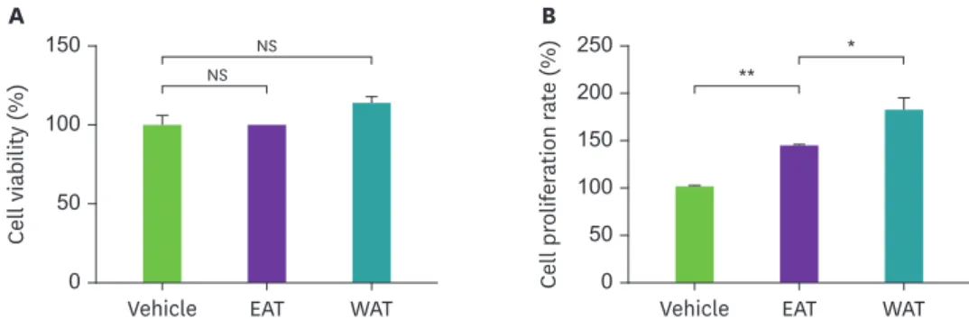
세포의생존율및분열에미치는영향AT
추출물의 세포 독성을 알아보기 위하여
THP-1세포에
DMSO와 에탄올 추출물
,물 추출물
을
50 μg/mL로 처리하고
24시간 후 세포의 생존율을 측정한 결과
,모든 샘플에서 세포 독성은
없는 것으로 나타났다
(Fig. 1A).이에 더해
,세포 증식에 영향을 미치는지 알아보기 위해 각 추
150
100
50
0 Vehicle EAT WAT
A B
Cell viability (%)
NS
250 200 150 100 50 0
Cell proliferation rate (%)
Vehicle EAT WAT
*
**
NS
Fig. 1. The effect of AT extract on cell-viability and proliferation of THP-1 cells. THP-1 cells (5 × 10⁴/well) were cultured with ethanol extract or water extract of AT (50 μg/mL), respectively, for 24 hours (A) or 72 hours (B). Cell viability and proliferation were analyzed as described in materials and methods. Results are present as the means
± SEM (n = 3) and asterisks indicate significant differences (t-tests). Representative data from 1 of 3 independent experiments is shown.
EAT, ethanol extracted Aster tataricus; WAT, water-extracted Aster tataricus; AT, Aster tataricus; SEM, standard error of mean; NS, not significant.
*p < 0.05; **p < 0.01.
출물을 처리하고
72시간 후
CCK-8을 이용하여
450 nm에서 흡광도를 측정한 결과
, DMSO를 처리한 대조군에 비해 에탄올 추출물은
1.5배
,물 추출물은
2배 높게 관찰되었다
(Fig. 1B).이
결과는
AT추출물이
THP-1세포의 증식을 증가시켰음을 의미한다
.AT
물추출물에의한THP-1
세포의cytokine
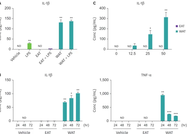
분비증가본 실험에서는
AT추출물의
THP-1세포의
cytokine분비에 미치는 영향을 조사하였다
. THP-1세포에 각 추출물을
48시간 동안 처리한 후
LPS를 첨가하여
24시간 동안 자극하였다
.배양 상
층액에서 대표적인 염증성
cytokine인
IL-1β의 양을
ELISA로 측정한 결과
,에탄올 추출물은 단
독으로 처리 시 아무런 영향이 없었으나
(검출되지 않음
),추출물을 전 처리 후
LPS를 처리한
경우
LPS만 처리한 그룹에 비해
cytokine의 생산이 감소하였다
.반면
,물 추출물의 경우 단독
처리 시
LPS를 처리한 그룹보다 더 많은 양의
IL-1β의 생산을 유도하였으며
LPS를 추가적으로
처리하여도 시너지 효과는 없었다
(Fig. 2A). IL-1β의 분비량은 추출물의 처리 시간과 농도에 의존적으로 증가하는 경향을 보였으며
, TNF-α의 경우 오히려
24시간 후 급격히 감소하였다
(Fig. 2B and C).이는
TNF-α가
autocrine으로 작용하였기 때문으로 여겨진다
.200 150 100 50 0
Vehicl
e LPS EAT
EAT + LPS WAT WAT + LPS
Conc (pg/mL)
IL-1β
ND ND
**
**
**
A
150
100
50
0 24 48 72 24 48 72 24 48 72
Vehicle EAT WAT
(hr)
Conc (pg/mL)
IL-1β
ND
* **
**
B
***
1,500
1,000
500
0 24 48 72 24 48 72 24 48 72
Vehicle EAT WAT
(hr)
Conc (pg/mL)
TNF-α
ND ND
***
**
400 300 200 100
0 0 12.5 25 50
Conc (pg/mL)
IL-1β
*
**
C
ND
ND ND ND
EAT WAT
Fig. 2. The water extract of AT induces cytokine secretion by THP-1 cells. (A) THP-1 cells were cultured with ethanol extract or water extract of AT (50 μg/mL) for 48 hours, and then further incubated in the presence or absence of LPS (10 ng/mL) for 24 hours. (B) THP-1 cells were cultured with ethanol extract or water extract of AT (50 μg/mL) for indicated times. (C) THP-1 cells were cultured in the presence of indicated doses of extracts of AT for 72 hours. Concentration of IL-1β and TNF-α in the culture supernatant were measured by enzyme-linked immunosorbent assay. Results are present as the means ± SEM (n = 3) and asterisks indicate significant differences compared to vehicle (t-tests). Representative data from 1 of 3 independent experiments is shown.
Conc, concentration; IL-1β, interleukin-1β; LPS, lipopolysaccharide; EAT, ethanol extracted Aster tataricus; WAT, water-extracted Aster tataricus; ND, not detected; TNF-α, tumor necrosis factor-α; AT, Aster tataricus; SEM, standard error of mean.
*p < 0.05; **p < 0.01; ***p < 0.001.
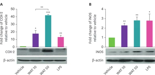
AT
물추출물에의한THP-1
세포의COX-2
와iNOS
발현조절면역 반응의 중요한 매개체인
prostaglandin과
nitric oxide (NO)생산에 관여하는
COX-2와
iNOS
단백질의 발현에 대한 영향을 알아보았다
. THP-1세포에
AT물 추출물을 농도별로 처
리하고
8시간 후
COX-2의 발현 정도와
24시간 후
iNOS의 발현 변화를
western blotting으로 확
인하였다
. Fig. 3에나타낸 것과 같이
COX-2의 경우
AT의 농도 의존적으로 발현이 유도되었
으며
, iNOS의 발현은 물 추출물을 처리한 세포에서 대조군에 비하여 유의하게 증가하는 것
을 확인 할 수 있었다
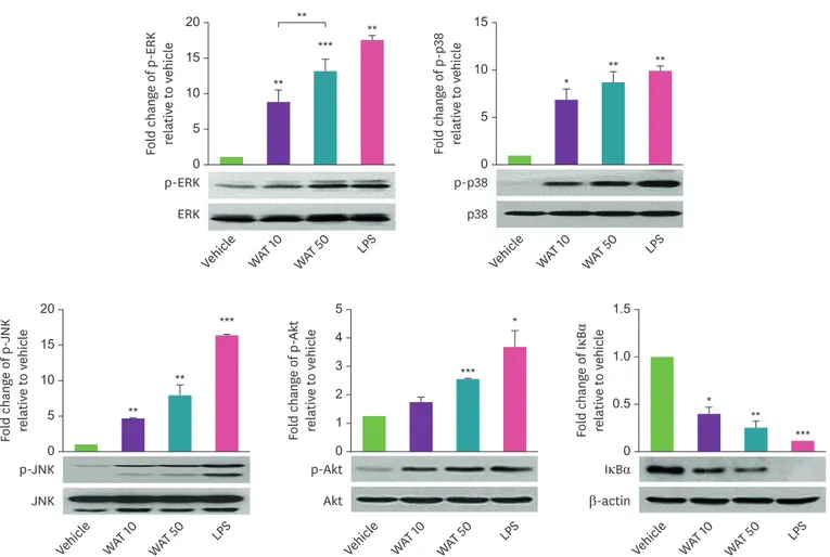
.AT
물추출물에의한THP-1
세포의신호전달분자활성화AT
추출물을
THP-1세포에 농도별로 처리하고
30분 후
MAPK와
Akt의 인산화 및
IκBα의 발현 변화를
western blotting으로 조사하였다
. AT물 추출물을 처리한 경우
, ERK, p38 MAPK, JNK를 비롯한
Akt의 인산화가 대조군에 비해 유의적으로 증가되었다
.뿐만 아니라
, AT물 추출
물에 의해
IκBα의 분해가 유도되는 것을 확인할 수 있었다
(Fig. 4).시험한 모든 신호전달 분
자의 변화가 관찰된 것은 순수 분리된 한 종류의 물질이 아닌
AT물 추출물 전체를 사용한 것에서 기인한 것으로 여겨진다
.여러 신호 전달 분자 중 어떤 분자가
AT물 추출물에 의한
cytokine
의 생산에 관여하는지 알아보기 위하여
3종의
MAPK와
Akt억제제를 이용하여 실험
을 수행하였다
. THP-1세포에
PD98059 (50 μM, MEK1억제제
), SB203580 (10 μM, p38 MAPK억 제제
), SP600125 (50 μM, JNK억제제
), wortmannin (10 nM, PI3K억제제
)을
30분 간 전처리하 고
AT물 추출물
50 μg/mL을 처리하였다
. 24시간 후 상층액에서
IL-1β와
TNF-α의 농도를 측정 한 결과
,두
cytokine모두
MAPK에 의해 조절되는 것을 확인하였다
(Fig. 5).흥미롭게도
, p38MAPK
의 억제제를 처리한 경우
IL-1β의 분비는 증가한 반면
, TNF-α의 분비는 감소되었다
.이
결과는
p38 MAPK가 두 종의
cytokine분비에 모두 관여하지만
IL-1β는 음성적으로
, TNF-α는 양성적으로 조절하는 반대의 역할을 가지고 있음을 의미한다
.** *
**
*
**
***
** 4
3 2 1 0
Vehicl e
WAT 10
WAT 50 LPS
A
Fold change of COX-2 relative to vehicle 50 40 30 20 10 0
Vehicl e
WAT 10
WAT 50 LPS
B
Fold change of iNOS relative to vehicle
COX-2 β-actin
iNOS β-actin
Fig. 3. The AT water extract increases the protein expression levels of COX-2 and iNOS in THP-1 cells. THP-1 cells were cultured with AT water extract (10 or 50 μg/mL) for 8 or 24 hours to detect COX-2 and iNOS, respectively.
LPS (10 ng/mL) was used as a positive control. The protein expression level of COX-2 (A) and iNOS (B) were detected by western blotting. The band intensity was measured and normalized from 3 independent experiments using imageJ. Results are present as the means ± SEM and asterisks indicate significant differences (t-tests).
COX-2, cyclooxygenase-2; WAT, water-extracted Aster tataricus; LPS, lipopolysaccharide; iNOS, inducible nitric oxide synthase; AT, Aster tataricus; SEM, standard error of mean.
*p < 0.05; **p < 0.01; ***p < 0.001.
20 15 10 5 0
Vehicl e
WAT 10
WAT 50 LPS
Fold change of p-JNK relative to vehicle ***
**
**
p-JNK JNK
5 4 3 2 1 0
Vehicl e
WAT 10
WAT 50 LPS
Fold change of p-Akt relative to vehicle *
***
p-Akt Akt 20
15 10 5 0
Vehicl e
WAT 10
WAT 50 LPS
Fold change of p-ERK relative to vehicle *****
**
p-ERK ERK
15
10
5
0
Vehicl e
WAT 10
WAT 50 LPS Fold change of p-p38 relative to vehicle
** **
*
p-p38 p38
**
*
***
**
1.5
1.0
0.5
0
Vehicl e
WAT 10
WAT 50 LPS Fold change of IκBα relative to vehicle
IκBα β-actin
Fig. 4. The AT water extract induces the phosphorylation of MAPK and Akt and the degradation of IκBα in THP-1 cells. THP-1 cells were cultured with AT water extract (10 or 50 μg/mL) for 30 minutes. Phosphorylated and unphosphorylated form of ERK, p-38 MAPK, JNK, and Akt and whole IκBα were detected by western blotting. The band intensity was measured and normalized from 3 independent experiments. Results are present as the means ± SEM and asterisks indicate significant differences compared to vehicle (t-tests).
ERK, extracellular-signal-regulated kinase; WAT, water-extracted Aster tataricus; LPS, lipopolysaccharide; JNK, c-Jun N-terminal kinase; IκBα, inhibitor of nuclear factor kappa B; SEM, standard error of mean.
*p < 0.05; **p < 0.01; ***p < 0.001.
250
150 200
100 50 0
Contr ol
PD98059 SB203580
SP600125 Wortmannin
Conc (pg/mL)
IL-1β
ND ND ND ND*** ND
*
**
Vehicle WAT
250
150 200
100 50 0
Contr ol
PD98059 SB203580
SP600125 Wortmannin
Conc (pg/mL)
TNF-α
ND ***
*** ***
Vehicle WAT
Fig. 5. The AT water extract regulates cytokine secretion in THP-1 through MAPK signaling pathway. THP-1 cells were pre-incubated with inhibitors of extracellular- signal-regulated kinase, p38 MAPK, c-Jun N-terminal kinase, and PI3K for 30 minutes, and then further cultured in the presence or absence of AT water extract for 24 hours. Concentration of IL-1β and TNF-α in the culture supernatant were measured by enzyme-linked immunosorbent assay. Results are present as the means ± SEM (n = 3) and asterisks indicate significant differences compared to control (t-tests). Representative data from 1 of 3 independent experiments is shown.
IL-1β, interleukin-1β; Conc, concentration; ND, not detected; WAT, water-extracted Aster tataricus; AT, Aster tataricus; TNF-α, tumor necrosis factor-α; MAPK, mitogen-activated protein kinase; SEM, standard error of mean.
*p < 0.05; **p < 0.01; ***p < 0.001.
고찰
AT
는
2000년간 중국 전통 의술에서 사용되어온 약초로
,뿌리는 자완이라 불리며 다양한 생
리활성물질들을 함유하고 있는 것으로 알려져 있다
.진해거담
,항바이러스
,항균
,항암
,지혈 등을 포함한 매우 다양한 효능을 가진다고 알려져 있으며
,자완을 말려 물로 달이거나 가루
로 빻아 복용하는 방법으로 이용된다
.꽃잎과 어린잎
,순도 식용으로 사용 가능하다
[1,18,19].대표적인 면역조절물질인 사포닌
(saponin)이 자완으로부터 분리되었고
,항염 작용이 있다
는 것이 발표된 바 있다
[11].본 논문에서는 뿌리를 제외한 꽃과 줄기
,잎 부분으로부터 추출
한 에탄올 추출물과 물 추출물을 이용하여
AT지상부 추출물에도 면역조절기능이 있는지 알 아보고자 하였다
.이전 연구들의 경우
, AT추출물의 세포 분열에 관한 영향은 신경 교종
,위암 세포
,구강 편평
상피 종 세포 등 암세포의 증식을 억제하여 항암 효과를 나타내는 것에 초점이 맞춰져 있다
[20-22].
본 연구에서는 면역 세포의 증식에 대한 영향을 확인하고자 하였다
.면역 세포는 세
포마다 특정한 자극에 의해 활성화되고 분열이 유도된다
. B세포의 경우
, B세포 수용체와
IL- 4, IL-2와 같은
cytokine에 의해
, T세포는
T세포 수용체와
CD28공동 자극 수용체에 대한 자
극과
IL-2와 같은
cytokine에 반응하여 활성화되고 분열이 유도된다
[23,24].선천 면역 세포의
경우도 림프구와 마찬가지로
NK세포는
IL-2, IL-15등 다양한
cytokine의 조합에 의해
,단핵구 는
macrophage colony-stimulating factor등에 의해
in vitro에서 분열이 유도된다
[25,26]. THP- 1세포에 추출물들을
72시간 처리하였을 때
,대조군에 비해 세포의 증식이 증가한 것을 확인
할 수 있었다
.이는
AT추출물이
THP-1세포의 활성을 증가시켰다고 해석될 수 있다
.이에 따
라
,본 저자는
THP-1세포에서
AT추출물이 염증성
cytokine분비에 미치는 영향을 조사하였
다
. THP-1세포에 각 추출물을 처리하고
LPS를 첨가한 결과
,에탄올 추출물 처리 시
LPS에 의
한
cytokine생산이 저해되었고 물 추출물의 경우 그와 반대로 단독 처리만으로도
cytokine의
생산을
LPS보다 높은 수준으로 유도하였다
.이미
, AT뿌리의 에탄올 또는 메탄올 추출물에
대한 연구는 많이 이루어져 있으며 염증성
cytokine의 생산을 감소시키는 결과가 보고된 바
있다
.그러나 지상부 추출물에 대한 연구와 추출 용매에 따른 효과에 대한 연구는 매우 미비
한 상태이다
.개미취에 대한 연구는 아니지만
, Han과
Kim [27]은 대파의 부위 별
,그리고 추
출용매에 따라 폴리페놀과 플라보노이드의 함량이 다르며
,그에 따라 항산화 효능이 다르게
나타나는 것을 보고한 바 있다
.또한
, Lee등
[28]의 사백산 물 추출물과 에탄올 추출물을 이용
하여 항염 효과를 비교한 연구에서는 추출물의 고성능 액체 크로마토그래피 분석 결과는 동 일한 패턴을 나타내지만 항염 효능에서 두 추출물 간 유의한 차이가 있음을 보여주었다
.개 미취 지상부 추출물이 추출 용매에 따라 활성이 다른 것 또한 추출 성분의 차이에서 비롯된 것일 수 있다
.추후
,물 추출물과 에탄올 추출물의 성분 분석 및 각 추출 성분의 면역 조절 활 성에 대한 연구가 필요할 것으로 사료된다
.Fig. 1에서
나타낸 것과 같이 에탄올 추출물은 물 추출물과 마찬가지로
THP-1의 분열을 증가
시켰다
.반면 염증성
cytokine의 분비는 저해하는 결과가 관찰되었다
.단핵구는 염증성 대식
세포인
M1또는 항염증성 대식세포인
M2서브타입으로 분화될 수 있는데
,항염 효능을 나타 내는 천연물질들의 경우
M1대식세포를
M2대식세포로 전환시키는 조절 능력이 있음이 보
고된 바 있다
[29].본 연구에서
AT추출물은
LPS처리 전
48시간 동안 수행되었다
.아마도 이
과정 동안
AT에탄올 추출물이
THP-1세포를
M2대식세포로 분화되도록 유도하여 염증성
cytokine
의 분비가 감소된 것일 수 있다
.이에 대한 추가적인 연구가 더 필요할 것으로 생각된 다
.이와 반대로
,물 추출물의 경우 염증성
cytokine인
IL-1β와
TNF-α의 생산 증가와 함께
,염
증 매개체인
PGE2와
NO의 생산에 관여하는 효소인
COX-2와
iNOS단백질의 발현을 유도하였
다
.염증은 크게 급성 염증과 만성 염증으로 구분할 수 있는데
,급성 염증은 감염체에 대한 면 역 반응에서 필수적인 반응으로 감염체를 인지한 선천 면역 세포에서 염증성 분자들이 분비
되면서 발생하며 감염체에 의한 자극이 제거되면 염증 반응도 사라진다
[11].이 결과들을 통
하여
AT물 추출물에 염증 반응을 증가시키는
,즉 면역 반응을 증가시키는 효과가 있는 것으 로 판단된다
.본 연구에서는
AT물 추출물을 그대로 사용한 것이므로 앞서 언급한 대로 추출 물에 포함된 각 성분 별 효능 분석이 필요할 것으로 사료된다
.이전 연구에 의하면
AT추출물은 다양한 신호 전달 경로를 통해 항염
,항암 효능을 나타낸다
.AT
로부터 추출된
polysaccharide는
Akt의 인산화를 저해하여 신경 교종 세포의 성장을 억제
하였고
,사포닌은
MAPK와
NF-κB신호 전달 경로를 억제함으로써
LPS에 의해 유도되는 염증
을 감소시켰다
[9,20].본 연구에서는
AT물 추출물에 의한 신호 전달 분자의 변화를 조사하
였다
. Fig. 4에나타낸 것과 같이
AT물 추출물은
Akt, MAPK의 인산화를 증가시키고
IκBα의
분해를 유도하였다
.이는 이전 연구들과는 상반되는 결과이지만 면역 반응을 증가시키는 현
상과 일치하는 결과이다
. AT물 추출물에 의한 염증성
cytokine의 분비를 조절하는 신호 전
달 경로를 알아보기 위해
PI3K와
MAPK억제제를 이용하여 실험을 진행한 결과
, 3종의
MAPK가
cytokine분비 조절에 관여하는 것을 확인하였다
.흥미롭게도
, p38 MAPK의 억제제를 처리
한 경우
IL-1β의 생산이 증가하였다
.이전 연구에 의하면
, p38 MAPK는 마우스 대식세포주인
RAW264.7
세포에서
LPS에 의한
IL-1β의 생산을 양성적으로 조절하며
TNF-α의 생산에는 관
여하지 않는 것으로 보고되었다
[30].반면
, Zhang등은
THP-1세포에서 두 종류의
LPS가 다
른
MAPK신호 전달 경로를 통하여
IL-1β와
TNF-α의 생산을 조절하며
p38 MAPK억제제를 처
리한 경우 대장균
LPS에 의한 염증성
cytokine들의 생산이 모두 저해됨을 보여주었다
[31].한
편
,또 다른 연구에서는
p38 MAPK의 억제제로 사용되는
SB253580이
p38 MAPK의 활성을 저
해하는 것뿐만 아니라
ERK신호 전달 경로를 활성화한다는 결과를 보여주었다
[32].그러나
Fig. 5에서
볼 수 있듯이
TNF-α의 생산은
SB253580처리 시 감소하였다
. TNF-α또한
ERK에 의 해 조절이 되므로 위에 언급한 결과를 적용한다면
IL-1β와 함께 증가할 것으로 예상된다
.이 결과는 아마도
AT물 추출물에 여러 성분들이 포함되어 있고 하나의 성분이 아닌 둘 이상의
물질이 각기 다른 신호 전달 경로를 통해
cytokine의 생산을 조절하기 때문일 가능성이 있다
.본 연구는
AT지상부 물 추출물과 에탄올 추출물의 면역 조절 효능을 검증하고자 수행되었 다
.연구 결과를 종합해보면
, AT물 추출물과 에탄올 추출물은
THP-1세포에 독성을 나타내 지 않으며 세포 분열을 증가시켰다
.에탄올 추출물은
LPS에 의한
IL-1β의 생산을 저해한 반면
,물 추출물은 단독으로
IL-1β와
TNF-α의 생산을 증가시켰다
.또한
,면역 반응에 관련된
COX-2와
iNOS의 발현을 유도하였으며 이와 함께
Akt, MAPK, NF-κB신호 전달 경로를 활성화하였
다
. ERK, p38 MAPK, JNK억제제가
AT물 추출물에 의한
cytokine생산을 감소시켰으며
, p38MAPK
의 경우 두
cytokine생산에 있어 반대로 작용함을 확인하였다
(Fig. 6).이러한 결과들
을 통하여
, AT지상부 물 추출물에 면역 증강 효능을 갖는 물질이 하나 이상 포함되어 있으
며 면역력 강화제의 천연 소재로써 이용될 수 있음을 제시한다
. AT의 뿌리는 항염제로써 사
용됨으로 지상부의 이용 시 뿌리와 분리하여 사용하는 것이 효능을 나타내기에 적합할 것으
로 사료된다
.요약
본 연구는
AT의 뿌리를 제외한 전체
AT의 에탄올 및 물 추출물의 면역 조절 효과를 비교하고
THP-1
의
cytokine분비를 조절하는 분자 메커니즘을 조사하였다
. AT의 물 추출물 및 에탄올
추출물은
THP-1세포에 독성이 없으며 세포 증식을 증가키는 것을 확인하였다
.에탄올 추출
물은 영향이 없는데 반해
,물 추출물은
THP-1의
IL-1β의 분비를 증가시켰으며
COX-2및
iNOS단백질의 발현을 증가시켰다
.또한
, MAPK및
Akt의 인산화와
IkBα의 분해를 유도하는 것을 확인하였다
. AT에 의한
IL-1β분비는
ERK및
JNK억제제에 의해 감소되었으며
, TNF-α의 분비 는
ERK, p38 MAPK및
JNK억제제에 의해 감소되었다
.흥미롭게도
, p38 MAPK억제제는
AT에
의한
IL-1β의 생성을 추가로 증가시켰다
.이 결과는
AT지상부의 물 추출물에
MAPK신호 전
달 경로를 통해 면역 세포를 자극하여
cytokine의 생산을 유도하는 생리활성물질이 존재한다
는 것을 의미한다
.따라서
, AT지상부는 면역력 강화제의 천연 소재로써 이용될 수 있을 것
으로 사료된다
.REFERENCES
1. Yu P, Cheng S, Xiang J, Yu B, Zhang M, Zhang C, et al. Expectorant, antitussive, anti-inflammatory activities and compositional analysis of Aster tataricus. J Ethnopharmacol 2015; 164: 328-333.
PUBMED | CROSSREF
iNOS, COX-2, Cytokine (IL-1β, TNF-α) Nucleus
Cytoplasm THP-1 cell
Degradation AT
p38 ERK JNK iNOS
p38 ERK JNK
Akt IκBα
IκBα
Akt COX-2
P P P P
P p65 p50
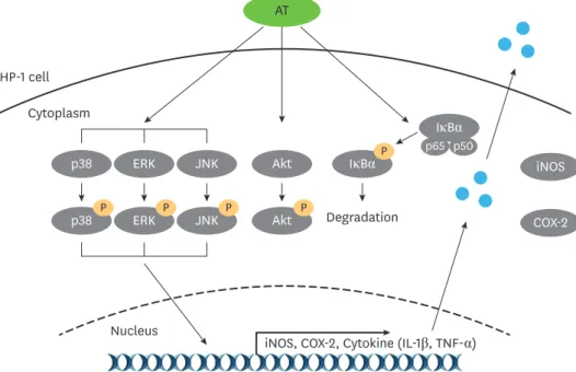
Fig. 6. Schematic diagram of signal transduction of the AT water extract in THP-1 cells. The AT water extract induces phosphorylation of MAPK and Akt and degradation of IκBα in THP-1 cells. It also increases the expression of COX-2 and iNOS and induces the secretion of IL-1β and TNF-α. AT regulates cytokine secretion through the MAPK signaling pathway. Among them, p38 MAPK plays an opposite role in the production of 2 cytokines that negatively regulates IL-1β production, while positively regulates TNF-α production.
AT, Aster tataricus; ERK, extracellular-signal-regulated kinase; JNK, c-Jun N-terminal kinase; IκBα, inhibitor of nuclear factor kappa B; iNOS, inducible nitric oxide synthase; COX-2, cyclooxygenase-2; IL-1β, interleukin-1β;
TNF-α, tumor necrosis factor-α; MAPK, mitogen-activated protein kinase.
2. Morita H, Nagashima S, Uchiumi Y, Kuroki O, Takeya K, Itokawa H. Cyclic peptides from higher plants.
XXVIII. Antitumor activity and hepatic microsomal biotransformation of cyclic pentapeptides, astins, from Aster tataricus. Chem Pharm Bull (Tokyo) 1996; 44(5): 1026-1032.
PUBMED | CROSSREF
3. Shao Y, Ho CT, Chin CK, Poobrasert O, Yang SW, Cordell GA. Asterlingulatosides C and D, cytotoxic triterpenoid saponins from Aster lingulatus. J Nat Prod 1997; 60(7): 743-746.
PUBMED | CROSSREF
4. Wang CZ, Yu DQ. Triterpenoid saponins from Aster auriculatus. Planta Med 1998; 64(1): 50-53.
PUBMED | CROSSREF
5. Akihisa T, Kimura Y, Koike K, Tai T, Yasukawa K, Arai K, et al. Astertarone A: a triterpenoid ketone isolated from the roots of Aster tataricus L. Chem Pharm Bull (Tokyo) 1998; 46(11): 1824-1826.
CROSSREF
6. Tanaka R, Nagao T, Okabe H, Yamauchi T. Studies on the constituents of Aster tataricus L. f. IV.
Structures of Aster saponins isolated from the herb. Chem Pharm Bull (Tokyo) 1990; 38(5): 1153-1157.
CROSSREF
7. Ng TB, Liu F, Lu Y, Cheng CH, Wang Z. Antioxidant activity of compounds from the medicinal herb Aster tataricus. Comp Biochem Physiol C Toxicol Pharmacol 2003; 136(2): 109-115.
PUBMED | CROSSREF
8. Du H, Zhang M, Yao K, Hu Z. Protective effect of Aster tataricus extract on retinal damage on the virtue of its antioxidant and anti-inflammatory effect in diabetic rat. Biomed Pharmacother 2017; 89: 617-622.
PUBMED | CROSSREF
9. Zhang HT, Tian M, He QW, Chi N, Xiu CM, Wang YB. Effect of Aster tataricus on production of inflammatory mediators in LPS stimulated rat astrocytoma cell line (C6) and THP-1 cells. Saudi Pharm J 2017; 25(3): 370-375.
PUBMED | CROSSREF
10. Su XD, Jang HJ, Li HX, Kim YH, Yang SY. Identification of potential inflammatory inhibitors from Aster tataricus. Bioorg Chem 2019; 92: 103208.
PUBMED | CROSSREF
11. Su XD, Jang HJ, Wang CY, Lee SW, Rho MC, Kim YH, et al. Anti-inflammatory potential of saponins from Aster tataricus via NF-κB/MAPK activation. J Nat Prod 2019; 82(5): 1139-1148.
PUBMED | CROSSREF
12. Janeway CA Jr, Travers P, Walport M, Shlomchik MJ. Immunobiology. 6th ed. New York (NY): Garland Science; 2005.
13. Janeway CA Jr, Medzhitov R. Innate immune recognition. Annu Rev Immunol 2002; 20(1): 197-216.
PUBMED | CROSSREF
14. Erwig LP, Rees AJ. Macrophage activation and programming and its role for macrophage function in glomerular inflammation. Kidney Blood Press Res 1999; 22(1-2): 21-25.
PUBMED | CROSSREF
15. Epelman S, Lavine KJ, Randolph GJ. Origin and functions of tissue macrophages. Immunity 2014; 41(1):
21-35.
PUBMED | CROSSREF
16. Rua R, McGavern DB. Elucidation of monocyte/macrophage dynamics and function by intravital imaging.
J Leukoc Biol 2015; 98(3): 319-332.
PUBMED | CROSSREF
17. Jakubzick CV, Randolph GJ, Henson PM. Monocyte differentiation and antigen-presenting functions. Nat Rev Immunol 2017; 17(6): 349-362.
PUBMED | CROSSREF
18. Bown D. Encyclopedia of herbs & their uses. London: Dorling Kindersley; 1995.
19. World Health Organization, Regional Office for the Western Pacific. Medicinal plants in the Republic of Korea: information on 150 commonly used medicinal plants. Manila: WHO Regional Office for the Western Pacific; 1998.
20. Du L, Mei HF, Yin X, Xing YQ. Delayed growth of glioma by a polysaccharide from Aster tataricus involve upregulation of Bax/Bcl-2 ratio, activation of caspase-3/8/9, and downregulation of the Akt. Tumour Biol 2014; 35(3): 1819-1825.
PUBMED | CROSSREF
21. Zhang Y, Wang Q, Wang T, Zhang H, Tian Y, Luo H, et al. Inhibition of human gastric carcinoma cell growth in vitro by a polysaccharide from Aster tataricus. Int J Biol Macromol 2012; 51(4): 509-513.
PUBMED | CROSSREF
22. Wang R, Xiao S, Niu Z. Anti-cancer activity of Aster tataricus on scc-9 human oral squamous carcinoma.
Afr J Tradit Complement Altern Med 2017; 14(2): 142-147.
PUBMED | CROSSREF
23. Choe J, Kim HS, Armitage RJ, Choi YS. The functional role of B cell antigen receptor stimulation and IL-4 in the generation of human memory B cells from germinal center B cells. J Immunol 1997; 159(8): 3757-3766.
PUBMED
24. Appleman LJ, Berezovskaya A, Grass I, Boussiotis VA. CD28 costimulation mediates T cell expansion via IL- 2-independent and IL-2-dependent regulation of cell cycle progression. J Immunol 2000; 164(1): 144-151.
PUBMED | CROSSREF
25. Caligiuri MA. Human natural killer cells. Blood 2008; 112(3): 461-469.
PUBMED | CROSSREF
26. Lari R, Kitchener PD, Hamilton JA. The proliferative human monocyte subpopulation contains osteoclast precursors. Arthritis Res Ther 2009; 11(1): R23.
PUBMED | CROSSREF
27. Han I, Kim JH. Antioxidant and physiological activities of water and ethanol extracts of diverse parts of welsh onion. J Korean Soc Food Sci Nutr 2017; 46(4): 426-434.
CROSSREF
28. Lee DS, Choi HG, Kim KS, Kim DC, Min HK, Li B, et al. The comparison between sabaek-san water and 30% EtOH extracts for anti-inflammatory effects. Yakhak Hoeji 2012; 56(4): 240-247.
29. Saqib U, Sarkar S, Suk K, Mohammad O, Baig MS, Savai R. Phytochemicals as modulators of M1-M2 macrophages in inflammation. Oncotarget 2018; 9(25): 17937-17950.
PUBMED | CROSSREF
30. Baldassare JJ, Bi Y, Bellone CJ. The role of p38 mitogen-activated protein kinase in IL-1 beta transcription.
J Immunol 1999; 162(9): 5367-5373.
PUBMED
31. Zhang D, Chen L, Li S, Gu Z, Yan J. Lipopolysaccharide (LPS) of Porphyromonas gingivalis induces IL- 1beta, TNF-alpha and IL-6 production by THP-1 cells in a way different from that of Escherichia coli LPS.
Innate Immun 2008; 14(2): 99-107.
PUBMED | CROSSREF
32. Numazawa S, Watabe M, Nishimura S, Kurosawa M, Izuno M, Yoshida T. Regulation of ERK-mediated signal transduction by p38 MAP kinase in human monocytic THP-1 cells. J Biochem 2003; 133(5): 599-605.
PUBMED | CROSSREF