41(8), 1057~1065(2012) http://dx.doi.org/10.3746/jkfn.2012.41.8.1057
천궁( Cnidium officinale ) 및 당귀( Angelica gigas ) 휘발성 향기추출물의 항염증효과
최상원
1†
․김은옥1
․임현희1
․김진경2
1
대구가톨릭대학교 식품영양학과
2
대구가톨릭대학교 의생명과학과
Anti-Inflammatory Effects of Volatile Flavor Extracts from Cnidium officinale and Angelica gigas
Sang-Won Choi
1†
, Eun-Ok Kim1
, Hyun-Hee Leem1
, and Jin-Kyung Kim2
1
Dept. of Food Science and Nutrition and2
Dept. of Biomedical Science, Catholic University of Daegu, Hayang, Gyeongbuk 712-702, KoreaAbstract
The effects of volatile flavor extracts of eight different herbal medicines, Juniperus rigida (JR), Saussurea lappa SL), Cnidium officinale (CO), Angelica gigas (AG), Eugenia caryophyllata (EC), Angelica tenuissima (AT), Mentha arvense (MA), and Artemisiae argyi (AA), were investigated on LPS-stimulated inflammation using Raw 264.7 cells. The volatile flavor extracts of CO and AG considerably inhibited LPS-stimulated NO, PGE
2, IL-6, and TNF-α (except AG) production, as well as iNOS expression. Major volatile components of CO were identified as ligustilide and of β-eudesmol as AG by GC-MS analysis. Thus, these results suggest that the volatile extracts of CO and AG may be useful as potential therapeutic agents for inflammation-associated disorders.
Key words: Cnidium officinale, Angelica gigas, volatile flavor extracts, anti-inflammation, GC-MS analysis
†
Corresponding author. E-mail: [email protected]
†
Phone: 82-53-850-3525, Fax: 82-53-850-3516
서 론
염증은 생체나 조직에 물리적 충격이나 화학물질, 세균감 염 등의 자극에 대한 생체방어기전이며, 지속적인 염증반응 은 점막손상을 촉진시켜 결과적으로 통증, 부종, 발적, 발열 등을 일으켜 기능장애를 유발하며 당뇨병, 동맥경화증, 관절 염 및 암 등의 발생과 깊은 연관을 갖고 있다(1,2). 체내 염증 과정에서 대식세포는 inducible nitric oxide synthase(iNOS) 에 의해서 만들어지는 일산화질소(NO)와 cyclooxygenase- 2(COX-2)에 의해서 과량 만들어지는 프로스타글란딘 E
2(PGE
2) 등과 같은 염증 촉진 인자들을 생성한다(3). 또한, tumor necrosis factor-α(TNF-α), interleukin-6(IL-6) 및 interleukin-1β(IL-1β)와 같은 사이토카인을 생산하여 다양 한 염증반응을 매개한다(4,5). 이와 같이 NO, PGE
2및 염증 성 사이토카인 등이 염증반응과 밀접하게 관련되어 있어, 이들의 생성과 생성에 관여하는 효소의 발현을 조절할 수 있는 물질들이 염증질환의 예방 및 치료제로서 각광을 받고 있다.
지금까지 개발된 합성 항염증제는 크게 스테로이드(히드 로코르티손, 프레드니솔론, 베타메타손)와 비스테로이드(아
스피린, 인도메타신, 이부프로펜)로 나눌 수가 있으며, 이들 은 대부분 위장, 신장 및 심장질환 등의 부작용을 나타내어 (6,7) 그 사용이 제한받고 있어 현재 보다 안전하고 효과 있 는 천연 유래 항염증 치료제의 개발이 필요하다. 최근에는 항염증활성을 갖는 식품 및 생약추출물을 이용한 각종 염증 성 질환의 치료제 개발이 활발히 이루어지고 있다(8,9).
천궁(
Cnidium officinale)은 미나리과(Umbelliferae)에 속 하는 다년생 초본으로 중국이 원산지로써 중국, 한국, 일본 등지에서 재배되고 있다. 한방에서는 그 뿌리줄기를 풍냉으 로 인한 두통, 동통, 빈혈, 월경불순, 불임 등과 같은 부인과 질환의 치료에 널리 이용하고 있다(10). 천궁에는 정유가 1
~2% 함유되어 있으며, 그 성분으로는 ligustilide, butyli-
dene phthalide, cnidilide 및 neocnidilide 등이 보고되어 있고
(11,12), 천궁의 생리적작용으로는 항산화(10) 및 소염·진통
작용(13) 그리고 혈소판응집 억제 활성(14) 등의 보고가 있
다. 한편, 당귀(
Angelica gigas)는 미나리과에 속하는 다년
생 초본으로 1∼2년 된 생근을 가을에 채취하여 건조한 것을
이용하고 있다(15). 당귀는 보혈제의 대표적인 생약으로 혈
을 보하여 주고 청열작용과 혈의 흐름을 좋게 함으로 빈혈이
나 어혈로 인한 혈행장애에 크게 활용하고 있으며 월경불순,
월경정지, 신체허약, 두통, 복통 및 변비에 널리 응용된다.
당귀의 주요 성분으로는 pyranocoumarin계 화합물인 de- cursin, decursinol 및 nodakenin과 휘발성 향기성분인 α- pinene, β-eudesmol 등을 함유하고 있다(16,17). 당귀의 약 리작용으로는 항산화(18), 항암(19), 항염증(20) 및 혈액순환 개선 등에 대해 보고된 바 있다. 이와 같이 천궁 및 당귀의 주요 생리활성성분 및 생리적 효능에 관한 많은 연구가 수행 되어져 왔으나, 그들의 휘발성 향기추출물의 생리적 효능 및 그를 이용한 한방화장품의 개발에 관한 연구는 거의 없는 실정이다.
최근 본 연구진들은 천궁 및 당귀를 주 베이스로 제조한 한방복합처방제의 항염증 효능을 검증한 결과 천궁 및 당귀 를 함유한 한방처방제의 휘발성 향기추출물이 우수한 항염 증 활성을 나타내었다(21). 따라서 천궁 및 당귀의 휘발성 향기추출물의 항염증 효능을 보다 상세히 조사할 필요가 있 었으며, 아울러 그들의 주된 휘발성 향기성분의 분석이 요구 되었다.
본 연구에서는 생약의 휘발성 향기추출물의 항염증활성 을 검증하기 위해 먼저 천궁 및 당귀를 포함한 8가지 생약을 수증기증류추출법(steamed distillation extraction, SDE)을 이용하여 휘발성 향기성분을 추출한 후 마우스의 대식세포 주인 Raw 264.7 세포를 이용한 모델시스템에서 생약 향기추 출물의 항염증 효과를 측정하였으며, 아울러 항염증 활성이 높은 천궁 및 당귀 향기추출물의 휘발성 향기성분을 GC- MS 분석을 통해 확인하였다.
재료 및 방법
실험재료 및 시약
본 실험에 사용한 8가지 생약[정향(
Eugenia caryophyllata), 고본(
Angelica tenuissima), 노간주(
Juniperus ridiga), 박 하(
Mentha arvense), 당귀(
Angelica gigeas), 목향(
Saus- surea lappa), 애엽(
Artemisiae argyi) 및 천궁(
Cnidium of- ficinale)] 중 정향(인도네시아산)을 제외하고는 전부 국산으 로 영천 약전시장에서 구입한 것을 사용하였으며, 건조된 상 태로 저온(-5
oC)에서 보관하면서 실험재료로 사용하였다. 실 험에 사용한 시약인 lipopolysaccharide(LPS), dimethyl sulfoxide(DMSO)는 Sigma사(St. Louis, MO, USA)로부터, Dulbecco's Modified Eagle's Medium(DMEM), Fetal Bovine Serum(FBS), penicillin 및 streptomycin은 Hyclone 사(Logan, UT, USA)로부터 구입하였다. CellTiter 96 AQueous One Solution과 Griess reagent system은 Pro- mega(Madison, WI, USA), PGE
2enzyme-linked immun- osorbent assay(ELISA) kit은 Parameter
TMR&D사(Min- neapolis, MI, USA), Tumor necrosis factor-α(TNF-α)와 Interleukin-6(IL-6) ELISA kit는 eBioscience사(San Diego, CA, USA)로부터 각각 구입하여 사용하였다. Inducible
NOS(iNOS)와 COX-2 항체는 Cell signaling사(Danvers, MA, USA)에서, β-actin 항체는 Sigma사로부터 구입하였 다. 그 외 시약 및 용매는 분석용 특급 또는 HPLC급 시약을 사용하였다.
천궁 및 당귀를 포함한 8가지 생약의 향기추출물의 제조 천궁 및 당귀를 포함한 8가지 생약의 휘발성 향기추출물 은 SDE법에 의해 다음과 같이 제조하였다. 먼저, Schultz 등(22)의 방법에 따라 분쇄한 생약 50 g에 초순수 1 L를 혼합 하고 SDE 장치에서 재증류한 diethyl ether 500 mL를 사용 하여 상압 하에서 4시간 동안 추출한 후 무수황산나트륨을 가하여 -4
oC에서 하룻밤 방치시켜 탈수시키고 유기용매층 은 rotary vacuum evaporator(Eyela, Tokyo, Japan) 및 질소 purger를 이용하여 농축 후 향기추출물을 제조하였다.
천궁 및 당귀 향기추출물의 휘발성 향기성분의 분석 천궁 및 당귀 향기추출물의 휘발성 향기성분의 분석은 GC-MS를 이용하여 다음과 같이 측정하였다. GC-MS 기기 는 HP 5973 Mass selective detector(Agilent technologies Inc., Chandler, AZ, USA)가 장착된 Agilent 5975C Series gas chromatography(Agilent technologies Inc.)를 사용하 였으며, 분리용 칼럼은 HP-5MS 5% phenyl methyl silox nonpolar(30 m×0.25 mm, i.d., 0.25 μm film thickness, J
& W, Folsom, CA, USA)를 사용하였고, 이때 oven의 온도 는 50
oC에서 3분간 유지한 다음 5
oC/min의 속도로 300
oC까 지 상승시켰으며, carrier gas의 유속은 1 mL/min(He)로 유 지하였다. 분리된 화합물의 이온화는 electron impact ion- ization(EI mode) 방법으로 행하였으며 ionization voltage와 ion source의 온도는 각각 70 eV와 230
oC로 설정하였고, 분 석할 분자량의 범위는 40∼400(m/z)으로 설정하였으며, GC injector의 온도는 250
oC로 설정하였다. SDE추출법으로 추출 된 향기추출물을 split ratio 1:20으로 주입하여 분석하였다.
GC-MS에 의해 total ionization chromatogram(TIC)에 분리된 각 peak의 휘발성향기성분 분석은 mass spectrum library(Wiley 275 & 7N, NBS 75K)(23)와 mass spectral data book의 spectrum과 비교하여 확인하였다.
세포배양 및 세포증식
실험에 사용된 마우스 대식세포주인 Raw 264.7 세포는 한국 세포주은행에서 분양 받아 사용하였으며, 10% FBS와 1% antibiotics(penicillin/streptomycin)를 첨가한 DMEM 배지를 이용하여 5% CO
2, 37
oC incubator에서 배양하였다.
8가지 생약의 휘발성 향기추출물의 세포증식 능력을 측정
하기 위해 Raw 264.7 세포를 96 well plate에 well당 1×10
4세포가 되도록 분주한 후 18시간 배양하였다. 이때 향기추출
물은 10∼200 μg/mL의 농도가 되도록 150 μL/well을 세포
에 처리하여 24시간 배양한 후, CellTiter 96 AQueous One
solution assay를 이용하여 20 μL/well의 one solution을 첨
가하고 2시간 반응시킨 후 490 nm에서 흡광도를 측정하여
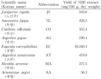
Table 1. Yield of volatile flavor extracts from eight different crude medicinal drugs by SDE apparatus
Scientific name
(Korean name) Abbreviation Yield of SDE extract (mg/100 g, dry weight) Juniperus rigida
(노간주) Saussurea lappa Cnidium officinale (목향) Angelica gigas (천궁) Eugenia caryophyllata (당귀) Angelica tenuissima (정향) Mentha arvense (고본) Artemisiae argyi (박하)
(애엽)
JS SL CO AG EC AT MA AA
105.6 428.3 352.4 196.4 16,340.4 419.8 227.5 56.3
Data represent average of duplicate determination.
Standard deviations and statistical analysis are omitted for simplicity.
세포증식에 미치는 효과를 측정하였다(24).
NO 및 PGE
2측정
NO의 농도는 배양액 내의 nitrite 농도를 Green 등(25)의 방법에 따라 측정하였다. Raw 264.7 세포를 48 well plate에 2×10
5cells/well이 되도록 분주하고 18시간 배양한 후, 천궁 및 당귀 휘발성 향기추출물 1∼50 μg/mL의 농도가 되도록 전처리하고 1시간 후에 LPS 100 ng/mL 처리하여 20시간 배양하였다. 배양액 중에 함유된 NO의 양을 Griess Reagent System을 이용하여 측정하였고, PGE
2는 PGE
2ELISA kit 을 사용하여 측정하였다.
Cytokine 측정
Raw 264.7 세포를 48 well plate에 2×10
5cells/well이 되 도록 분주한 후 18시간 배양한 후 천궁 및 당귀 휘발성 향기 추출물 1∼50 μg/mL의 농도가 되도록 전처리하고 1시간 후 에 LPS 100 ng/mL 처리하여 배양하였다. 24시간 배양한 후 배양액에 함유된 IL-6 및 TNF-α를 ELISA kit를 이용하 여 측정하였다.
Western blot assay
iNOS 및 COX-2의 단백질 발현 양을 측정하기 위해, Raw 264.7 세포를 100 mm 배양접시에 1×10
6cells/well이 되도 록 분주한 후 18시간 배양하였다. 천궁 및 당귀 향기추출물 1, 10, 25, 50 μg/mL와 LPS(100 ng/mL)를 동시처리 또는 LPS 단독처리 하여 20시간 배양한 후 세포를 수집하여 PBS (phosphate buffered saline)로 세척한 다음, 용해 완충액 (lysis buffer)을 첨가하여 세포를 용해시킨 후 원심분리 하 여 세포막 성분 등을 제거하고 세포 내 단백질을 얻었다.
BCA assay reagent(Pierce, Rockford, IL, USA)로 단백질을 정량한 후 20∼50 μg의 lysate를 8% sodium dodecylsulfate (SDS)가 포함되어 있는 polyacrylamide gel에서 전기영동 후, nitrocellulose transfer membrane으로 옮겼다. 항체의 비특이적 결합을 억제시키기 위해 membrane을 5% 탈지 분 유액이 포함된 TBST(50 mM Tris-HCl, pH 7.5, 150 mM NaCl, 0.1% Tween-20)로 1시간 실온에서 배양한 후 iNOS 와 COX-2의 항체를 이용하여 4
oC에서 하룻밤 반응시켰다.
iNOS와 COX-2 발현 양은 horseradish peroxidase(HRP)가 붙어있는 2차 항체로 실온에서 2시간 반응 후 chemilumin- escence reagents(Amersham Biosciences, Piscataway, NJ, USA)를 이용하여 가시화하였다. 각 단백질 밴드의 강도는 GelDoc-It TS imaging system(Ultra-Violet Products Ltd, Cambridge, UK)을 사용하여 수치화하였다.
통계처리
본 연구의 실험 결과들은 3회 반복 측정하여 평균값±표 준편차로 나타내었으며, 모든 자료의 통계분석은 SAS (Statistical Analysis System) software package(SAS 9.1.3, SAS Institute Inc., Cary, NC, USA)를 사용하여 실시하였다.
이때 분산분석은 ANOVA test를, 각 시험구간의 평균차이에 대한 유의성 검정은 Duncan multiple range test를 이용하여 p<0.05 수준일 때 유의한 차이가 있는 것으로 간주하였다.
결과 및 고찰
수증기증류추출법에 의해 제조된 8가지 생약의 휘발성 향기추출물의 수율
수증기증류추출법에 의해 제조된 8가지 생약 휘발성 향기 추출물의 수율을 조사한 결과는 Table 1과 같다. 먼저 정향 향기추출물의 수율이 16,340.4 mg%로 8가지 생약 중 가장 높게 나타났으며, 그 다음으로 목향(428.3 mg%)>고본(419.8 mg%)>천궁(352.4 mg%)>박하(227.5 mg%)>당귀(196.4 mg%)>노간주(105.6 mg%) 순으로 낮은 수율을 나타내었 고, 애엽의 경우 56.3 mg%으로 가장 낮은 수율을 나타내었다.
마우스 대식세포에서의 세포증식에 미치는 영향 대식세포는 염증반응 시에 NO와 같은 활성산소종 및 IL- 6, TNF-α와 같은 염증성 사이토카인을 생산하여 감염초기 에 생체방어에 중요한 역할을 하므로 항염증 효과를 검증하 는 염증모델로 많이 사용된다(26,27). 먼저 8가지 생약(노간 주, 목향, 천궁, 당귀, 정향, 고본, 박하, 애엽)의 향기추출물의 처리에 의한 Raw 264.7 대식세포에의 증식에 미치는 영향을 관찰하기 위해, 생약 향기추출물을 0, 10, 50, 100, 200 μg/mL 의 농도로 처리하여 24시간 배양 후 Raw 264.7 세포의 증식 을 측정하였다. 그 결과 Fig. 1에서 보는 바와 같이, 8가지 생약의 향기추출물 중 노간주, 당귀, 박하 및 애엽은 본 실험 에 사용한 어떤 농도에서도 세포독성을 나타내지 않았다.
반면, 천궁 및 정향은 200 μg/mL 이상의 농도에서 세포독성
0 5 10 15 20 25
Control LPS 10 μg/mL 50 μg/mL
N O p ro d u c ti o n ( μ M ) .
JR CO AG MA AA Control LPS
a
c b
d cd b c c c c b
Fig. 2. Inhibitory effects on NO production of the volatile flavor extracts from five different crude drugs extracted by SDE in LPS-induced Raw 264.7 cells. Cells were treated with 10 and 50 μg/mL of the volatile extracts from five different crude drugs in the presence of 100 ng/mL LPS or with LPS alone for 18 hr. The results are expressed as mean±SD from three in- dependent experiments. Bars with different letters are signifi- cantly different at p<0.05 by Duncan multiple range test when compared with LPS-stimulated cells. JR: Juniperus ridiga, CO:
Cnidium officinale, AG: Angelica gigeas, MA: Mentha arvense, AA: Artemisiae argyi.
0.0 0.5 1.0 1.5 2.0
Control JR SL CO AG ET AT MA AA
C e ll vi a b ili ty ( 4 9 0 n m ) .
10 ug/mL 50 ug/mL 100 ug/mL 200 ug/mL Control
NS
a
a a a a ab ab ab a
c b a a
c b ab a
b a a
b
c b a
b
b b b
EC
Fig. 1. Cytotoxicity of the volatile flavor extracts from eight different crude drugs extracted by SDE against Raw 264.7 cells.
Cells were treated with 0∼200 μg/mL of the volatile extracts from eight different crude drugs for 24 hr. Cell viability was determined using the CellTiter 96 AQueous One Solu- tion Assay. The results are expressed as mean±SD from three independent experi- ments. Bars with different letters are sig- nificantly different at p<0.05 by Duncan multiple range test. JR: Juniperus ridiga, SL:
Saussurea lappa, CO: Cnidium officinale, AG:
Angelica gigeas, EC: Eugenia caryophyllata, AT: Angelica tenuissima, MA: Mentha ar- vense, AA: Artemisiae argyi.
을 나타내었고, 특히 목향 및 고본은 각각 50 μg/mL 및 100 μ g/mL 이상의 낮은 농도에서도 세포독성을 나타내었다. 따 라서 8가지 생약 중 세포독성이 있는 목향 및 고본과 100 μ g/mL의 농도에서 NO 생성 저해효과가 낮은 정향(데이터 생략)을 제외한 5가지 생약 즉 노간주, 천궁, 당귀, 박하 및 애엽의 향기추출물을 0∼50 μg/mL 농도로 조정하여 실험을 진행하였다.
NO 및 PGE
2생성에 미치는 영향
그람음성세균의 내독소(endotoxin)로 알려진 LPS를 대 식세포에 처리하게 되면 NO, PGs, 염증성 사이토카인과 같 은 물질들이 생성되어 염증반응을 조절하는 다양한 병리학 적 반응이 유도된다(28). 따라서 본 연구에서는 생약 향기추 출물의 항염증작용을 검토하고자, Raw 264.7 세포에 염증 유발물질인 LPS(100 ng/mL)와 함께 다양한 농도의 생약 향기추출물을 동시에 처리한 후 NO 및 PGE
2의 생성 억제효 과를 관찰하였다. Fig. 2에서 보는 바와 같이 Raw 264.7 세포 에 LPS를 처리하여 증가된 NO의 생성은 5가지 생약 향기추 출물 10 μg/mL 및 50 μg/mL 농도를 첨가함으로써 LPS 단 독처리 군에 비해 유의적으로 억제되었다. 10 μg/mL 농도에 서의 NO 생성은 천궁, 당귀, 박하 및 애엽이 유사하였으며 이는 노간주에 비해 낮은 수준이었다. 한편, 50 μg/mL의 농 도에서는 천궁이 가장 큰 NO 생성 저해효과를 나타내었으 며, 다음으로 당귀, 애엽, 박하 및 노간주 순으로 나타났다.
이전의
in vitro결과에서는 당귀가 COX-2 및 15-LO(15-li- poxygenase) 저해활성이 상당히 큰 것으로 보고한 바 있다 (29). 따라서 5가지 생약 중 항염증 활성이 클 것으로 사료되 는 천궁 및 당귀 향기추출물을 농도별로 처리한 결과, 모두 농도의존적으로 NO의 생성을 억제하였다(Fig. 3A).
한편, 염증반응의 또 다른 체내 매개물질 중 하나인 PGE
2생성에 미치는 천궁 및 당귀 향기추출물의 억제 효과를 측정 하였다. Fig. 3B에서 보는 바와 같이, LPS를 처리하여 증가 된 PGE
2의 생성은 천궁 및 당귀 향기추출물을 농도별로 첨 가함으로써 모든 처리 농도에서 LPS 단독처리 군에 비해
유의적으로 억제하였으며, 농도의존적으로 PGE
2의 생성이 억제되었다. 이와 같은 결과는 염증반응 시 천궁 및 당귀 향 기추출물(1∼50 μg/mL)의 처리가 주요 염증성 매개물질인 NO 및 PGE
2의 생성을 억제함으로써 항염증 활성이 나타남 을 알 수 있었다.
iNOS 및 COX-2 발현에 미치는 영향
염증반응 매개물질인 NO와 PGE
2는 각각 iNOS와 COX라
는 효소에 의해 생합성 된다. NOS는 iNOS, neuronal NOS,
그리고 endothelial NOS(eNOS)의 3종류가 존재하며, 이 중
iNOS는 LPS, cytokine과 같은 자극에 의해 급격하게 유도
되어 과량의 NO를 생성한다. 한편, cyclooxygenase는 COX-
1과 COX-2 두 종류가 있으며, COX-1은 생체 내의 대부분
의 조직에 존재하며 일정하게 발현되는 반면, COX-2는 염
증자극원에 의해 macrophage와 같은 염증성 세포에서 유도
0 5 10 15 20
Control LPS CO AG
N O p ro d u c ti o n ( μ M ) .
1 μg/mL 10 μg/mL 25 μg/mL
50 μg/mL Control LPS
a
d c b a
d c b a (A)
0 700 1400 2100 2800
Control LPS CO AG
P G E
2( p g /m L ) .
1 μg/mL 10 μg/mL 25 μg/mL
50 μg/mL Control LPS
a
cd d c b
cd d c b (B)
Fig. 3. Inhibitory effects on NO (A) and PGE
2(B) production of the volatile flavor extracts from Cnidium officinale (CO) and Angelica gigas (AG) in LPS-induced Raw 264.7 cells. Cells were treated with 1, 10, 25 or 50 μg/mL of volatile extracts from CO and AG in the presence of 100 ng/mL LPS or with LPS alone for 18 hr. The results are expressed as mean±SD from three independent experiments. Bars with different letters are significantly different at p<0.05 by Duncan multiple range test when compared with LPS-stimulated cells.
되며, 다량의 PG를 생성함으로써 염증관련 질병을 유발하는 것으로 밝혀져 있다(30).
앞서 관찰된 천궁 및 당귀 향기추출물의 NO와 PGE
2의 생성억제 효과가 이들을 생성하는 iNOS와 COX-2의 단백 질 발현억제에 기인되는지의 여부를 이들 단백질에 대한 western blot 방법으로 조사하였다. Raw 264.7 세포에 LPS 단독처리 또는 LPS와 천궁 및 당귀 향기추출물을 동시에 처리한 후 iNOS와 COX-2 단백질의 발현에 미치는 영향을 관찰한 결과, Raw 264.7 세포에 LPS를 처리함으로써 iNOS 와 COX-2 단백질의 발현이 급격히 증가되었다. 그러나 천 궁 및 당귀 향기추출물을 처리함으로써 iNOS 단백질 발현 을 농도의존적으로 감소시킴을 관찰하였다(Fig. 4). COX-2 단백질 발현 양의 변화를 관찰한 결과, 천궁 및 당귀 향기추 출물의 첨가가 LPS 처리에 의해 유도된 COX-2 단백질 발 현을 50 μg/mL의 농도에서만 저하시키는 것을 관찰하였다 (Fig. 4). 이상의 결과들을 종합해 보면, 염증반응 시 천궁 및 당귀 향기추출물의 처리가 iNOS와 COX-2 단백질 발현 을 감소시켜 NO 및 PGE
2생성을 억제함으로써 항염증 효과 를 나타냄을 확인할 수 있었다.
염증성 사이토카인 분비에 미치는 영향
IL-6 및 TNF-α와 같은 염증성 사이토카인은 염증반응을 매개하는 물질로 초기 염증반응에 관여하고 있는 것으로 알 려져 있다(31). 천궁 및 당귀 향기추출물이 염증반응 시 대식 세포가 분비하는 염증성 사이토카인의 생성에 미치는 영향 을 관찰한 결과, IL-6의 경우 LPS 단독처리에 의해 크게 증가하였으나 10∼50 μg/mL 농도의 천궁 및 당귀 향기추출 물 처리에 의해 그 생성이 유의적으로 감소하였으며 천궁이 당귀보다 감소 효과가 더 크게 나타났다(Fig. 5A). 그러나 Fig. 5B에 나타낸 것과 같이 LPS 단독처리군에 비하여 천궁 향기추출물 50 μg/mL의 처리에 의해 TNF-α 분비가 억제된 반면, 당귀 향기추출물의 처리에 의한 TNF-α 분비억제는
관찰되지 않았다. 따라서 천궁 휘발성 향기추출물이 당귀보 다 높은 IL-6 및 TNF-α의 억제효과를 나타내는 것은 천궁 향기추출물에 존재하는 특정 향기성분의 보다 큰 항염증효 과에 의한 것으로 생각된다.
GC-MS에 의한 천궁 및 당귀의 향기성분 분석
앞서 보는 바와 같이 항염증 활성이 우수한 천궁 및 당귀 향기추출물의 휘발성 향기성분을 조사하기 위해 SDE법으 로 추출한 후 GC-MS로 분석한 결과는 Fig. 6 및 Fig. 7과 같다. 먼저 Fig. 6에서 보는 바와 같이 천궁의 경우의 주요 향기성분은 eugenol(peak 1, M.W.=164.20), 3-butylphthalide (peak 2, M.W.=190.24), butylidene phthalide(peak 3, M.W.=
188.23) 및 ligustilide(peak 4, M.W.=190.24)를 각각 확인하 였으며, 이들 향기성분 중 ligustilide이 천궁의 주된 휘발성 향기성분임을 확인하였다. 이러한 결과는 butylidene phtha- lide, butyl phthalide, senkyunolide 및 ligustilide 등이 천궁 의 주된 향기성분임을 보고한 Kim 등(11)과 Lee 등(12)의 연구결과와 유사하였다. 또한, 천궁의 주된 휘발성 향기성분 인 ligustilide는 당귀, 백지, 고본 등과 같은 Angelica 속 생약 의 정유성분으로 잘 알려져 있으며(32), 항염증, 항산화 및 혈소판 응집 저해효과가 있는 것으로 보고된 바가 있다(33- 35). 한편, 당귀의 경우(Fig. 7), eugenol(peak 1, M.W.=
164.20), β-maaliene(peak 2, M.W.=204.36), β-eudesmol
(peak 3, M.W.=222.37) 및 9-tetradecenoic acid(peak 4,
M.W.=226.36)를 확인하였으며 이들 향기성분 중 β-eudesmol
이 당귀의 주된 향기성분임을 확인하였다. 이러한 결과는
α -pinene, limonene 및 β-eudesmol 등이 당귀의 주된 향기
성분임을 보고한 Seo 등(17)의 연구결과와 유사하였다. 당
귀의 향기성분인 eugenol 및 β-eudesmol은 항염증 및 항암
효과가 있는 것으로 이미 보고된 바 있다(36,37). 따라서 천
궁 및 당귀 향기추출물의 항염증 활성은 그들의 주된 향기성
분인 ligustilide 및 β-eudesmol에 기인되었을 것으로 사료
iNOS
COX-2
β-Actin
LPS (100 ng/mL) - + + + + + CO (μg/mL) - - 1 10 25 50 (A)
0 20 40 60 80 100 120
iNOS and COX-2 expression (% control) .
iNOS COX-2
- + + + + + LPS (100 ng/mL) - - 1 10 25 50 CO (μg/mL)
a
b
e a
d c
a a b a
iNOS
COX-2
β-Actin
LPS (100 ng/mL) - + + + + + AG (μg/mL) - - 1 10 25 50 (B)
0 20 40 60 80 100 120
iN O S a n d C O X -2 e xp re ssi o n (% co n tr o l) . iNOS COX-2 - + + + + + LPS (100 ng/mL) - - 1 10 25 50 AG (μg/mL)
a
b
e d
a a
c a b a
Fig. 4. Suppressive effects on iNOS and COX-2 expression of the volatile flavor extracts from Cnidium officinale (CO) (A) and Angelica gigas (AG) (B) in LPS-induced Raw 264.7 cells. Cells were treated with 1, 10, 25 or 50 μg/mL of volatile extracts from CO and AG in the presence of 100 ng/mL LPS or with LPS alone for 18 hr. The cells were lysed, and the lysates were analyzed by immunoblotting with an anti-iNOS and anti-COX-2 antibody. The blot was stripped and reproved with an anti-β-actin antibody to confirm equal loading. Photographs of chemiluminescent detection of the blot, which the representative of three independent experi- ments, are shown. The data were presented as mean±SD from three independent experiments. Bars with different letters are significantly different at p<0.05 by Duncan multiple range test when compared with LPS-stimulated cells.
0 1000 2000 3000 4000 5000 6000
Control LPS CO AG
IL -6 ( p g /m L ) .
1 μg/mL 10 μg/mL 25 μg/mL
50 μg/mL Control LPS
a
d c b a
d c b a (A)
0 4 8 12
Control LPS CO AG
T N F -α ( n g /m L ) .
1 μg/mL 10 μg/mL 25 μg/mL
50 μg/mL Control LPS
(B)
a
NS
b a ab a
Fig. 5. Inhibitory effects on IL-6 (A) and TNF-α (B) production of the volatile flavor extracts from Cnidium officinale (CO) and Angelica gigas (AG) in LPS-induced Raw 264.7 cells. Cells were treated with 1, 10, 25 or 50 μg/mL of volatile extracts from CO and AG in the presence of 100 ng/mL LPS or with LPS alone for 24 hr. The results are expressed as mean±SD from three independent experiments. Bars with different letters are significantly different at p<0.05 by Duncan multiple range test when compared with LPS-stimulated cells.
된다.
이상의 결과로부터 천궁 및 당귀 향기추출물은 세포독성 없이 낮은 농도에서 다양한 염증반응물질의 생성 억제효과 가 우수하였기에 향후 염증 치료용 기능성소재로서 개발할 가치가 있을 것으로 사료된다.
요 약
본 연구는 생약의 휘발성 향기추출액을 이용한 염증치료
용 기능성 신소재를 개발하기 위해 대식세포주인 Raw 264.7
세포를 이용한 모델시스템에서 천궁 및 당귀의 휘발성 향기
1
2
3 4
2
3-Butylphthalide (m.w.=190.24) Eugenol (m.w.=164.20)
1
3
Butylidene phthalide (m.w.=188.23)
4
Ligustilide (m.w.=190.24) (A)
(B)
Fig. 6. GC chromatograms (A) and MS spectra (B) of the volatile flavor extracts from Cnidium officinale (CO).
추출물의 항염증 효과를 검정하였으며, 아울러 이들 향기추 출물의 향기성분을 GC-MS 분석을 통해 확인하였다. 그 결 과 8가지 생약(노간주, 목향, 천궁, 당귀, 정향, 고본, 박하, 애엽)의 향기추출물 중 천궁 및 당귀가 LPS 처리에 의한 NO의 생성을 가장 크게 억제하였으며 PGE
2의 생성도 현저 히 억제시키는 것으로 나타났다. 또한, 천궁 및 당귀의 향기 추출물은 NO의 생합성 효소인 iNOS 단백질의 발현 또한 농도의존적으로 감소시킴을 확인할 수 있었으며, 염증성 사 이토카인인 IL-6의 생성 억제효과도 우수하였다. 천궁 및 당귀 향기추출물의 휘발성 향기성분을 GC-MS로 분석한 결 과, 천궁은 eugenol, 3-butylphthalide, butylidene phthalide 및 ligustilide를 확인하였으며 이들 향기성분 중 ligustilide 가 천궁의 주된 휘발성 향기성분임을 확인하였고, 당귀는
eugenol, β-maaliene, β-eudesmol 및 9-tetradecenoic acid
를 확인하였으며 이들 향기성분 중 β-eudesmol이 당귀의
주된 향기성분임을 확인하였다. 이러한 연구결과로 미루어
볼 때 천궁 및 당귀의 휘발성 향기추출물이 대식세포에 의해
생성되는 염증반응의 매개물질인 NO, PGE
2, IL-6의 생성을
저해함으로써 항염증 활성을 가지는 것으로 생각되며, 이러
한 항염증 활성은 천궁 및 당귀에 함유된 항염증성 향기성분
인 ligustilide 및 β-eudesmol이 크게 기여했을 것으로 사료
된다. 따라서 천궁 및 당귀의 휘발성 향기추출물은 천연 유
래의 항염증 물질로서 잠재적인 가치가 있어 향후 염증질환
예방 및 치료용 기능성식품 및 화장품 소재로 활용할 수 있
을 것으로 생각된다.
1 2 3
4
Eugenol (m.w.=164.20) 1
β-Maaliene (m.w.=204.36) 2
β-Eudesmol (m.w.=222.37) 3
9-Tetradecenoic acid (m.w.=226.36) 4 (A)
(B)
Fig. 7. GC chromatograms (A) and MS spectra (B) of the volatile flavor extracts from Angelica gigas (AG).
감사의 글
본 연구는 2011년도 대구가톨릭대학교 교내연구비의 지 원에 의하여 수행되었으며 이에 감사드립니다.
문 헌