두시 하태독법이 시행된 비만 생쥐에서 Th2 분화조절을 통한 알러지성 비염 유발 감소 효과
안상현1․김기봉2,3
1세명대학교 한의과대학 해부학교실, 2부산대학교한방병원 한방소아과, 3부산대학교 한의학전문대학원
Received: April 24, 2018 ∙ Revised: May 14, 2018 ∙ Accepted: May 14, 2018 Corresponding Author: Kibong Kim
Department of Korean Pediatrics, Pusan National University Korean Medicine Hospital, Geumo-ro 20, Mulgeum-eup, Yangsan-si, Gyeongsangnam-do, 50612, Republic of Korea
Tel: +82-55-360-5952, Fax: +82-55-360-5952 E-mail: [email protected]
ⓒ The Association of Pediatrics of Korean Medicine. All rights reserved. This is an open-access article distributed under the tenus of the Creative Commons Attribution Non-Commercial License (http://creativecommons.org/licenses/by-nc/3.0/), which permits unrestricted non-commercial use, distribution, and reproduction in any medium, provided the original work is properly cited.
Abstract
Effects of Douchi Hataedock Treatment on Induction of Allergic Rhinitis in Obese Induced NC/Nga Mice
Ahn Sang Hyun1․Kim Ki Bong2,3
1Dept. of Anatomy, College of Korean Medicine, Semyung University,
2Dept. of Korean Pediatrics, Korean Medicine Hospital of Pusan National University,
3School of Korean Medicine, Pusan National University
Objectives
This study investigated the effects of Hataedock treatment with Douchi on induction of allergic rhinitis in obese induced NC/Nga mice.
Methods
NC/Nga mice were divided into control group (Ctrl), allergic rhinitis induced obese mice group (ARE), and allergic rhinitis induced obese mice group with Douchi Hataedock treatment (FGT). The 3-week-old mice of the FGT group were given one 10 mg/kg dose of Douchi Hataedock extract and sensitized with allergic antigens at weeks 4, 5, and 6. After 1 week of final sensitization, allergic rhinitis was induced primarily in mice nasal cavities for five days. After one week of the completion with the first induction, the second induction was introduced by the same method. After 1 week, few samples of the nasal mucosal tissues of each group were prepared. The factor of Th2 differentiation and inflammation control such that IL-4, STAT6, CD40, FcεRI, substance P, MMP-9, NF-κB p65, p-IkB, iNOS and COX-2 were observed by immunohistochemistry. Also, the difference in nasal mucosal injury was observed by histochemical method (PAS staining).
Results
The FGT group showed that reduced IL-4 production, STAT6 expression and CD40 expression by regulating excessive Th2 differentiation. Also, production of substance P and MMP-9 and activity of FcεRI in mast cells were decreased. Inhibition of NF-kB p65 activity was induced by inhibition of p-IkB, and the production of inflammatory enzymes iNOS and COX-2 were decreased. In addition, the damage of intramural respiratory epithelium was low and excessive mucin secretion in goblet cells was low.
Conclusions
This study confirmed the possibility of controlling the allergic rhinitis in obese children who are expected to have an overactive inflammation.
Key words: Hataedock, Douchi, Obesity, Allergic rhinitis, Th2 differentiation
J Pediatr Korean Med. May, 2018;32(2):01-10 ISSN 1226-8038(Print), 2287-9463(Online), https://doi.org/10.7778/jpkm.2018.32.2.001
Ⅰ. Introduction
소아 비만은 세계적으로 현저하게 증가하고 있다.
한국의 영유아 및 취학 전 아동의 비만 유병률은 2008 년 1.4%에서 2015년 2.8%로 증가했으며, 7세-18세에 서는 2008년 8.36%에서 2016년에 14.3%로 증가했다1). 최근 연구에서는 비만이 아토피피부염, 알러지성 비염, 천식과 같은 염증성 질환의 발병률을 증가시킨다는 사 실이 밝혀졌다2,3). 몇몇 연구에서는 비만이 tumor ne- crosis factor-α (TNF-α), nuclear factor-kappa B (NF-κB)4), Th2 cytokines 등을 포함한 염증 인자와 경로를 촉진시 킴으로써 염증을 유발한다는 연구 결과를 제시하고 있 다5-7). 소아 비만이 증가하면 염증성 질환의 위험이 높 아진다는 인구조사 결과가 이러한 연구결과를 뒷받침 하고 있다8-10).
소아의 알러지성 비염은 태아 때의 면역체계가 형 성한 Th2 skewed condition과 연관이 있다11,12). 임신 중 비자기 (non-self)인 태아에 대한 거부반응을 낮춤으로 써 임신을 유지하기 위해 모체 면역체계가 Th2 중심 으로 전환되는데, 이로 인해 모체 내 Th2 cytokine이 Th1 cytokine보다 과발현된다13). 이러한 변화는 자연 스럽게 태아 면역체계도 Th2 skewed condition을 형성 시키며, 이러한 면역불균형은 신생아에서 외부 자극에 따른 Th2 분화 유도로 알러지성 비염을 일으킨다14). 특히 과다 지방섭취에 따른 비만 신생아 내 omega 6의 축적 증가는 과도한 염증 반응 발생 가능성을 높이게 된다15).
한의학에서도 과도한 지방 섭취와 같은 임신 중 잘 못된 식생활 습관을 신생아와 소아에서 발병하는 아토 피피부염, 알러지성 비염 같은 염증성 질환의 원인 중 하나로 본다16). 임신 중 잘못된 식생활 습관으로 인해 면역체계의 불균형이 더 나빠지고, 이로 인해 태독 (胎 毒)이 발병한다. 이러한 태독에 따른 질환 발병을 예방 하기 위해 전통적으로 하태독법 (下胎毒法)을 사용하 였다. 하태독법은 출생 직후 한약재를 달인 약물에 부 드러운 천을 적셔 신생아의 입 안 더러운 것을 닦아주 고 소량 먹이는 방법이다17). 하태독법에는 주로 황련, 감초, 두시 등의 약재가 사용되었으며, 이 중 두시는 신량해표약 (辛凉解表藥)으로 체표의 열을 발산시킨다.
두시는 서리태를 발효하는 과정에서 isoflavone 성분 즉, genistein과 daidzein이 증가되는데18), 특히 isoflavone 성 분은 Th2 분화 조절을 통한 염증이상에 효과가 있는
것으로 보고되었다19-21).
하태독법의 알러지성 비염 완화 효과에 관한 연구22) 에서 태령 3주에 하태독법을 시행한 NC/Nga 생쥐에 알러지성 비염을 유발한 후 관찰한 결과, 코 점막 손상 이 완화되었으며, 하태독법을 실시한 군에서 inter- leukin (IL)-4, signal transducer and activator of tran- scription 6 (STAT6), cluster of differentiation 40 (CD40), high-affinity IgE receptor (FceRI), substance P, matrix metallopeptidase 9 (MMP-9), NF-κB, p-IkB, inducible nitric oxide synthase (iNOS), cyclooxygenase (COX)-2 양 성반응이 현저히 낮았다.
본 연구는 Th2 skewed condition 상태에서 발병한 소 아의 알러지성 비염에 대한 발병 조절 효과가 있는 하 태독법이 비만이라는 염증성 조직손상을 가중시키는 요인이 포함된 상태에서 발병 조절 효과가 있는지를 조사하기 위해서 실시되었다. 하태독법을 자발적 Th2 skewed condition이 일어나는 NC/Nga 생쥐에 실시한 후 알러지성 비염을 유발시킨 후 관찰하였다. 비만은 실험기간 (10주) 동안 고지방식이의 섭식을 통해 유도 하였다. 면역조직화학법으로 Th2 분화 조절에 따른 과 다 염증조절을 조사하였고, 조직화학적으로 점막손상 차이를 비교하였다. 본 연구는 하태독법이 비만 소아 의 증가에 따른 알러지성 비염 발병의 증가를 조절할 수 있는 근거로서 의미가 있다고 생각되며, 이에 그 결 과를 보고하고자 한다.
Ⅱ. Materials and Methods
1. 두시추출물의 제조
두시 (Douchi, fermented Glycine max Merr.; Okchundang, Ulsan, Korea)는 서리태 (Glycine max Merr.)를 청호 (Artemisia Apiacea Herba)와 상엽 (Mori Folium) 2가지 약재 를 1:1로 한 전탕액에 수침한 후 4시간 동안 약한 불로 삶는다. 이후 삼베 천 위에 삶은 콩을 잘 펴고 같이 끓 였던 청호와 상엽을 섞어 덮은 후 2일간 발효시킨 다음 열풍으로 건조시켰다. 두시 열수추출물은 두시 100 g 을 파쇄하여 증류수 1000 ㎖에 넣고 3시간 동안 전탕 한 후 여과하였다. 그 여액을 rotary evaporator에서 100
㎖으로 농축한 후 동결 건조하여 추출물 32 g (수득률 32%)을 얻었다. 두시추출물의 HPLC을 통해 isoflavone (genistin, genistein, daidzine, daidzein)을 확인하였다.
2. 실험동물
실험동물은 태령 3주령의 NC/Nga 수컷 생쥐 (13~
15 g, Orient, Korea)를 사용하였다. 대조군 (Ctrl), 알러 지성 비염 유발 비만생쥐군 (ARE), 두시 하태독법 시행 후 알러지성 비염 유발 비만생쥐군 (FGT)으로 나누었 으며, 각 군에 10마리씩 배정하였다. ARE과 FGT에는 실험기간동안 high fat diet (fat, 60%; carbohydrate, 20%; protein, 20%; DIO DIET, USA)를 자율 섭식시켰 다. FGT는 태령 3주에 두시추출물 10 ㎎/㎏을 1회 경 구 투여하는 하태독법을 실시하였다.
3. 알러지성 비염 유발
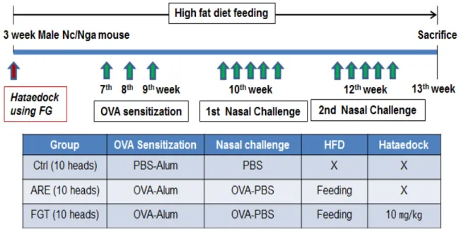
알러지성 비염 유발을 위해 ovalbumin (OVA: chick- en egg albumine, grade V, Sigma)을 항원으로 사용하였 다. OVA 25 ㎍ : 수산화알루미늄겔 (Al(OH)3 gel) 1 ㎎ : 인산완충용액 (PBS: phosphate buffered saline) 300 ㎕ 을 7주령, 8주령, 9주령에 0.1% OVA용액을 복강주사 하여 감작시켰다. 최종 감작 후 7일이 경과된 10주령에 5일 동안 OVA 100 ㎍ : 인산완충용액 (PBS: phosphate buffered saline) 20 ㎕을 생쥐 비강에 점적하여 알러지 성 비염을 1차 유발하였다. 1차 유발 후 7일이 경과된
12주령에 동일한 방법으로 2차 유발하였다 (Fig. 1). 유 발은 콧물과 코 긁기 행동으로 확인하였다. 본 연구과 정은 부산대학교 IACUC 승인을 받아 시행되었으며 (IACUC number: PNU-2016-1187), 실험실 동물의 관 리와 사용에 대해서는 NIH 가이드라인에 따라 시행되 었다.
4. 조직절편제작
각 군을 13주령에 sodium pentobarbital 용액으로 마 취하여 처치한 후 vascular rinse와 10% 중성 포르말린 용액 (NBF: neutral buffered formalin)으로 심장관류고 정을 실시하였다. 머리 분리 후 비강 주변 구조물을 제 거한 다음 10% NBF에 실온에서 24시간 동안 고정하 였다. 이후 탈회액 (decalcification solution, BBC, UK)에 12시간 처리하고 세척한 후 통상적인 방법으로 파라핀 에 포매하여 5 ㎛ 두께로 연속절편을 만들었다.
5. 조직화학
알러지성 비염에 따른 코 점막상피의 손상을 조사 하기 위해 조직화학을 실시하였다. 중성점액질 (neutral mucin)을 분비하는 점액분비세포의 분포 변화를 peri-
Fig. 1. Protocol for allergen sensitization and challenge. Mice were sensitized on week 7, 8, and 9 by intraperitoneal injection of ovalbumin (OVA) emulsified in alum hydroxide (Alum) (OVA 25 ㎍ : Alum 1 ㎎ : PBS 300 ㎕). On weeks 4 after initial sensitization, the mice were challenged with OVA (OVA 100 ㎍ : PBS 20 ㎕) intranasally. On weeks 6 after initial sensitization, the mice were challenged with OVA (OVA 100 ㎍ : PBS 20 ㎕) intranasally. Hataedock using fermented Glycine (FG) was orally administered on weeks 3.
Abbreviations. Ctrl, no treatment group; ARE, obese mouse with allergic rhinitis (AR); FGT, Hataedock using FG treated obese mouse with AR; PBS, phosphate buffered saline; HFD, high fat diet.
odic acid Schiff (PAS) stain 실시 후 관찰하였다. 우선 periodic acid에서 5분간 반응시킨 후 Schiff reagent에서 15분 동안 처리한 다음 sulfurous rinse에서 각 2분씩 3회 세척한 후 hematoxylin에서 1분 동안 대조염색 하 였다.
6. 면역조직화학
Th2 분화조절과 이에 따른 비만세포 활성조절과 항 염증효과를 조사하기 위해 면역조직화학을 실시하였 다. 코 조직절편을 proteolysis 하기 위해 proteinase K (20 ㎍/㎖)에 5분 동안 처리한 후 10% normal mouse serum에서 2시간 동안에서 blocking 시켰다. 그리고 1 차 항체인 goat anti-IL-4 (1:200, Santa Cruz Biotec, USA), goat anti-STAT6 (1:50, Santa Cruz Biotec), goat anti-CD40 (1:50, Santa Cruz Biotec), goat anti-Fc ε re- ceptor (1:50, Santa Cruz Biotec), goat anti-substance P (1:100, Santa Cruz Biotec), goat anti-MMP-9 (1:100, Santa Cruz Biotec), goat anti-NF-kB p65 (1:500, Santa Cruz Biotec), goat anti-p-IκB (1:500, Santa Cruz Biotec), goat anti-iNOS (1:250, Santa Cruz Biotec), goat an- ti-COX-2 (1:100, Santa Cruz Biotec)에 4 ℃ humidified chamber에서 120시간 동안 반응시켰다. 그런 다음 2차 항체인 biotinylated mouse anti-goat IgG (1:100, Santa Cruz Biotec)에 실온에서 48시간 link 하였고, avidin bio- tin complex kit (Vector Lab, USA)에 1시간 동안 실온에 서 반응시켰다. 0.05% 3,3'-diaminobenzidine과 0.01%
HCl이 포함된 0.05M tris-HCl 완충용액 (pH 7.4)에서 발색시킨 후, hematoxylin으로 대조염색 하였다.
7. 영상분석
면역조직화학 결과는 image Pro Plus (Media cy- bernetics, USA)를 이용한 영상분석을 통해 수치화 (means ± standard error) 하였다. 각 군의 표본에서 임의 로 선정된 코점막 10부위를 x400 배율에서 촬영한 다 음 10,000,000 pixels를 영상분석 하였다.
8. 통계
영상분석 결과의 통계는 SPSS software (SPSS 23, SPSS Inc., USA)를 통해 이루어졌으며, one-way ANOVA 시행 을 통해 유의성 (P<0.05)을 검증하고 Duncan’s multiple range test로 사후 검증하였다.
Ⅲ. Results
1. Th2 분화 조절
IL-4는 세포질에서 강한 양성반응을 보였으며, ARE 에 비해 FGT에서 79% 유의성 있는 감소가 나타났다.
핵 주변 세포질에서 강한 양성반응을 보인 STAT6은 ARE에 비해 FGT에서 88% 유의성 있는 감소가 나타 났다. CD40 양성반응은 세포막 주변 세포질에서 약한 양성반응을 보였으며, ARE에 비해 FGT에서 85% 유의 성 있는 감소가 나타났다 (Fig. 2).
2. 비만세포 활성 조절
FcɛRI 양성반응은 ARE에 비해 FGT에서 97% 유의 성 있는 반응 감소가 나타났다. substance P 양성반응은 ARE에 비해 FGT에서 68% 유의성 있는 감소가 나타 났다. MMP-9 양성반응은 ARE에 비해 FGT에서 71%
유의성 있는 감소가 나타났다 (Fig. 3).
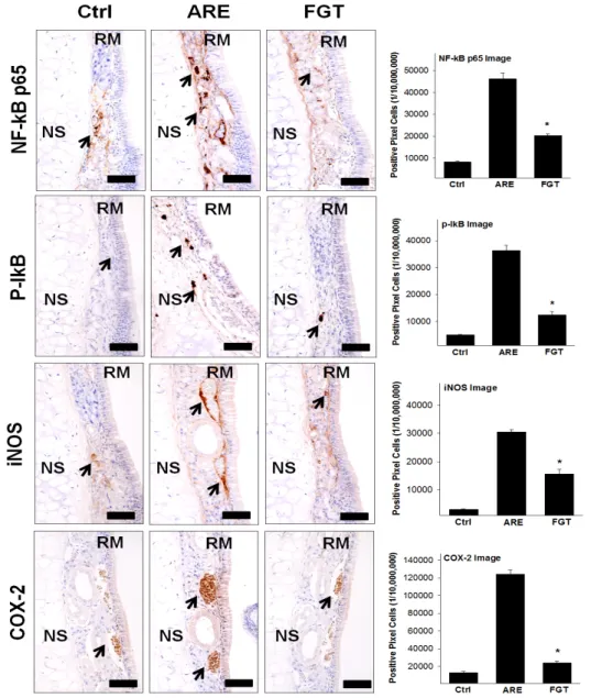
3. 항염증효과
염증효소전사인자인 NF-kB p65 양성반응은 ARE에 비해 FGT에서 56% 유의성 있는 반응 감소가 나타났 다. p-IkB 양성반응도 ARE에 비해 FGT에서 66% 유의 성 있는 감소를 보였다. 염증효소 iNOS 양성반응은 ARE에 비해 FGT에서 49% 유의성 있는 감소가 나타 났다. 염증효소 COX-2 양성반응도 ARE에 비해 FGT 에서 81% 유의성 있게 감소하였다 (Fig. 4).
4. 코 점막 손상
PAS 염색 결과, 알러지성 비염 유발 후 아래코선반 에서 호흡상피의 손상이 증가하였는데 분비소낭이 없 는 술잔세포의 다수 출현과 부종에 따른 점막고유층의 비대가 관찰되었다. FGT는 ARE에 비해 손상이 적은 것으로 관찰되었다 (Fig. 5).
Fig. 2. The regulation of Th2 skewed condition in obese mouse with AR by HTD. The IL-4, STAT6, and CD40 positive reaction decreased in the FGT compared with the ARE group (immunohistochemistry; Bar size, 50 ㎛). Data of image analysis was also shown same result (*: p<0.05, compared with the ARE).
Abbreviations. Ctrl, no treatment group; ARE, obese mouse with allergic rhinitis (AR); FGT, Hataedock using FG (HTD) treated obese mouse with AR; NS, nasal septum; RM, respiratory mucosa; arrow, positive reaction cells.
Fig. 3. The regulation of mast cell's activation in obese mouse with AR by HTD. The FcɛRI, substance P, and MMP-9 positive reaction decreased in the FGT compared with the ARE group (immunohistochemistry; Bar size, 50 ㎛). Data of image analysis was also shown same result (*: p<0.05, compared with the ARE). Abbreviations as same Fig. 2.
Fig. 4. The anti-inflammation effect in obese mouse with AR by HTD. The NF-kB p65, p-IkB, iNOS, and COX-2 positive reaction decreased in the FGT compared with the ARE group (immunohistochemistry; Bar size, 50 ㎛). Data of image analysis was also shown same result (*: p<0.05, compared with the ARE). Abbreviations as same Fig. 2.
Fig. 5. The alleviation of respiratory mucosa's damages (arrow) in obese mouse with AR by HTD. The damages of respiratory mucosa as vacated goblet cell and edema decreased in the FGT compared with the ARE group (PAS histochemistry; Bar size, 50 ㎛). Abbreviations as same Fig. 2.
Ⅳ. Discussion
최근 환경오염의 증가와 다양한 스트레스 등으로 인하여 소아에서 알러지성 비염이 증가하고 있다1). 이 러한 소아 알러지성 비염은 면역불균형으로 인하여 발 병하는 것으로 보고되고 있다23,24). 영유아에서 발생하 는 면역계통 이상은 면역체계의 비정상적 발달로 Th1 과 Th2 간의 불균형이 발생하고 Th2 반응에 치우친 과 도한 면역반응으로 볼 수 있으며25), 소아 비만이 이러 한 소아 염증성 질환 발병 증가의 가중요인으로 인식 되고 있다8-10).
인체 내 Th1 반응과 Th2 반응은 상호간의 길항작용 을 통하여 균형을 유지한다. Th1과 Th2 중 어느 한쪽 이 과도한 반응을 보이면 상호간의 균형이 깨어지고 질병을 유발하게 된다26). IFN-γ은 Th1 유형 cytokine으 로 Th2 분열을 억제하는 반면에 IL-4는 Th2 유형 cyto- kine으로 대식세포를 억제하고 Th1 생성 역시 억제시
킨다26,27). B 세포는 체내에서 알레르겐 항체를 만드는
데, IL-4가 분비되면 이 B 세포에서 IgE 발현이 증가하 게 된다. 또한 T 세포가 활성화되면서 B 세포에 의해 발현된 peptide를 인식하고 B 세포의 CD40와 T 세포의 CD40이 결합하여 B 세포를 활성화하게 된다. 이러한 과정에서 T 세포는 IL-4 같은 cytokine을 생산하여 B 세포에 자극을 주게 되고, 이러한 자극으로 B 세포는 분화되어 IgE 발현이 증가하게 된다28).
두시의 Th2 분화 조절을 통한 알러지성 비염 완화 효과를 확인하기 위한 이전 연구22)에서 IL-4, STAT6, CD40, FcɛRI, substance P, MMP-9, NF-kB p65, p-IkB, iNOS 양성반응이 모두 유의하게 적은 것을 확인하였 다. 이러한 선행 연구결과를 통하여 두시 하태독법이 알러지성 비염 유발을 감소시키는 효과가 있다는 것은 확인하였으나, 비만으로 인하여 염증 유발이 가중되는 상태에서도 효과를 기대할 수 있는지 알아보고자 본 연구를 시작하였다.
Th2 분화 조절을 확인하기 위하여 IL-4, STAT6, CD40의 양성반응을 조사한 결과, IL-4는 FGT에서 유 의성 있는 감소를 보였으며, STAT6 역시 FGT에서 유 의성 있는 감소가 나타났다. CD40 양성반응도 ARE에 비해 FGT에서 유의성 있는 감소를 보였다. 이것은 두 시 하태독법이 비만 상태에서 유발된 Th2 분화도 제어 함으로써 비만 상태의 알러지성 비염에도 유발 억제
효과가 있음을 의미한다.
또한 비만세포의 활성 조절에 관여하는 FcɛRI는 FGT에서 유의성 있는 감소를 보였으며, 소양증에 관여 하는 substance P 역시 유의성 있는 감소가 나타났다.
코점막 내 부종을 유도하는 MMP-9 양성반응도 FGT에 서 유의성 있는 감소가 나타났다. 비만세포는 표면에 FcεRI 수용체를 발현하는데29), 이러한 FcεRI와 IgE가 결합하여 알레르겐 수용체가 된다30). 항원이 체내로 들 어오면 세포들을 활성화시켜 histamine, prostaglandin, serotonin 등을 분비하여 염증 반응을 유도한다31). 비만 세포에서 분비된 substance P는 소양증을 유발하여 긁 기 행동을 하게 만든다32,33). MMP-9은 cytokine에 의해 합성되거나 분비되는데34), 점막고유층의 유리섬유를 파괴하여 염증관련 세포의 이동을 쉽게 만들어준다
35,36)
. 따라서 FcɛRI, substance P, MMP-9 양성반응이 유 의성 있게 감소하였다는 것을 보면, 두시 하태독법이 비만세포에서 탈과립을 억제하고 비만 상태의 알러지 성 비염에서 비만세포 활성 알러지 반응을 조절할 수 있을 것으로 생각된다. 또한 substance P의 발현이 억제 되었다는 것은 알러지성 비염에서 나타나는 소양증을 완화시켜 긁기 행동에 따른 2차적 점막 손상 가능성의 감소를 의미한다.
염증효소전사인자인 NF-kB p65 양성반응은 FGT에 서 유의성 있는 감소를 보였으며, p-IkB 양성반응도 유 의성 있는 감소를 보였다. 염증효소 iNOS 양성반응 역 시 FGT에서 유의성 있는 감소가 나타났다. 염증효소 COX-2 양성반응도 유의성 있는 감소를 보였다. 염증 질환이나 알러지 질환에서 비만세포와 대식세포가 자 극을 받으면 NF-κB, nitric oxide (NO)를 분비한다. NF- κB는 염증 반응 전사인자로, hypoxia-inducible factor (HIF)-1이나 COX-2의 유전자 발현을 조절한다37). 또한 iNOS에 의해 지나치게 과생산된 NO는 염증을 심화시 킨다38). NF-kB p65, p-IkB, iNOS, COX-2 양성반응이 유의성 있게 감소하였다는 것은 두시 하태독법이 비만 상태의 알러지성 비염에서 염증 억제 효과가 있음을 의미한다.
본 연구에서는 두시 하태독법을 시행한 비만 생쥐 에서 발병하는 알러지성 비염 완화 효과를 알아보기 위해 PAS 염색 결과, 알러지성 비염 유발 후 아래코선 반에서 호흡상피의 손상이 증가하지만 FGT는 ARE에 비해 손상이 적은 것으로 관찰되었다. 이는 두시 하태 독법이 실제로 비만 상태의 알러지성 비염으로 인한
호흡상피 손상 유발을 억제하는 효과가 있음을 보여주 는 결과이다.
이상의 연구결과로 볼 때, 두시 하태독법은 비만 상 태에서도 염증 유발 물질 분비를 감소시키고 알러지 질환의 염증 반응을 감소시키는 것으로 생각된다. 따 라서 출생 시 두시 하태독법의 시행은 비만 소아에서 알러지성 비염의 발병을 충분히 감소시킬 수 있을 것 으로 기대된다. 향후 추가연구를 통해 하태독법 실시 후 비강 점막 내의 구체적인 Th2 condition 기전 규명 이 필요할 것으로 생각된다.
Ⅴ. Conclusion
두시 하태독법이 시행된 비만 생쥐에서 Th2 분화 조절을 통한 알러지성 비염 유발 감소 효과를 확인하 기 위한 본 연구는 태령 3주의 약물 처리된 NC/Nga 생쥐의 아래코선반 호흡상피를 면역조직학적 변화를 통해 관찰한 결과 다음과 같은 결과를 얻었다.
1. IL-4, STAT6, CD40 양성반응은 두시 하태독법 시 행군에서 유의하게 감소하였다.
2. FcɛRI, substance P, MMP-9 양성반응은 두시 하태 독법 시행군에서 유의하게 감소하였다.
3. NF-kB p65, p-IkB, iNOS, COX-2 양성반응은 두 시 하태독법 시행군에서 유의하게 감소하였다.
4. 두시 하태독법 시행군에서 알러지성 비염 유발 후 아래코선반에서 손상된 호흡상피와 술잔세포 내 점액분비가 적었다.
Acknowledgement
이 논문은 2017학년도 세명대학교 교내학술연구비 지원에 의해 수행된 연구임.
References
1. Kang KS. Nutritional counseling for obese children with obesity-related metabolic abnormalities in Korea. Pediatr Gastroenterol Hepatol Nutr. 2017;20(2):71-8.
2. Lim MS, Lee CH, Sim S, Hong SK, Choi HG. Physical activity, sedentary habits, sleep, and obesity are associated with asthma, allergic rhinitis, and atopic dermatitis in Korean adolescents. Yonsei Med J. 2017;58(5):1040-6.
3. Kelishadi R, Roufarshbaf M, Soheili S, Payghambarzadeh F, Masjedi M. Association of childhood obesity and the immune system: a systematic review of reviews.
Child Obes. 2017;13(4):332-46.
4. Mraz M, Haluzik M. The role of adipose tissue immune cells in obesity and low-grade inflammation. J Endocrinol.
2014;222(3):R113-27.
5. Simpson EL. Comorbidity in atopic dermatitis. Curr Dermatol Rep. 2012;1(1):29-38.
6. Guttman-Yassky E, Krueger JG, Lebwohl MG. Systemic immune mechanisms in atopic dermatitis and psoriasis with implications for treatment. Exp Dermatol. 2018;
27(4):409-17.
7. Kim BB, Kim JR, Kim JH, Kim YA, Park JS, Yeom MH, Lee HJ, Lee KW, Kang NJ. 7,3',4'-Trihydroxyiso- flavone ameliorates the development of dermatopha- goides farinae-induced atopic dermatitis in NC/Nga mice. Evid Based Complement Alternat Med. 2013;
2013:636597.
8. Murray CS, Canoy D, Buchan I, Woodcock A, Simpson A, Custovic A. Body mass index in young children and allergic disease: gender differences in a longitudinal study. Clin Exp Allergy. 2011;41(1):78-85.
9. Silverberg JI, Becker L, Kwasny M, Menter A, Cordoro KM, Paller AS. Central obesity and high blood pressure in pediatric patients with atopic dermatitis. JAMA Dermatol. 2015;151(2):144-52.
10. Weinmayr G, Forastiere F, Buchele G, Jaensch A, Strachan DP, Nagel G. Overweight/obesity and respira- tory and allergic disease in children: international study of asthma and allergies in childhood (ISAAC) phase two. PLoS One. 2014;9(12):e113996.
11. Leung DY. Atopic dermatitis: new insights and oppor- tunities for therapeutic intervention. J Allergy Clin Immunol. 2000;105(5):860-76.
12. Mosmann TR, Cherwinski H, Bond MW, Giedlin MA, Coffman RL. Two types of murine helper T cell clone.
I. Definition according to profiles of lymphokine activ- ities and secreted proteins. J Immunol. 1986;136(7):
2348-57.
13. Saini V, Arora S, Yadav A, Bhattacharjee J. Cytokines in recurrent pregnancy loss. Clin Chim Acta. 2011;412 (9-10):702-8.
14. Son JH, Jeong JH, Yoo DY. Effects of Pal-Mul-Tang on cytokines production from immune cells during the late stage of pregnancy in BALB/c mice. J Orient Gynecol.
2000;13(1):342-75.
15. Patterson E, Wall R, Fitzgerald GF, Ross RP, Stanton C. Health implications of high dietary omega-6 poly- unsaturated fatty acids. J Nutr Metab. 2012;2012:
539426.
16. Im GM, Jeong HW, Kim HS, Jeong WY. Oriental medical approach on the allergic disease. Korean J Orient Physiol Pathol. 2002;16(5):831-9.
17. Kang MS, Chang GT, Kim JH. A study on fetal toxicosis removal therapy. J Pediatr Korean Med. 2003;17(1):
29-51.
18. Jung AR, Ahn SH, Park IS, Park SY, Jeong SI, Cheon JH, Kim K. Douchi (fermented Glycine max Merr.) alle- viates atopic dermatitis-like skin lesions in NC/Nga mice by regulation of PKC and IL-4. BMC Complement Altern Med. 2016;16(1):416.
19. Zhang JH, Tatsumi E, Ding CH, Li LT. Angiotensin I-converting enzyme inhibitory peptides in Douchi, a Chinese traditional fermented soybean product. Food Chem. 2006;98(3):551-7.
20. Lin J, Xu Y, Zhao T, Sun L, Yang M, Liu T, Sun H, Zhang L. Genistein suppresses smooth muscle cell-de- rived foam cell formation through tyrosine kinase pathway. Biochem Biophys Res Commun. 2015;463(4):
1297-304.
21. Bao ZS, Hong L, Guan Y, Dong XW, Zheng HS, Tan GL, Xie QM. Inhibition of airway inflammation,
hyperresponsiveness and remodeling by soy isoflavone in a murine model of allergic asthma. Int Immunopharmacol.
2011;11(8):899-906.
22. Choi JY, Ahn SH, Kim K. Reduction of allergic rhinitis by controlling the Th2 differentiation of Douchi Hataedock.
J Int Korean Med. 2017;38(4):468-78.
23. Kim JW. Atopic dermatitis from an allergic and im- munological perspective. Korean J Dermatol. 2003;
41(6):687-9.
24. Park YM. Advances in the pathophysiology of atopic dermatitis. Pediatr Allergy Respir Dis. 2006;16(3):
189-96.
25. Romagnani S. Regulation of the development of type 2 T-helper cells in allergy. Curr Opin Immunol. 1994;
6(6):838-46.
26. Kidd P. Th1/Th2 balance: the hypothesis, its limitations, and implications for health and disease. Altern Med Rev. 2003;8(3):223-46.
27. Romagnani S. T-cell subsets (Th1 versus Th2). Ann Allergy Asthma Immunol. 2000;85(1):9-18, 21.
28. Akdis M, Akdis CA. IgE class switching and cellular memory. Nat Immunol. 2012;13(4):312-4.
29. Amin K. The role of mast cells in allergic inflammation.
Respir Med. 2012;106(1):9-14.
30. Metcalfe DD, Baram D, Mekori YA. Mast cells. Physiol Rev. 1997;77(4):1033-79.
31. Stone KD, Prussin C, Metcalfe DD. IgE, mast cells, basophils, and eosinophils. J Allergy Clin Immunol.
2010;125(2Suppl2):S73-80.
32. Ebner K, Singewald N. The role of substance P in stress and anxiety responses. Amino Acids. 2006;31(3):
251-72.
33. Yamaoka J, Kawana S. Rapid changes in substance P signaling and neutral endopeptidase induced by skin-scratching stimulation in mice. J Dermatol Sci.
2007;48(2):123-32.
34. Woessner JF. MMPs and TIMPs-an historical perspective.
Mol Biotechnol. 2002;22(1):33-49.
35. Burrage PS, Mix KS, Brinckerhoff CE. Matrix metal- loproteinases: role in arthritis. Front Biosci. 2006;11:
529-43.
36. Onodera S, Kaneda K, Mizue Y, Koyama Y, Fujinaga M, Nishihira J. Macrophage migration inhibitory factor up-regulates expression of matrix metalloproteinases in synovial fibroblast of rheumatoid arthritis. J Biol Chem.
2000;275(1):444-50.
37. Jung YJ, Isaacs JS, Lee S, Trepel J, Neckers L.
IL-1beta-mediated up-regulation of HIF-1alpha via an
NFkappaB/COX-2 pathway identifies HIF-1 as a critical link between inflammation and oncogenesis. FASEB J.
2003;17(14):2115-7.
38. Kroncke KD, Fehsel K, Kolb-Bachofen V. Nitric oxide:
cytotoxicity versus cytoprotection-how, why, when, and where?. Nitric Oxide. 1997;1(2):107-20.