■투 고 : 2009년 11월 25일, 수 정 : 2009년 12월 18일, 채 택 : 2009년 12월 21일
■교신저자 : 이남열, 서울시 서대문구 창천동 르메이에르 5차 825호 (Tel : 010-2355-5124, E-mail : [email protected])
아토피樣 피부염 NC/Nga 생쥐에서 滋陰除濕湯加減과 아토피 크림-紫雲膏의 병용투여가 피부염에 미치는 영향
이남열․김윤희․한재경
대전대학교 한의과대학 소아과학교실
Abstract
Effects of Concurrent Administration of
JaUmJeSeupTangKaKam(JUJSTK) and Atopy Cream,
Jawoongo(AJ) on Atopic Dermatitis-like Skin Lesions in NC/Nga Mouse
Lee Nam Yerl, Kim Yun Hee, Han Jae Kyung
Department of Pediatrics, College of Oriental Medicine, Dae Jeon University Objectives
The purpose of this study is to examine the effect of a concurrent administration of JUJSTK and AJ on atopic dermatitis in an in-vivoexperiment. Thus, this study is expressed by using NC/Nga atopic dermatitis mice which have histological and clinical similarities to that of humans have been used.
Methods
Clinical skin score, hematology, serum total IgE and IgG1 of the mouse was evaluated, and cytokine levels, total number of the cells, immunohistochemical staining, histological features of axillary lymph node(ALN), peripheral blood mononuclear cells(PBMCs), and a dorsal skin tissue of the mouse were analyzed.
Results
Oral administration of JUJSTK and concurrent administration of JUJSTK and AJ lowered the clinical skin score, total cell number of WBC, eosinophils in blood, and serum total of IgE & IgG1, IFN-γ, IL-5, IL-13, IL-17. In addition, total cell number of ALN and dorsal skin tissue, absolute cell number of CD3e+ T cell, CD4+ Th cell, CD8+ c/sT cell, CD3+CCR3+ cell, CCR3+ cell, CD3+CD69+, CD4+CXCR5+ in ALN, PBMCs, absolute cell number of CCR3+, CD3+/CD69+, CD11b+/Gr-1+, CD11b+/Gr-1+ in dorsal skin tissue, Eotaxin2 mRNA, CCR3 mRNA in dorsal skin tissue and gene expression of IL-5 mRNA, IL-13 mRNA in ALN were significantly decreased. Furthermore, thickness of epidermis infiltrated inflammatory immune cell & mast cell in dermis, histological infiltration of mast cell, the size of inflammatory lymphocytes cells & plasma cells in ALN and histological infiltration of CD4+
& CCR3+in ALN and dorsal skin tissue were significantly decreased as well.
Conclusions
Concurrent administration of JUJSTK and AJ on atopic dermatitis in an in-vivoexperimentby using an NC/Nga atopic dermatitis mouse was very effective as an atopic dermatitis treatment.
Key words: atopic dermatitis, JaUmJeSeupTangKaKam, atopy cream, Jawoongo, NC/Nga mouse
Ⅰ. 緖 論
아토피 피부염은 심한 소양증을 동반한 만성 재발성 피부염으로 개인적 혹은 가족적인 아토 피 병력을 가지는 유전적 소인을 보인다1,2).
2002년 보고에 의하면 선진국에서는 아토피 질환의 이환율이 상당히 증가하는 추세에 있 으며3) 국내에서도 알레르기 질환의 급증과 함 께 아토피 피부염 환자가 증가하고 있고 2000 년 전국 초등 및 중학생을 대상으로 한 연구에 따르면 초등학생의 24.9 %, 중학생의 12.8 % 가 아토피 피부염을 진단받은 것으로 조사되 었다4). 이는 과거 영아 습진이나 태열 등으로 대표되며 자연 경과를 기대했던 아토피 피부 염이 최근에는 연령이 증가하여도 증상이 지 속되거나 심해지는 경향이 있음을 나타내는 것으로 아토피 피부염에 대한 조기 진단과 치 료의 필요성을 보여주고 있다.
한의학에서 아토피 피부염은 浸淫瘡, 乳癬, 奶癬, 胎熱, 胎癬, 胎斂瘡, 濕疹, 四彎風, 旋耳 瘡, 臍瘡 등의 범주에 속하는데5-9), 이들의 발 생원인은 血熱, 濕熱, 血燥 등으로 보고 淸熱, 解毒, 除濕, 祛風, 養血 등의 治法을 주로 사용 하고 있다10-12). 치료에는 內外兼治法이 다용되 었는데13) 최근에는 外治法의 사용은 점차 줄 고 內治法인 내복약에 치우친 경향이 있어 연 령이 낮은 소아에게 치료의 효과를 높이기 위 해서는 內外治法의 병용이 필요한 실정14)이다.
滋陰除濕湯加減은 ≪皮膚病中醫診療學≫15)
에 수록되었으며 血虛風燥로 인한 피부질환에 사용되는 처방으로 아토피 피부염을 포함한 습진 및 소양증에 응용되고 있다. 아토피 크림 은 아로마 오일을 이용한 외용제로 부작용이 거의 없고16) 항염작용과 소독작용을 하여 아토 피 피부염을 완화시키는 것으로 알려져 있으며17) 紫雲膏는 ≪外科正宗≫의 潤肌膏8)에서 유래 된 것으로18) 生肌創傷 및 습진, 건선, 탈모, 아 토피성 피부염 등 광범위한 피부질환에 사용 되는 연고제로 임상적인 연구19-22)등을 통하여 유의한 효과가 있는 것으로 보고되고 있다.
최근 한 등23)이 임상에서 한약과 외용제를 병용하여 치료한 사례를 보고한 바 있고 內外 治療法을 병행한 아토피의 연구24-26)가 활발하 게 이루어지고 있으나, 그 면역학적 기전을 살 펴본 실험적 연구는 부족한 실정이다.
이러한 배경으로 본 연구에서는 滋陰除濕湯 加減 (JaUmJeSeupTangKaKam, 이하 JUJSTK)이 Biostir Mite Antigen Cream (이하 BMAC) 도포 로 아토피 피부염을 유발시킨 NC/Nga 생쥐의 Th17 세포 분화억제에 미치는 영향을 실험적 으로 규명하고자 하였다. In vivo 실험에서 JUJSTK 투여군과 아토피 크림, 紫雲膏 (Atopy cream, Jawoongo, 이하 AJ)와 JUJSTK (이하 AJ+JUJSTK) 병용투여군의 아토피 피부염 치 유에 미치는 효과를 평가하기 위해 12 주령의 피부염이 유발된 NC/Nga 생쥐의 혈청 중 IgE 수준과 clinical skin score, total IgG 수준을 분석 하였고 Axillary Lymph Node (이하 ALN)와
Peripheral Blood Mononuclear Cell (이하 PBMC), 등피부조직에서 세포수, 유세포 형광분석 및 염증 유전자 발현 분석을 하였다. 그리고 ALN과 등피부조직에서 H&E 염색을 통하여 epidermis 의 두께와 toluidine blue 염색을 통하여 비만세 포의 침윤정도를 관찰하였다. 마지막으로 ALN 과 등피부조직에서 면역조직화학 염색을 통하 여 ALN에 침윤된 CCR3+ 세포와 등피부조직 에 분포하는 CD4+ 세포를 관찰하여 JUJSTK, AJ+JUJSTK 투여 후 아토피 피부염 치료에 유 의한 결과를 얻었기에 보고하는 바이다.
Ⅱ. 實 驗
1. 材料
1) 시약 및 기기 (1) 시약
시약은 diethyl pyrocarbonate(DEPC), chloroform, trichloroacetic acid, isopropanol, Tris-HCl, KCl, MgCl2, 적혈구 용혈액 (ACK lysis solution), DMEM 배양액, dulbecco's phosphate buffered saline (D-PBS), Sulforhodamin B(SRB), 2-isopropanol, sodium dodecyl sulfate (SDS), PMA, ionomycin, FK506, antibiotics는 Sigma사 (USA) 제품을 사용 하였으며, 우태아혈청 (fetal bovine serum, FBS)은 Hyclone사 (Logan, USA) 제품을, anti-CD3-PE (phycoerythrin), anti-CD4-FITC (fluorescein isothiocyanate), anti-CCR3-PE, anti-B220-PE, anti-CD8-FITC, anti- B220-FITC, anti-CD49b-FITC, anti-CD40 mAb, rmIL-4, rmIL-10, BD Cytofix/Cytoperm plus kit, anti-CD3 mAb, anti-foxp3-PE, anti-IFN-γ-PE, anti- CD28 mAb 등은 Pharmingen사 (Torreyana, USA) 제품을, CD4+ T cell isolation kit와 B cell isolation kit는 Miltenyi Biotec (Bergisch Gladbach, Germany) 제품을, IL-4, IFN-γ, IL-5, IL-13 ELISA kit는
BioSource사 (California, USA) 제품을, IgE, IgG1 ELISA kit는 SHIBAYAGI 사 (Shibukawa, Japan) 제품을, anti-mouse CCR3 mAb와 anti- mouse CD4 mAb는 Santa-Cruz사 (California, USA) 제품 을, LSAB kit는 DAKO사 (Glostrup, Denmark) 제 품을 사용하였으며, 기타 일반 시약은 특급 시 약을 사용하였다.
(2) 기기
기기는 열탕추출기 (대웅, Korea), rotary vaccum evaporator (Büchi B-480, Switzerland), freeze dryer (EYELA FDU-540, Japan), CO2 incubator (Forma scientific Co., USA), clean bench (Vision scientific Co., Korea), autoclave (Sanyo, Japan), micro-pipet (Gilson, France), water bath (Vision scientific Co., Korea), vortex mixer (Vision scientific Co., Korea), spectrophotometer (Shimazue, Japan), centrifuge (Sigma, USA), deep-freezer (Sanyo, Japan), 자동혈구측정기 (MS9-5, France), Quantitative Real- Time RT-PCR (Applied Biosystems, USA), 형광유 세포분석기 (Cytometry, BD, USA), ice-maker (Vision scientific Co., Korea), homogenizer (OMNI, USA), plate shaker (Lab-Line, USA), VarioMACS (Bergisch Gladbach, Germany), FACScalibur (BD, USA) 및 ELISA leader (Molecular Devices, USA) 등을 사 용하였다.
2) 동물
수컷 7 주령 SPF(Specific Pathogen Free) NC/Nga 생쥐 (15~20g)는 C-harles River Japan 사 (Yokohama, Japan)에서 공급받았다. 동물은 실험 당일까지 고형사료 (항생제 무첨가, 삼양 사료 Co.)와 물을 충분히 공급하고 온도 22±2
℃, 습도 55±15 %, 12 시간 (light-dark cycle)의 환경에서 1 주간 적응시킨 후 실험에 사용하 였다.
Herb Name Scientific Name Amount(g)
地 骨 皮 Lycii Radicis Cortex 15
益 母 草 Leonuri Herba 15
知 母 Anemarrhenae Rhizoma 10
澤 瀉 Alismatis Rhizoma 10
防 風 Ledebouriellae Radix 10
何 首 烏 Polygoni Multiflori Radix 10
甘 草 Glycyrrhizae Radix 10
熟 地 黃 Rehmanniae Radix Preparat 10
柴 胡 Bupleuri Radix 6
黃 芩 Scutellariae Radix 6
白 芍 藥 Paeoniae Radix Alba 6
當 歸 Angelicae Gigantis Radix 6
Total 114
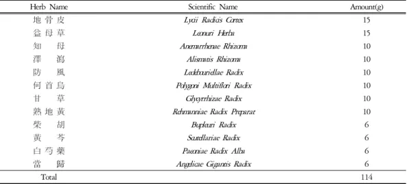
Table 1. Composition of JaUmJeSeupTangKaKam
Aroma-oil
Tea-tree 10 drops
Lavender 7 drops
Chamomile-roman 3 drops
Base Cream 20 ㎖
Table 2. Composition of Atopy cream
Herb Name Scientific Name Amount(g)
紫 草 Lithospermi Radix 0.074
當 歸 Angelicae Gigantis Radix 0.049
胡 麻 油 Sesameseed Oil 0.613
蜜 蠟 Bees Wax 0.245
豚 脂 Swine Oil 0.019
Total 1.000
Table 3. Composition of Jawoongo
3) 약물
(1) 滋陰除濕湯加減 조제
본 실험에 사용한 滋陰除濕湯加減의 구성 은 ≪皮膚病中醫診療學≫15)에 준하였으며 사 용한 약재들은 대전대학교 둔산 한방병원에서 구입 정선하여 사용하였고 그 한 첩의 내용과 분량은 다음과 같다(Table 1).
(2) 아토피 크림과 紫雲膏 조제
본 실험의 아토피 크림에 사용된 아로마 오 일과 베이스 크림은 Fine Korea. Co. (Inchon, Korea)에서 구입하여 사용하였고 그 배합은 다 음과 같으며(Table 2), 紫雲膏의 구성은 ≪大田 大學校 韓方病院 處方集≫27)에 준하였으며 사 용한 약재들은 대전대학교 둔산 한방병원에서 구입 정선하여 사용하였고 1g 당 구성은 다음 과 같다(Table 3).
Scheme 1. Experimental design for the induction of atopic dermatitis-like skin NC/Nga mouse (3) 滋陰除濕湯加減 추출물 분리
滋陰除濕湯加減 2 첩 분량에 증류수 2,000
㎖를 가하여 열탕 추출기에서 3 시간 추출하 여 얻은 액을 흡입 여과하여 이를 감압 증류장 치로 농축하여, 다시 동결 건조기를 이용하여 완전 건조한 滋陰除濕湯加減 추출물 23.8g을 냉동 보관 (- 84℃)하면서 적당한 농도로 희석 하여 사용하였다.
2. 方法 1) In vivo
(1) 피부염 유도 및 시료처리
7 주령의 NC/Nga 생쥐를 1 주일 동안 적응 시키고 이미 피부염이 발생된 18 주령의 NC/
Nga 생쥐 (Atopy NC/Nga mouse)와 2 주간 같 은 공간에서 동시 사육하여 항원감작을 시킨 후 눈에서 capillary 관을 이용하여 100 ㎕의 혈 액을 채혈한 후 마취제인 chloral hydrate (10
%)로 마취한 후 귀와 등 쪽 목 부위를 깨끗하 게 제모한 후 피부의 미세 상처가 치유되도록 24 시간 방치하였다. 그리고 중앙실험동물실 에서 제공하는 BMAC는 Dermatophagoides farinae crude extract (mite antigen, lyophilized)를 항원으 로 사용하였으며 0.5 % TWeen 20이 포함된 ointment base로 제작되었다28). BMAC는 1 주에 2 회 3 주간 등 부위 및 목 부분에 고르게 도 포하였고 도포 2~3 시간 전에 4 % SDS 용액 을 분무하여 피부염이 잘 유발되도록 피부층 을 파괴하였으며 2 주 후 등부분에 피부염이
충분히 유발되면서 긁는 행동이 심화되면 육 안 평가를 실시하였다. 정상군 (NC/Nga- wild type, 이하 NC/Nga-WT)은 7 주령 NC/Nga 생 쥐를 15 주령까지 SPF 조건에서 사육하였다.
(2) 약물처리 및 치료평가
실험은 7 주령 NC/Nga 생쥐를 15 주령까지 SPF 조건에서 사육한 정상군(Normal, Nr)과 BMAC를 도포한 대조군 (Control, CT), BMAC 를 도포하고 FK506(tacrolimus)을 도포한 양성 대조군 (Positive Control, PC), JUJSTK (714 ㎎/
㎏)와 AJ (200 ㎕/마리, 각각 100 ㎕씩)+JUJSTK (714 ㎎/㎏)를 투여한 실험군으로 나누어 실시 하였다.
BMAC는 총 3 주간 (10 주령~13 주령) 월, 목에 4 % SDS 용액을 분무하고 2 시간 경과한 다음 등에 200 ㎕씩 도포하였다. 그리고 12 주 령부터 15 주령까지 3 주간 매일 1 회 오후 3
~4시 사이에 JUJSTK만 단독투여 하거나 AJ+
JUJSTK(아토피 크림 도포 후 자운고 도포, 이 후 JUJSTK 투여)를 병용투여 하였다. 또한 양 성대조군에는 양성대조약물 FK506 0.3 %도 12 주령부터 15 주령까지 3 주간 매일 1 회 등 부분에 골고루 도포하였다. 최종 15 주령에 관 능평가를 실시한 다음 혈액을 채혈하고 등 부 위의 피부를 절제하여 10 % 포르말린 용액에 담아 보관하였다.
(3) Clinical skin score
NC/Nga 생쥐의 피부염은 아토피성 피부염 에서 일반적으로 사용되는 임상적 육안 평가 법을 이용하였다. BMAC를 도포하고, AJ 도포 와 JUJSTK (714 ㎎/㎏) 투여 종료 후 15 주령 에서 육안평가를 실시하였다. 육안평가 항목은 Yamamoto의 평가항목29)을 참고하여 erythema / hemorrhage, dryness / scarring, edema, excoriation / erosion, lichenification 5 가지 항목으로 하고 육 안평가 결과는 각각 평가한 점수의 총 합으 로 나타냈다. 각각의 항목은 없음 (0), 약함 (1), 중증도 (2), 심함 (3)으로 채점하였으며 최 소 0 점에서, 최고 15 점 사이의 점수를 측정 하였다30).
(4) Hematology
최종 관능평가를 실시한 후 EDTA 처리된 튜브형 주사기로 심장 혈액 0.5 ㎖를 채취하였 다. 전혈을 바이오톡스텍(주)(청주, 충청북도)에 의뢰하여 백혈구 중 호중구, 호산구, 호염기구, 단핵구, 림프구의 총 세포수를 측정하였다. 백 혈구 수는 심장천자법으로 채취한 혈액을 자 동혈구측정기 (MS9-5, MELET SCHLOESING, France)로 Fonio법31)에 준하여 Minos-ST로 측정 하였다.
(5) 채혈 및 IgE와 IgG1 측정
NC/Nga 생쥐의 눈에서 8 주령, 12 주령, 15 주령에 capillery 관을 이용하여 약 100 ㎕의 혈 액을 채혈한 후 원심분리기 6,500 rpm에서 20 분간 원심분리한 후 30 ㎕의 혈청을 분리하여 IgE 수준을 측정하였고, IgG1 수준은 15 주령 의 NC/Nga 생쥐를 ethylether로 흡입 마취한 다 음 심장천자법으로 혈액을 분리한 후, 각각의 혈청을 취하여 -70 ℃에 냉동 보관하였고 혈청 내 IgE와 IgG1 농도는 enzyme-linked immuno-
sorbent assay로 측정하였다. 각각 well에 IgE는 8 주령, 12 주령, 15 주령에 채혈한 혈청 5 ㎕ (1/10 dilution)와 dilution buffer 45 ㎕를 혼합하 여 96 well plate의 각 well에 분주하였고 IgG1 은 15 주령에서 채혈한 혈청 50 ㎕ (1/10 dilution) 과 dilution buffer 50 ㎕를 혼합하여 각 well에 분주하였다. 각각 2 시간 동안 25 ℃ 실온에서 방치한 후 2 회 washing 완충용액으로 세척한 다음 각각 antibody biotin-IgE conjugated와 antibody biotin-IgG1 conjugated를 넣고 2 시간 방치하였 다. 다시 2 회 세척 후 완충용액으로 세척한 다음 antibody Avidin-HRP conjugeted 100 ㎕를 처리하고 1 시간 실온에서 방치한 후 다시 세 척하였다. TMB 기질을 100 ㎕씩 분주하고 암 소에서 30 분간 방치한 후 100 ㎕의 stop 용액 을 처리한 후 ELISA leader 450 ㎚에서 각각 IgE와 IgG1에 대한 흡광도를 측정하였다32).
(6) ALN 세포분리 및 cytokine 측정 약물투여 종료 후 (15 주령) ALN을 적출하 여 100 mesh로 ALN 세포를 분리하였다. 전날 BMAC 10 ㎍/㎖을 96 well plate에 coating하여 4 ℃ 냉장고에서 overnight한 다음 D-PBS로 2 회 수세하였다. 분리한 ALN 세포는 ACK 용액 으로 RBC를 제거한 후 BMAC가 coating된 각 각의 well에 5 × 10⁵ 세포씩 5 % FBS-DMEM 배양액에서 48 시간 동안 배양한 후 원심분리 기 2,000 rpm에서 3 분간 원심분리한 후 200
㎕의 배양상층액을 얻었다. 배양상층액내의 IL-4 (BioSource, USA), IFN-γ (BioSource, USA), IL-5 (BioSource, USA), IL-13 (R&D system, USA) 의 수준 측정은 enzyme-linked immuno-sorbent assay로 측정하였다. 각 well에 배양상층액 50
㎕를 분주하고 2 시간 동안 25 ℃ 실온에서 방 치한 후 2 회 washing 완충용액으로 세척한 다 음 각각 antibody biotin-IL-4 conjugated, antibody
biotin-IL-5 conjugated, antibody biotin-IL-13 conjugated를 넣고 2 시간 방치하였다. 다시 2 회 세척 후 완충용액으로 세척한 다음 antibody Avidin-HRP conjugeted 100 ㎕를 처리하고 1 시 간 실온에서 방치한 후 다시 세척하였다. TMB 기질을 100 ㎕씩 분주하고 암소에서 30 분간 방치한 후 100 ㎕의 stop 용액을 처리한 후 ELISA leader 450 ㎚에서 흡광도를 측정하였다32).
(7) ALN, PBMCs 및 등피부조직에서 형광 유세포 분석
NC/Nga 생쥐에서 ALN을 적출하여 100 mesh 로 세포를 분리하여 D-PBS로 5 분간 원심분리 (1,700 rpm)하고 2 회 세척한 후 cell strainer (FALCON)에 통과시켜 세포 이외의 분해되지 않은 조직이나 불순물을 제거하였다. PBMCs 는 실험을 종료한 후 NC/Nga 생쥐에서 heparin 을 처리한 3 ㎖ 주사기로 채혈한 후 미리 준비 한 10 ㎖의 ACK용액 (8.3 g NH4Cl, 1 g KHCO3, in 1 ℓ of demineralized water + 0.1 mM EDTA) 에 혼합하여 실온에서 5 분 동안 처리하여 적 혈구를 제거하였다. 2 회 1 %의 FBS가 함유된 PBS (FACS buffer)로 세척한 후 cell strainer (FALCON)에 통과시켜 세포 이외의 불순물을 제거하였다. 등피부조직은 잘게 chopping한 후 collagenase 1 ㎎/㎖ (in 2 % FBS + RPMI 1640) 를 넣고 37 ℃ shaker (180 rpm, 20 min.) 배양 기에서 배양한 후 상층액을 회수하는 방법으 로 4 회 반복하였다. 분리한 ALN, PBMCs, 등 피부조직 침윤세포의 총세포수를 측정한 다음 모든 조직의 세포등을 5 × 10⁵ 세포로 조정한 후 4 ℃에서 면역 형광염색(immuno-fluorescence staining)을 실시하였다. 각각에 anti-CD3e-PE, anti-CD19-FITC, anti-CD4- FITC, anti-CD8-FITC, anti-CD23-FITC, anti-CD49b-FITC, anti-CCR3-PE, anti-B220-PE를 넣고 30 분간 얼음에서 반응시
킨 후 3 회 이상 PBS로 세척한 후 flow cytometry 의 Cell Quest 프로그램을 이용하여 CD3+, CD19+, CD4+, CD8+, CCR3+, B220+CD23+세 포수를 백분율(%)로 분석한 후 총 세포수를 적 용하여 각 조직에서의 절대 세포수(absolute number)를 산출하였다.
(8) Quantitative Real-Time-PCR in dorsal skin
& ALN
① NC/Nga 생쥐의 등 피부조직과 ALN에서 RNA 분리
Atopic dermatitis-like skin NC/Nga 생쥐의 등 피부조직과 ALN을 적출하여 각각에 RNAzolB 500 ㎕를 넣고 용해될 때까지 homogenizer로 분 쇄하였다. 이 조직분쇄 혼합 부유액에 chloroform (CHCl3) 50 ㎕를 첨가한 후 15 초간 다시 혼합 하였다. 이를 얼음에 15 분간 방치한 후 13,000 rpm에서 원심 분리한 후 약 200 ㎕의 상층액 을 회수하여 2-propanol 200 ㎕와 동량 혼합 후 천천히 흔들고 얼음에서 15 분간 방치하였다.
이를 다시 13,000 rpm에서 원심 분리한 후 80
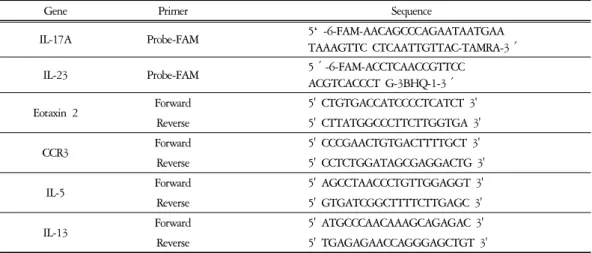
% EtOH로 수세하고 3 분간 vaccum pump에서 건조하여 RNA를 추출하였다. 추출한 RNA는 DEPC를 처리한 20 ㎕의 증류수에 녹여 heating block 75 ℃에서 불활성화 시킨 후 first strand cDNA합성에 사용하였다. Quantitative Real Time PCR은 7500 Real-Time PCR system을 이용하여 수행하였다33). Mouse Olionucleotid의 염기배열 은 다음과 같다(Table 5).
Cytokine 유전자 발현은 SYBRⓇ Green PCR Master mix를 사용하였고, internal standard는 GAPDH로 Taqman probe를 사용하였으며, primer 의 최종 농도가 200 nM이 되게 반응시켰다.
Eotaxin2와 CCR3 mRNA 발현은 등피부조직에 서 관찰하였고, Th2 mediate인 IL-5와 IL-13 mRNA 유전자 발현량 분석은 ALN에서 cDNA
Gene Primer Sequence
IL-17A Probe-FAM 5‘ -6-FAM-AACAGCCCAGAATAATGAA
TAAAGTTC CTCAATTGTTAC-TAMRA-3′
IL-23 Probe-FAM 5′-6-FAM-ACCTCAACCGTTCC
ACGTCACCCT G-3BHQ-1-3′
Eotaxin 2 Forward 5' CTGTGACCATCCCCTCATCT 3'
Reverse 5' CTTATGGCCCTTCTTGGTGA 3'
CCR3 Forward 5' CCCGAACTGTGACTTTTGCT 3'
Reverse 5' CCTCTGGATAGCGAGGACTG 3'
IL-5 Forward 5' AGCCTAACCCTGTTGGAGGT 3'
Reverse 5' GTGATCGGCTTTTCTTGAGC 3'
IL-13 Forward 5' ATGCCCAACAAAGCAGAGAC 3'
Reverse 5' TGAGAGAACCAGGGAGCTGT 3'
Table 5. Primer Sequence
를 합성하여 분석하였다. 또한 Taqman법으로 IL-17과 IL-23 mRNA 발현은 등피부조직에서 관찰하였다. Quantitative Real Time PCR의 조 건은 pre-denaturation은 2 min at 50 ℃, 10 min at 94 ℃, 40 cycles을 0.15 min at 95℃, 1 min at 60℃에서 수행하였다. 대조군, FK506 도포 군, JUJSTK 투여군, AJ+JUJSTK 투여군은 internal standard로 GAPDH를 사용하여 target group의 Quantitative Real Time PCR은
y = x(1 + e)n x = starting quantity y = yield
n = number of cycles e = efficiency
로 계산하여 RQ(relative quantitative)값을 측 정하였다.
(9) Histology
ALN과 등 쪽 목 부분의 피부를 떼어내어 10 % paraform-aldehyde에서 24 시간 동안 포르 말린에 고정한 후 그 조직을 파라핀으로 포 매하였고 5 ㎛ 두께로 block을 만들었다. 조 직 부분은 염증을 일으키는 epidermis, dermis,
keratinocytes, neutrophils / eosinophils 그 외 다른 세포와 부종을 식별하는 hematoxyline / eosin (H&E) 염색과 비만세포를 염색하는 toluidine blue 염색으로 비만세포의 침윤을 광학현미경 (Nikon, Japan, × 200)으로 관찰하였다34).
(10) Immunohistochemical staining
모든 NC/Nga 생쥐는 15 주령에서 면역화학 조직염색을 위하여 등쪽 피부를 적출하여 10% 포르말린 용액에 고정한 다음, 파라핀 블 록을 만든 후 rat anti-mouse CCR3 mAb와 rat anti-mouse CD4 mAb를 사용하였다. 조직절편 을 4 ㎛ 두께로 세절하고 probe-on plus slide에 부착시켜 건조시키고 탈 파라핀(deparaffinized) 한 후 함수시켜 0.01 M citrate buffer (pH 6.0)를 이용해 microwave oven에 15 분간 전 처리하였 다. 조직 내 과산화효소의 작용을 억제하기 위 하여 3 % H2O2에 10 분간 처리한 후 조직 내 의 항원과 비 특이적 단백결합을 억제하기 위 해 정상 혈청으로 단백질을 차단시키고 일차 단일항체에 1 시간 동안 부착시킨 다음 완충 액으로 세척하였다. LSAB kit를 이용하여 PE- conjugated goat anti-rat IgG에 30 분간 반응시
Fig. 1. Topical application of JUJSTK & AJ+JUJSTK treatment of atopic dermatitis-like skin NC/Nga mouse induced by BMAC for 3 weeks.
Shown are back of BMAC-ointment NC/Nga mouse, BMAC plus JUJSTK (714 ㎎/㎏)-orally adminstration in NC/Nga mouse and BMAC plus AJ (200 ㎍/mouse) plus JUJSTK (714 ㎎/㎏)-orally adminstration in NC/Nga mouse for 3 weeks.
켰다. 3 회 Tris-buffered saline with 0.1 % Tween 20 (TBST)용액으로 수세한 후 잘 건조하였다.
현미경은 형광위상차현미경을 사용하여 × 400 배율로 관찰하였다35).
3. 통계처리
실험으로부터 얻은 결과는 mean±standard error로 기록하였고 유의성 검증은 Student's T-test 분석방법을 이용하였으며36) P<0.05인 경우 유의성이 있는 것으로 판정하였다.
Ⅲ. 成 績
1. In vivo
1) BMAC를 이용한 아토피 피부염 유발 및 JUJSTK, AJ 투여 3 주 후 피부변화 BMAC를 이용한 대조군에서 긁는 행동을 동반한 홍반, 부종, 인설, 가피, 태선화 등의 증 상이 확인되었고 3 주 후 JUJSTK 투여군보다 AJ+JUJSTK 투여군에서 긁는 행동을 동반한 홍반, 부종, 인설, 가피, 태선화 등의 증상이 현 저하게 감소하였다(Fig. 1).
2) Clinical skin score
Clinical skin score는 Control군이 NC/Nga-WT 군에 비하여 증가하였고, FK506 도포군은 Control군에 비해 감소를 나타내었으며 (p<
0.001), 단독 JUJSTK (p<0.01) 투여군은 유의 성 있는 감소가 없었으나 AJ+JUJSTK 투여군 은 유의성 있는 감소를 나타냈다 (p<0.01) (Fig. 2).
3) Hematology
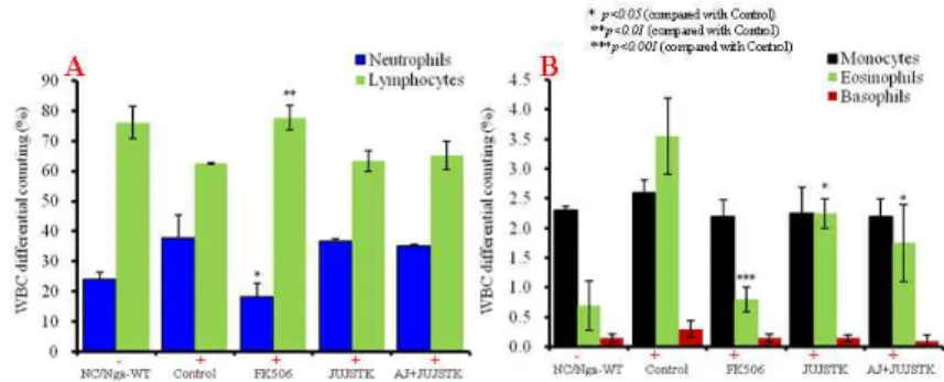
실험종료 후 WBC 비율 중 neutrophil의 비 율은 NC/Nga-WT군에 비하여 Control군이 증 가하였고 FK506 도포군의 비율은 Control군에 비하여 유의성 있게 감소하였으며 (p<0.05) JUJSTK 투여군과 AJ+JUJSTK 투여군에서는 차이가 없었다. Lymphocytes의 비율은 NC/
Nga-WT군에 비하여 Control군이 약간 감소하 였고 FK506 도포군에서 유의성 있는 증가를 보였으나 (P<0.01) JUJSTK 투여군과 AJ+
JUJSTK 투여군은 대조군과의 차이가 나타나 지 않았다(Fig. 3A).
Eosinophil은 NC/Nga-WT군에 비하여 Control 군이 현저하게 증가하였고 FK506 도포군은 대
Fig. 2. Clinical skin features and severity of atopic dermatitis skin lesions in atopic dermatitis-like skin NC/Nga mouse induced by BMAC.
Atopic dermatitis NC/Nga mouse was induced by BMAC treatment in the dorsal skin, not treated BMAC (SPF normal, NC/Nga-WT), BMAC treatment for 3 weeks (Control), BMAC treatment for 3 weeks with FK506 (0.3%)-ointment (FK506), BMAC treatment for 3 weeks with JUJSTK (714 ㎎/㎏) orally administration (JUJSTK), and BMAC treatment for 3 weeks with AJ (200 ㎕/mouse)-ointment plus JUJSTK (714 ㎎/㎏) orally administration (AJ+JUJSTK) for 3 weeks. A total clinical skin score for atopic dermatitis-like lesions was defined as the sum of the individual scores graded as 0 (none), 1 (mild), 2 (moderate) and 3 (severe) for each of five signs and symptoms (erythema/hemorrhage, scarring/dryness, edema, excoriation/erosion and lichenification) on the three parts of the body: ear, FSCe and head and back. Each point represents the mean±SE of six mouse.
Fig. 3. WBC differential counting in atopic dermatitis like skin in NC/Nga mouse induced by BMAC.
Atopic dermatitis NC/Nga mouse was induced by BMAC treatment in the dorsal skin, not treated BMAC (SPF-normal, NC/Nga-WT), BMAC treatment for 3 weeks (Control), BMAC treatment for 3 weeks with FK506 (0.3%)-ointment (FK506), BMAC treatment for 3 weeks with JUJSTK orally administration (JUJSTK), and BMAC treatment for 3 weeks with AJ (200 ㎕/mouse)-ointment plus JUJSTK orally administration (AJ+JUJSTK) for 3 weeks. Blood was collected from the retro-orbital plexus under ether anesthesia and heparinized immediately thereafter. Cell contents were measured by hematology (BD, U.S.A.).
조군에 비하여 유의성 있게 감소하였으며 (p<
0.001) JUJSTK 투여군과 AJ+JUJSTK 투여군 에서도 Control 군에 비하여 유의성 있는 감소 를 보였고 (p<0.05), monocyte와 basophil은 NC/
Nga-WT군에 비하여 Control군이 약간 증가하
였으나 JUJSTK 투여군과 AJ+JUJSTK 투여군 에서는 큰 차이가 없었다(Fig. 3B).
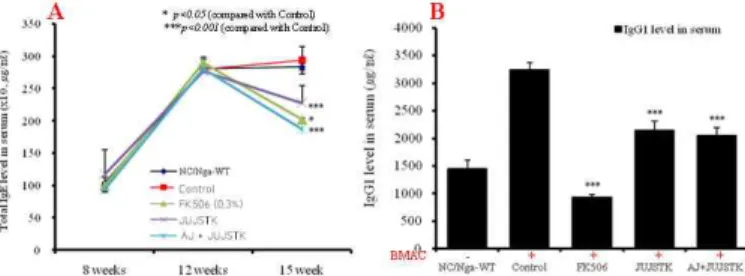
4) 혈청 IgE와 IgG1에 미치는 영향 IgE의 양은 NC/Nga-WT군에서 8 주령에서
Fig. 4. Serum IgE & IgG1 elevation and development of atopic dermatitis like skin in NC/Nga mouse induced by BMAC.
Atopic dermatitis NC/Nga mouse was induced by BMAC treatment in the dorsal skin, not treated BMAC (SPF-normal, NC/Nga-WT), BMAC treatment for 3 weeks (Control), BMAC treatment for 3 weeks with FK506 (0.3%)-ointment (FK506), BMAC treatment for 3 weeks with JUJSTK orally administration (JUJSTK), and BMAC treatment for 3 weeks with AJ (200 ㎕/mouse)-ointment plus JUJSTK orally administration (AJ+JUJSTK) for 3 weeks. Blood was collected from the retro-orbital plexus under ether anesthesia and heparinized immediately thereafter. Serum samples were obtained by centrifugation and stored at -20°C until use. Total IgE and IgG1 levels were measured by a sandwich ELISA using an ELISA kit (Shibayagi, Japan). Each point represents the mean±SE of six mouse.
Fig. 5. Culture supernatant IL-4, IL-5, IL-13, IL-17 and IFN-γ level in ALN in atopic dermatitis like skin in NC/Nga mouse induced by BMAC.
Atopic dermatitis NC/Nga mouse was induced by BMAC treatment in the dorsal skin, not treated BMAC (SPF-normal, NC/Nga-WT), BMAC treatment for 3 weeks (Control), BMAC treatment for 3 weeks with FK506 (0.3%)-ointment (FK506), BMAC treatment for 3 weeks with skin apply of JUJSTK orally administration (JUJSTK), and BMAC treatment for 3 weeks with skin apply of AJ (200 ㎕/mouse)-ointment plus JUJSTK orally administration (AJ+JUJSTK) for 3 weeks. ALN from mouse at 15 weeks of age were re-stimulated with BMAC (10 ㎍/㎖) for 48 hrs. IL-4, IL-5, IL-13, IL-17, and IFN-γ levels were measured by a sandwich ELISA using an mouse ELISA kit I (Biosource, U.S.A.).
12 주령 사이에 자연적으로 증가되었고 Control 군은 15 주령에서 NC/Nga-WT군과 비슷하게 증가되었으며, FK506 도포군, JUJSTK 투여군,
AJ+JUJSTK 투여군은 15 주령에서 Control군 에 비해 유의성 있게 감소하였다 (p<0.05, p<
0.001)(Fig 4A).
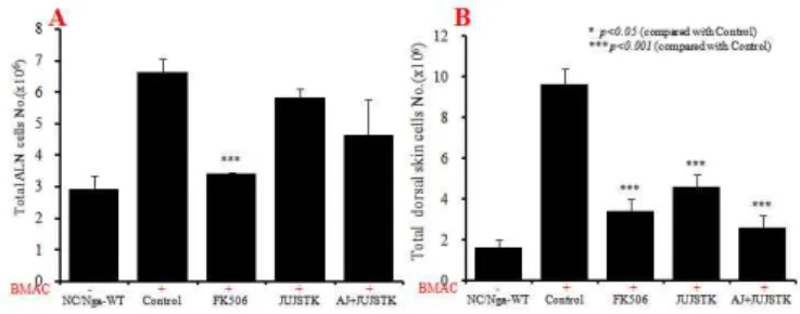
Fig. 6. Total cell number of ALN and dorsal skin in atopic dermatitis like skin NC/Nga mouse induced by BMAC.
Atopic dermatitis NC/Nga mouse was induced by BMAC treatment in the dorsal skin, not treated BMAC (SPF-normal, NC/Nga-WT), BMAC treatment for 3 weeks (Control), BMAC treatment for 3 weeks with FK506 (0.3%)-ointment (FK506), BMAC treatment for 3 weeks with skin apply of JUJSTK orally administration (JUJSTK), and BMAC treatment for 3 weeks with skin apply of AJ (200 ㎕/mouse)-ointment plus JUJSTK orally administration (AJ+JUJSTK) for 3 weeks.
IgG1의 수준은 FK506 도포군이 Control군에 비해 유의성 있게 감소하였고(p<0.001) JUJSTK 투여군, AJ+JUJSTK 투여군도 Control군에 비 해 유의성 있게 감소하였다 (p<0.001) Fig 4B).
5) Cytokine level in ALN
IFN-γ의 생산량은 NC/Nga-WT군에 비하여 Control군이 증가하였고, FK506 도포군은 Control 군에 비하여 유의성 있게 감소를 나타내었으 며 (p<0.001), JUJSTK 투여군, AJ+ UJSTK 투 여군은 Control군에 비하여 각각 유의성 있게 감소하였다 (p<0.01, p<0.001) Fig. 5A).
IL-4 생산량은 모든 실험군에서 차이가 나타 나지 않았다(Fig. 5B).
IL-5와 IL-13의 생산량은 NC/Nga-WT군에 비하여 Control군이 증가한 결과를 얻었고 FK 506 도포군은 Control군에 비하여 유의성 있게 감소를 나타내었으며 (p<0.001), JUJSTK 투여 군, AJ+JUJSTK 투여군은 Control군에 비하여 유의성 있게 감소하였다 (p<0.001)(Fig. 5C).
IL-17의 생산량은 NC/Nga-WT군에 비하여 Control군이 증가한 결과를 얻었고 FK506 도포 군은 Control군에 비하여 유의성 있게 감소를
나타내었으며 (p<0.001), JUJSTK 투여군, AJ+
JUJSTK 투여군은 Control군에 비하여 유의성 있게 감소하였다 (p<0.05, p<0.01)(Fig. 5D).
6) ALN, PBMCs 및 등피부조직에서의 유 세포형광분석
(1) ALN과 등피부조직의 세포수 측정 ALN 세포는 NC/Nga-WT군에 비하여 Control군이 증가하였고 FK506 도포군은 Control 군에 비하여 유의성 있게 감소하였으며 (p<
0.001) JUJSTK 투여군과 AJ+JUJSTK 투여군 에서는 Control군에 비하여 감소하였으나 유의 성은 없었다(Fig. 6A).
등피부조직의 세포도 NC/Nga-WT군에 비하 여 Control군이 증가하였고 FK506 도포군은 Control군에 비하여 유의성 있게 감소하였으며 (p<0.001) JUJSTK 투여군과 AJ+JUJSTK 투여 군에서도 Control군에 비하여 유의성 있게 감 소하였다 (p<0.001)(Fig. 6B).
(2) ALN에서 cell content 변화
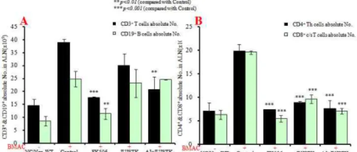
① CD3+&CD19+, CD4+&CD8+ 절대 세포수 ALN에서 활성 CD3+ T 세포의 절대 세포수
Fig. 7. Effects of JUJSTK & AJ+JUJSTK treatment on T and B cell changes of absolute numbers in ALN cells in NC/Nga mouse.
Atopic dermatitis NC/Nga mouse was induced by BMAC treatment in the dorsal skin, not treated BMAC (SPF-normal, NC/Nga-WT), BMAC treatment for 3 weeks (Control), BMAC treatment for 3 weeks with FK506 (0.3%)-ointment (FK506), BMAC treatment for 3 weeks with skin apply of JUJSTK orally administration (JUJSTK), and BMAC treatment for 3 weeks with skin apply of AJ (200 ㎕/mouse)-ointment plus JUJSTK orally administration (AJ+JUJSTK) for 3 weeks. NC/Nga mouse ALN cells (2 × 10⁵cells/㎖) were isolated from ALN, and the ALN cells were washed twice and analyzed by flow cytometry. Absolute number of CD3+, CD19+ (A), and CD4+, CD8+ (B) in NC/Nga mouse.
는 NC/Nga-WT군에 비하여 Control군이 증가 하였고 FK506 도포군은 유의성 있는 감소를 나타내었으며 (p<0.001) AJ+JUJSTK 투여군 은 Control군에 비하여 유의성 있는 감소를 나 타내었다 (p<0.01). CD19+ B 절대 세포수는 NC/Nga-WT군에 비하여 Control군이 현저하게 증가하였고 FK506 도포군은 Control군에 비하 여 유의성 있는 감소를 나타내었으나(p<0.01) JUJSTK 투여군, AJ+JUJSTK 투여군은 모두 Control군과 차이가 없었다(Fig. 7A).
CD4+ T 세포와 CD8+ c/sT 세포에 대한 절 대 세포수는 NC/Nga-WT군에 비하여 Control 군이 증가한 결과를 얻었고 FK506 도포군, JUJSTK 투여군, AJ+JUJSTK 투여군에서 Control 군에 비하여 유의성 있는 감소를 나타내었다 (p<0.001)(Fig. 7B).
② CD3+CD69+, B220+CD23+, CD3+CCR3+ 절대 세포수
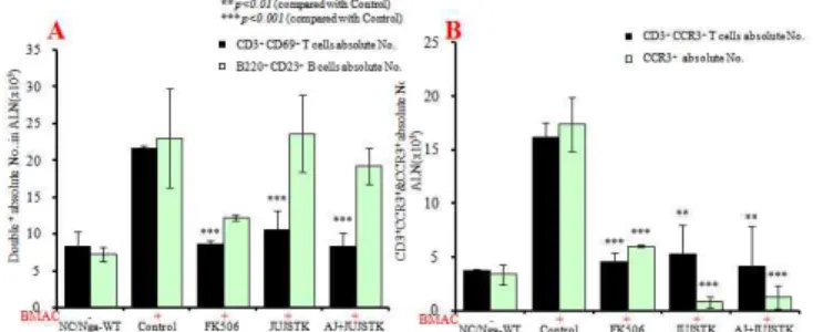
CD3+CD69+ T 세포와 B220+CD23+ B 세포 의 절대 세포수는 NC/Nga-WT군에 비하여 Control 군이 증가하였고 FK506 도포군, JUJSTK 투여
군, AJ+JUJSTK 투여군의 CD3+CD69+ 세포 의 절대 세포수는 Control군에 비하여 유의성 있는 감소를 나타내었다 (p<0.001)(Fig. 8A).
CD3+CCR3+ T 세포와 CCR3+ 세포의 절대 세포수는 NC/Nga-WT군에 비하여 Control군이 증가하였고 FK506 도포군, JUJSTK 투여군, AJ+JUJSTK 투여군은 Control군에 비하여 유 의성 있는 감소를 나타내었다 (p<0.01, p<
0.001)Fig. 8B).
③ CD4+CXCR5+ 절대 세포수
CD4+CXCR5+ 세포의 절대 세포수는 NC/
Nga-WT군에 비하여 Control군이 증가하였고, FK506 도포군, JUJSTK 투여군, AJ+JUJSTK 투여군은 Control군에 비하여 유의성 있는 감 소를 나타내었다 (p<0.001)(Fig. 9).
(3) PBMCs에서 cell content 변화
① Granuolcyte의 변화
Granulocytes의 빈도(%)는 NC/Nga-WT군에 비하여 Control군이 증가하였고, FK506 도포군 은 Control군에 비하여 유의성 있는 감소를 나
Fig. 8. Effects of JUJSTK & AJ+JUJSTK treatment on CD3+CD69+, B220+CD23+, CD3+CCR3+, CCR3+ changes of absolute numbers in ALN cells in NC/Nga mouse
Atopic dermatitis NC/Nga mouse was induced by BMAC treatment in the dorsal skin, not treated BMAC (SPF-normal, NC/Nga-WT), BMAC treatment for 3 weeks (Control), BMAC treatment for 3 weeks with FK506 (0.3%)-ointment (FK506), BMAC treatment for 3 weeks with skin apply of JUJSTK orally administration (JUJSTK), and BMAC treatment for 3 weeks with skin apply of AJ (200 ㎕/mouse)-ointment plus JUJSTK orally administration (AJ+JUJSTK) for 3 weeks. NC/Nga mouse ALN cells (2 × 10⁵ cells/㎖) were isolated from ALN, and the ALN cells were washed twice and analyzed by flow cytometry. Absolute number of CD3+CD69+, CD3+CD69+, B220+CD23+ (A), and CD3+CCR3+, CCR3+ (B) in NC/Nga mouse.
Fig. 9. Effects of JUJSTK & AJ+JUJSTK treatment on CD4+CXCR5+ gated cells and changes of absolute numbers in ALN cells in NC/Nga mouse.
Atopic dermatitis NC/Nga mouse was induced by BMAC treatment in the dorsal skin, not treated BMAC (SPF-normal, NC/Nga-WT), BMAC treatment for 3 weeks (Control), BMAC treatment for 3 weeks with FK506 (0.3%)-ointment (FK506), BMAC treatment for 3 weeks with skin apply of JUJSTK orally administration (JUJSTK), and BMAC treatment for 3 weeks with skin apply of AJ (200 ㎕/mouse)-ointment plus JUJSTK orally administration (AJ+JUJSTK) for 3 weeks. NC/Nga mouse ALN cells (2 × 10⁵ cells/㎖) were isolated from ALN, and the ALN cells were washed twice and analyzed by flow cytometry. Absolute number of CD4+CXCR5+ in NC/Nga mouse.
타내었으며(p<0.001), JUJSTK 투여군, AJ+
JUJSTK 투여군은 Control군에 비하여 각각 감 소하여 유의성 있는 결과를 나타냈다 (p<0.001) (Fig. 10).
② T & B cell content 변화
CD3+T 세포, CD4+Th 세포, CD8+c/s T 세 포, CD3+CD69+세포, B220+CD23+, CD3+CCR3+ 세포, CCR3+ 세포는 NC/Nga-WT군에 비하여
Control군이 총활성세포 빈도(%) 각각에서 모 두 증가하였고, FK506 도포군에서 모두 감소 를 나타내어 유의성 있는 결과를 나타내었다 (p<0.01, p<0.001). 또한 AJ+ UJSTK 투여군 은 Control군에 비하여 유의성 있게 감소하였 으며 (p<0.001, p<0.01) JUJSTK 투여군의 CD3+ CD69+, B220+CD23+, CCR3+ 세포의 활성세포 도 유의성 있는 감소를 나타내었다.
Fig. 10. Effects of JUJSTK & AJ+JUJSTK treatment on the percentage of granulocytes gated cells in PBMCs in NC/Nga mouse.
Atopic dermatitis NC/Nga mouse was induced by BMAC treatment in the dorsal skin, not treated BMAC (SPF-normal, NC/Nga-WT), BMAC treatment for 3 weeks (Control), BMAC treatment for 3 weeks with FK506 (0.3%)-ointment (FK506), BMAC treatment for 3 weeks with skin apply of JUJSTK orally administration (JUJSTK), and BMAC treatment for 3 weeks with skin apply of AJ (200 ㎕/mouse)-ointment plus JUJSTK orally administration (AJ+JUJSTK) for 3 weeks. NC/Nga mouse PBMCs (2 × 10⁵ cells/㎖) were isolated from Blood and the PBMCs were washed twice and analyzed by flow cytometry. Total cell content (%) of granulocytes gated cells in NC/Nga mouse.
Fig. 11. Effects of JUJSTK & AJ+JUJSTK treatment on the percentage of T & B gated cells in PBMCs in NC/Nga mouse.
Atopic dermatitis NC/Nga mouse was induced by BMAC treatment in the dorsal skin, not treated BMAC (SPF-normal, NC/Nga-WT), BMAC treatment for 3 weeks (Control), BMAC treatment for 3 weeks with FK506 (0.3%)-ointment (FK506), BMAC treatment for 3 weeks with skin apply of JUJSTK orally administration (JUJSTK), and BMAC treatment for 3 weeks with skin apply of AJ (200 ㎕/mouse)-ointment plus JUJSTK orally administration (AJ+JUJSTK) for 3 weeks. NC/Nga mouse PBMCs (2 × 10⁵cells/㎖) were isolated from Blood and the PBMCs were washed twice and analyzed by flow cytometry. Total cell content (%) of CD3+ & CD19+ (A), CD4+ & CD8+ (B), CD3+CD69+ & B220+CD23+ (C), CD3+CCR3+ & CCR3+ (D) in NC/Nga mouse.
CD19+B 세포에서는 NC/Nga-WT군에 비하 여 Control군이 총활성세포 빈도(%)가 감소한 결과를 얻었고 FK506 도포군은 Control군에 비 하여 유의성 있게 증가한 결과를 나타내었으
며 (p<0.001), JUJSTK 투여군과 AJ+JUJSTK 투여군도 유의성 있는 증가를 나타내었다 (p<
0.001)(Fig. 11).
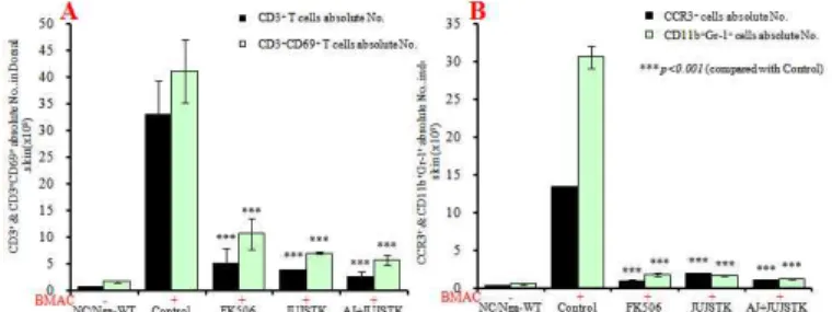
Fig. 12. Effects of JUJSTK & AJ+JUJSTK treatment on changes of absolute numbers in dorsal skin cells in NC/Nga mouse.
Atopic dermatitis NC/Nga mouse was induced by BMAC treatment in the dorsal skin, not treated BMAC (SPF-normal, NC/Nga-WT), BMAC treatment for 3 weeks (Control), BMAC treatment for 3 weeks with FK506 (0.3%)-ointment (FK506), BMAC treatment for 3 weeks with skin apply of JUJSTK orally administration (JUJSTK), and BMAC treatment for 3 weeks with skin apply of AJ (200 ㎕/mouse)-ointment plus JUJSTK orally administration (AJ+JUJSTK) for 3 weeks. NC/Nga mouse dorsal skin cells (2 × 10⁵ cells/㎖) were isolated from dorsal skin, and the dorsal skin cells were washed twice and analyzed by flow cytometry. Absolute number of CD3+ & CD3+CD69+ (A), CCR3+ & CD11b+Gr-1+ (B) in NC/Nga mouse.
Fig. 13. Effects of JUJSTK & AJ+JUJSTK treatment on IL-17 and IL-23 mRNA expression in dorsal skin tissue in NC/Nga mouse.
Atopic dermatitis NC/Nga mouse was induced by BMAC treatment in the dorsal skin, not treated BMAC (normal, NC/Nga-WT), BMAC treatment for 3 weeks (Control), BMAC treatment for 3 weeks with FK506 (0.3%)-ointment (FK506), BMAC treatment for 3 weeks with skin apply of JUJSTK (714 ㎎/㎏) orally administration (JUJSTK), and BMAC treatment for 3 weeks with skin apply of AJ (200 ㎕/mouse)-ointment plus JUJSTK (714 ㎎/㎏) orally administration (AJ+JUJSTK) for 3 weeks. IL-17 and IL-23 mRNA synthesized by real-time PCR was analyzed. The amount of Taqman probe was measured at the end of each cycle. The cycle number at which the emission intensity of the sample rises above the baseline is referred as to the RQ (relative quantitative) and is proportional to the target concentration. Real time PCR was performed in duplicate and analyzed by a Applied Biosystems 7500 Real-Time PCR system. Each point represents the mean±SE of six mouse.
(4) 등피부조직에서 T & B cell content 변화 CD3+ T 세포, CD3+CD69+ 세포, CCR3+, CD11b+Gr-1+ 세포를 측정한 결과 NC/Nga-WT 군에 비하여 Control군이 증가한 결과를 얻었 고 FK506 도포군은 Control군에 비하여 각각 유의성 있는 감소를 나타내었으며 (p<0.001), JUJSTK 투여군, AJ+JUJSTK 투여군은 Control
군에 비하여 유의성 있는 감소를 나타내었다 (p<0.001)(Fig. 12A, B).
7) 등피부조직에서 IL-17과 IL-23 mRNA 유전자 발현 분석
IL-17과 IL-23 mRNA는 NC/Nga-WT군에 비 하여 Control군이 증가하였고, FK506 도포군은
Fig. 14. Effects of JUJSTK & AJ+JUJSTK treatment on eotaxin 2, CCR3, IL-5, and IL-13 mRNA expression in NC/Nga mouse.
Atopic dermatitis NC/Nga mouse was induced by BMAC treatment in the dorsal skin, not treated BMAC (SPF-normal, NC/Nga-WT), BMAC treatment for 3 weeks (Control), BMAC treatment for 3 weeks with FK506 (0.3%)-ointment (FK506), BMAC treatment for 3 weeks with skin apply of JUJSTK orally administration (JUJSTK), and BMAC treatment for 3 weeks with skin apply of AJ (200 ㎕/mouse)-ointment plus JUJSTK orally administration (AJ+JUJSTK) for 3 weeks. Total RNAs were extracted in dorsal skin tissue or ALN, and eotaxin 2, CCR3, IL-5, and IL-13 mRNA synthesized by real-time PCR was analyzed. The amount of SYBR Green was measured at the end of each cycle. The cycle number at which the emission intensity of the sample rises above the baseline is referred as to the RQ (relative quantitative) and is proportional to the target concentration. Real time PCR was performed in duplicate and analyzed by a Applied Biosystems 7500 Real-Time PCR system.
Control군에 비하여 유의성 있게 억제되었으며 (p<0.001), JUJSTK 투여군과 AJ+JUJSTK 투 여군에서도 대조군에 비하여 각각 유의성 있 게 억제되었다 (p<0.01, p<0.001)(Fig. 13).
8) 등피부조직과 ALN에서 염증 유전자발 현 분석
Eotaxin2 mRNA와 CCR3 mRNA의 유전자발 현은 NC/Nga-WT군에 비하여 Control군이 각 각 증가한 결과를 얻었고 FK506 도포군은 Control군에 비하여 유의성 있는 억제효과를 나타내었으며 (p<0.001), JUJSTK 투여군, AJ+
JUJSTK 투여군은 Control군에 비하여 각각 유 의성 있는 억제효과를 나타내었다 (p<0.001) (Fig. 14A).
IL-5 mRNA와 IL-13 mRNA의 유전자발현은 NC/Nga-WT군에 비하여 Control군이 각각 증 가한 결과를 얻었고 FK506 도포군은 Control군 에 비하여 유의성 있는 억제효과를 나타내었 으며(p<0.001), JUJSTK 투여군, AJ+JUJSTK
투여군은 IL-13 mRNA에서 Control군에 비하여 각각 유의성 있는 억제효과를 나타내었다 (p<0.001)(Fig. 14B).
9) Histology
(1) 등피부 조직의 조직검사 및 분석 등피부조직에 H&E 염색과 toluidine blue 염 색을 실시한 결과, NC/Nga-WT군(A)의 피부조 직에 비해 Control군(B)은 epidermis의 두께가 과형성, 확장되어 (long red arrow) 그 주변에 과각화, 색소침착, 과립증가, 부전각화증, 비만 세포의 침윤등이 NC/Nga-WT군에 비하여 현 저하게 증가되었고, FK506 도포군(C)의 일부 는 Control군에 비하여 NC/Nga-WT군에 가깝 게 epidermis의 두께가 줄어들었고 그 주변에 세포변형과 각화증상, 비만세포의 침윤 등이 감소를 나타내었다. JUJSTK 투여군(D)은 Control 군에 비하여 epidermis의 두께가 약간 줄어들었 고 그 주변에 과각화, 색소침착, 과립증가, 부 전각화증, 비만세포의 침윤 등이 감소를 나타
Fig. 15. Histological features of dorsal skin group in atopic dermatitis-like skin NC/Nga mouse induced by BMAC
Atopic dermatitis NC/Nga mouse induced by BMAC treatment in the dorsal skin, A; not treated BMAC (SPF-normal, NC/Nga-WT), B; BMAC treatment for 3 weeks (Control), C; BMAC treatment for 3 weeks with FK506 (0.3%)-ointment (FK506), D; BMAC treatment for 3 weeks with JUJSTK (714 ㎎/㎏) orally administration (JUJSTK), and E; BMAC treatment for 3 weeks with AJ (200 ㎕/mouse)-ointment plus JUJSTK (714 ㎎/㎏) orally administration (AJ+JUJSTK) for 3 weeks. NC/Nga skin biopsy were stained with hematoxylin and eosin (H&E) and shows the thickening of the epidermis (red arrow) by bright microscope (× 200). Data represent individual values and the average value of four individual mouse in each group.
Fig. 16. Histological status of the skin stained with toluidine blue of dorsal skin group in atopic dermatitis-like skin NC/Nga mouse induced by BMAC.
Atopic dermatitis NC/Nga mouse induced by BMAC treatment in the dorsal skin, A; not treated BMAC (SPF-normal, NC/Nga-WT), B; BMAC treatment for 3 weeks (Control), C; BMAC treatment for 3 weeks with FK506 (0.3%)-ointment (FK506), D; BMAC treatment for 3 weeks with JUJSTK orally administration (JUJSTK), and E; BMAC treatment for 3 weeks with AJ (200 ㎕/mouse)-ointment plus JUJSTK orally administration (AJ+JUJSTK) for 3 weeks. NC/Nga skin biopsy were stained with toluidine blue staining and shows the degranulated mast cells in the dermis (red arrow) by bright microscope (× 200). Data represent individual values and the average value of four individual mouse in each group.
내었다. AJ+JUJSTK 투여군(E)은 Control군에 비하여 epidermis의 두께가 NC/Nga-WT군에 가깝게 현저하게 줄었고 그 주변에 과각화, 색 소침착, 과립증가, 부전각화증, 비만세포의 침 윤 등도 현저하게 감소하였다(Fig. 15, 16).
(2) ALN 조직 검사 및 분석
NC/Nga-WT군(A)에 비하여 Control군(B)에 서 inflammatory lymphocytes cells(ILC)과 plasma cells(PC) 침윤등이 현저하게 증가된 것이 관찰 되었고 FK506 도포군(C)은 Control군에 비하여 크기가 현저하게 줄어들었다. 그러나 JUJSTK 투여군(D)에서는 Control군에 비하여 약간 줄
Fig. 17. Histological features of ALN group in atopic dermatitis like skin NC/Nga mouse induced by BMAC.
Atopic dermatitis NC/Nga mouse induced by BMAC treatment in the dorsal skin, A; not treated BMAC (SPF-normal, NC/Nga-WT), B; BMAC treatment for 3 weeks (Control), C; BMAC treatment for 3 weeks with FK506 (0.3%)-ointment (FK506), D; BMAC treatment for 3 weeks with JUJSTK (714 ㎎/㎏) orally administration (JUJSTK), and E; BMAC treatment for 3 weeks with AJ (200 ㎕/mouse)-ointment plus JUJSTK (714 ㎎/㎏) orally administration (AJ+JUJSTK) for 3 weeks. NC/Nga ALN biopsy were stained with hematoxylin and eosin (H&E), and the NC/Nga control (B) shows ALN in the infiltration of the Inflammatory Lymphocytes Cells (ILC, blue arrow) and plasma Cells (PC) (arrows) by bright microscoph (Nikon, Japan, orignal magnification, × 200).
Fig. 18. Histological status of the tissue stained with toluidine blue of ALN group in atopic dermatitis-like skin NC/Nga mouse induced by BMAC.
Atopic dermatitis NC/Nga mouse was induced by BMAC treatment in the dorsal skin, A; not treated BMAC (SPF-normal, NC/Nga-WT), B; BMAC treatment for 3 weeks (Control), C; BMAC treatment for 3 weeks with FK506 (0.3%)-ointment (FK506), D; BMAC treatment for 3 weeks with JUJSTK (714 ㎎/㎏) orally administration (JUJSTK), and E; BMAC treatment for 3 weeks with AJ (200 ㎕/mouse)-ointment plus JUJSTK (714 ㎎/㎏) orally administration (AJ+JUJSTK) for 3 weeks. NC/Nga ALN biopsy were stained with toluidine blue staining and shows the degranulated mast cells in the dermis (red arrow) by bright microscope (× 200).
Data represent individual values and the average value of four individual mouse in each group.
어들었고 AJ+JUJSTK 투여군(E)은 Control군에 비하여 NC/Nga-WT군에 가깝게 감소하였다 (Fig. 17).
NC/Nga-WT군(A)에 비하여 Control군(B)의 ALN에서 mast 세포들이 cluster를 형성된 상태 로 관찰되었고 FK506 도포군(C)은 Control군에 비하여 현저하게 감소되었다. 그리고 JUJSTK
투여군(D), AJ+JUJSTK 투여군(E)은 Control군 과 비슷하게 감소되었다(Fig. 18).
10) Immunohistochemical staining
NC/Nga-WT군(A)에 비하여 Control군(B)의 등피부조직에서 CD4+ Th 세포들이 epidermis 아래 부분에 cluster를 형성된 상태로 현저하게
Fig. 19. Immunohistochemical staining of the skin stained with CD4+ Th cells of dorsal skin in atopic dermatitis-like skin NC/Nga mouse induced by BMAC.
Atopic dermatitis NC/Nga mouse was induced by BMAC treatment in the dorsal skin, A; not treated BMAC (SPF-normal, NC/Nga-WT), B; BMAC treatment for 3 weeks (Control), C; BMAC treatment for 3 weeks with FK506 (0.3%)-ointment (FK506), D; BMAC treatment for 3 weeks with JUJSTK (714 ㎎/㎏) orally administration (JUJSTK), and E; BMAC treatment for 3 weeks with skin apply of AJ (200 ㎕/mouse)-ointment plus JUJSTK (714 ㎎/㎏) orally administration (AJ+JUJSTK) for 3 weeks. Following 3 weeks, mouse dorsal skin biopsy were stained with anti-mouse CD4mAb respectively. Dorsal skin biopsy were stained with anti-mouse CD4mAb, used LSAB2 HRP. Rabbit/mouse(DAB) kit and shows the CD4+ T cells in the dermis (red arrow) by bright microscope (× 400).
Fig. 20. Immunohistochemical staining of the tissue stained with CCR3+ cells of ALN in atopic dermatitis-like skin NC/Nga mouse.
Atopic dermatitis NC/Nga mouse was induced by BMAC treatment in the dorsal skin, A; not treated BMAC (SPF-normal, NC/Nga-WT), B; BMAC treatment for 3 weeks (Control), C; BMAC treatment for 3 weeks with FK506 (0.3%)-ointment (FK506), D; BMAC treatment for 3 weeks with skin apply of JUJSTK (714 ㎎/㎏) orally administration (JUJSTK), and E; BMAC treatment for 3 weeks with skin apply of AJ (200 ㎕ /mouse)-ointment plus JUJSTK (714 ㎎/㎏) orally administration (AJ+JUJSTK) for 3 weeks. Following 3 weeks, mouse ALN biopsy were stained with anti-mouse CD4mAb respectively. ALN biopsy were stained with anti-mouse CCR3mAb, used LSAB2 HRP. Rabbit/mouse(DAB) kit and shows the CCR3+ cells in the dermis (red arrow) by bright microscope (× 400).
증가된 것을 관찰하였다. FK506 도포군(C)은 Control군에 비하여 감소되었고 AJ+JUJSTK 투여군(E)과 JUJSTK 투여군(D)은 CD4+ Th 세 포들이 NC/Nga-WT군에 가깝게 감소되었다 (Fig. 19).
NC/Nga-WT군(A)에 비하여 Control군(B)의
ALN에서 CCR3+ 세포들이 ALN 조직사이에 침윤되어 현저하게 증가된 것을 관찰하였다.
FK506 도포군(C)과 AJ+JUJSTK 투여군(E)에서 Control군에 비하여 NC/Nga-WT군에 가깝게 현저하게 감소되었고 JUJSTK 투여군(D)도 감 소되었다(Fig. 20).
Ⅳ. 考 察
아토피 피부염은 홍반, 부종, 소양증, 삼출 과 부스럼 딱지와 인설을 특징으로 하며1) 알 레르기성 습진, 소아습진, 굴측부 습진, 범발신 경피부염, Besnier 소양증으로 불리워지는 만성 재발성 염증성 피부 질환이다37,8). 아토피 피부 염의 발병 기전은 면역학적 기전과 함께 유전 적 요인, 환경적 요인이 복잡하게 관여한다.
또한 원인 항원으로 식품 항원과 흡입성 항원 을 들 수 있는데, 식품 항원은 영아와 어린 소 아에서, 흡입성 항원은 식품 항원보다는 좀 더 연장아에서 연관이 있다39).
아토피 피부염의 서양의학적 치료는 염증과 소양증의 조절, 2차 병변의 발생 방지를 목적 으로 한다. 스테로이드 외용제는 보습제, 식이 조절, 항히스타민제, 항생제와 함께 흔히 사용 된다. 그러나 이를 장기간 사용했을 때 피부의 위축이나 소아 환자에서 성장 지연의 가능성 등 각종 부작용이 문제되고 있어 새로운 치료 에 대한 필요성이 증가하고 있다41,42).
Naive T 세포는 항원적 자극에 의해 반응할 때 cytokine 환경이 T-cell을 적절히 분화시키는 데 예를 들어 국소적 APC(All Purpose Cure) 들 이 IL-12를 생산하는데 특히 IFN-γ가 존재할 때 CD4+T 세포들이 세포내 병원균에 대한 세 포매개성 면역반응을 촉진하는 IFN-γ 분비 Th1 세포로 우선적으로 분화된다43). 그리고 IL-4가 존재할 때 CD4+T 세포가 우선적으로 IL-4, IL-5, IL-13을 만드는 Th2 세포로 발달되 고 Th2는 세포외 병원균에 대한 체액성 면역 반응을 촉진시킨다44). 이런 T 세포들은 IL-17A, IL-17F (Th1, Th2, CD4+ T 세포에 의해 생성 되지 않는 cytokine들)이라는 생산물에 의해 Th17이라고 이름 붙여졌다. 또한 Th17 세포들 은 면역반응을 일으킨다고 알려져 있는 다른
범위의 요소들도 생산하는데 이들은 TNF-α, IL-6, GM-CSF, CXCL-1, CCL-20을 포함한다. T cell의 한부분의 주된 기여로 일반적인 면역반 응이 이루어진다는 것은 아직 규명되지 않은 채 남아 있지만 그들이 어떤 감염매개물의 제 거에 관한 역할을 할 수 있다고 보인다45). 최적 으로 조절된 T-cell subset(부분집합)반응의 결 과는 병원균의 제거와 memory T 세포의 생성 이다. 부적절하거나 지속적으로 활성을 띤 T- cell subset들은 자가면역이나 자연적 알러지와 같은 질병을 유발시킬 수 있으며46) IL-17은 염 증성 자가면역 반응을 일으키는 병원균인 T 세포를 생산하며 IL-23은 Th17 세포들의 증식 과 생존에 있어서의 중요한 요소이다47).
아토피 피부염은 한의학적으로는 乳癬, 胎 熱, 苔癬, 奶癬, 胎斂瘡, 濕疹 등의 범위에 속 하는 것48)으로 ≪巢氏諸病源候論≫7)에서는 “小 兒面上癬皮如甲錯起乾燥, 謂之乳癬.”이라 하 였고 ≪保嬰全書≫49)에서 “胎熱者 初生旬日之 間 自閉 色赤 眼胞腫 啼煩壯熱 溺黃”이라 하 여 아토피성 피부염과 유사한 원인 및 증상을 찾아볼 수 있으며 ≪醫宗金鑒․外科心法要訣
․嬰兒部․胎斂瘡≫50)에서는 胎斂瘡이 奶癬 과 동일 질병임을 밝히면서 乾斂과 濕斂으로 분류하여 치료하였다.
최근 한의학에서도 아토피 피부염 연구가 활발하게 이루어지고 있으며51-60) 임상에서의 연구로는 金銀花 추출물을 이용한 Kim 등61)의 연구 및 當歸飮子加減이 아토피 피부염의 발 진 억제에 미치는 영향에 대한 연구62)등이 있 었고, 아토피 피부염 환자에 仙方敗毒湯을 투 여하여 말초혈액의 단핵세포에서 IL-4, IL-5, IL-13, IgE의 수준이 대조군에 비하여 현저하 게 감소하였다는 연구가 있었다63).
滋陰除濕湯加減은 ≪皮膚病中醫診療學≫15) 에 수록된 처방으로 滋陰除濕, 潤燥止痒하는
효능이 있어 血燥皮疹을 치료한다고 하였다.
本方은 益氣시키고 補血潤燥하는 甘草, 當歸, 白芍藥, 熟地黃, 何首烏에 活血利水 작용이 있 는 益母草, 虛熱을 退하고 瀉火시키는 知母, 黃芩, 地骨皮, 風濕을 없애고 風熱을 發散시키 는 防風과 柴胡, 利水滲濕, 泄熱작용이 있는 澤瀉로 조성되어 血燥濕熱하여 피부가 건조하 고 가려우며 습진이 형성되고 태선화와 각질, 피부 비후, 결찰 후에 소량의 출혈이 발생하거 나 血痂가 생기며 특히 야간에 증상이 심해지는 것을 치료할 목적으로 사용되는 처방이다64).
아토피 크림은 아로마 오일을 이용한 외용 제로 강한 항염작용과 소독작용을 하는 알데 히드(Aldehyde), 에시드(Acid), 페놀(Phenol), 에테 르(Ether)등의 성분을 포함하고 있어 아토피성 피부의 염증을 줄여주는 것으로 알려져 있다.
Tea Tree는 면역기능을 강화, 염증과 가려움을 제거해 주고 독소배출을 도와주며 Lavender는 피부 재생 효과가 있어 세포 성장을 촉진시키 고 진정의 효능이 있으며65) Chamomile roman은 상처를 아물게 하고 강력한 안정효과와 항염 증 작용을 가지고 있다16). 베이스 크림은 천연 식물성 오일의 함유로 보습효과를 주며 알로 에베라와 비타민 E(초산토코페롤)가 함유되어 항산화작용을 한다66).
紫雲膏는 紫草, 當歸, 胡麻油, 蜜蠟 및 豚脂 의 5 가지 약재로 구성된 膏藥으로 陳實功의
≪外科正宗≫8)에 潤肌膏라 하여 피부를 滋潤 하여 깨끗하게 하는 膏藥을 華岡이 豚脂를 加 하여 ≪春林軒膏方便覽≫에 紫雲膏라 처음 命 名하였고67)≪醫宗金鑑․外科心法要訣≫68)에
“若肌膚燥裂者 用潤肌膏擦之甚效”라 하여 현 재 임상에서 습진, 건선, 탈모, 백전풍, 알러지 성 피부염, 아토피성 피부염 등 광범위한 피부 질환에 치료 연고로 사용되고 있다.
역대 문헌과 중의 잡지에서는 아토피성 피
부염 치료에 대한 內治法과 外治法이 다소 소 개된 바가 있다13). 그러나 아직까지 한의학 임 상에서 外治法을 보편적으로 활용하는데 한계 가 있으며 약물복용에만 치중하여 內治法에 의존하는 경향이 주류를 이루고 있는 실정이 다23). 특히 소아의 아토피성 피부염은 장기간 내복약을 투여해야 하는 어려움이 있어 단순 한 內治法을 활용하기보다 內外治法을 병용하 면 치료효과를 높이는 데 기여할 것69)으로 기 대된다.
이러한 배경에서 본 연구는 JUJSTK와 AJ+
JUJSTK 투여가 IL-17 생성억제를 촉진하여 아 토피질환을 치료하는데 효과적인 효능을 나타 내는지 알아보고자 하였다.
실험의 결과를 살펴보면, In vivo 실험에서 는 NC/Nga 생쥐에 BMAC를 적용하여 피부발 진을 유발시킨 후 양성대조군으로는 FK506 (Tacrolimus) 연고70)를 사용하였고 JUJSTK, AJ+
JUJSTK를 각각 투여하여 피부발진 및 활성화 된 면역질환이 억제되는지를 관찰하였다. BMAC 를 NC/Nga 생쥐에 3 주간 도포하고 최종 5 주 후 등피부에 나타난 소양행동을 동반한 홍반, 부종, 인설, 가피, 태선화 등의 단계를 clinical skin score로 측정한 결과 양성대조군인 FK506 과 AJ+JUJSTK 투여군은 대조군에 비하여 유 의성 있는 감소 효과를 나타내었다(Fig. 2). 실 험 종료 후 혈액을 채취하여 분석한 결과 WBC 내의 neutrophils과 eosinophils 비율(%)은 정상군 에 비해 대조군이 증가한 결과를 얻었고 FK506 도포군의 neutrophils, eosinophils, basophils 비율 은 대조군에 비하여 유의성 있는 감소를 나타 내었다. 또한 AJ+JUJSTK 투여군의 eosinophils 의 비율은 유의성 있는 감소를 나타내었고 (p<0.01), neutrophils와 basophils 비율은 대조군 에 비하여 감소를 나타내었지만 유의성은 나타 나지 않았다(Fig. 3). 이는 NF-κ B decoy ODNs
의 투여로 CD4 CD25 regulatory T cells (Treg)의 확장으로 혈액상의 eosinophils, basophils의 비율 이 감소되었다는 연구 결과71)와 일치한다. 또 한 염증 반응을 유도하는 IgE와 IgG1의 수치를 측정한 실험에서 대조군 수치 결과는 급격한 증가를 나타내었는데 반하여 FK506, JUJSTK 투여군, AJ+JUJSTK 투여군에서는 대조군에 비하여 감소하였다(Fig. 4). 이러한 결과는 AJ+
JUJSTK 투여군이 NC/Nga 생쥐에서 Th17 세포 의 분화를 억제하여 B 세포 분화에서 수반되는 IgE와 IgG1의 수치를 억제하는 것으로 보인다.
ALN는 만성아토피질환 동물에서 중요한 역 할을 하는 면역기관으로 아직 임상에서는 아 토피와의 연관성에 대한 연구와 임상보고가 부족한 상태이다. 그러나 일부 심한 만성아토 피환자의 경우 겨드랑이가 부어올라 ALN가의 크기가 증가되어 있다는 보고가 있으며72)최근 NC/Nga 아토피 연구보고에서 ALN을 target으 로 하여 연구결과를 보고하고 있다73). 본 실험 에서도 BMAC로 유발된 만성 아토피 피부발진 NC/Nga 생쥐의 ALN이 2개 이상인 경우로 아 토피 피부발진에 대한 NC/Nga 생쥐의 임파절 target을 ALN으로 정하였다.
BMAC 도포로 등에 피부염을 유발한 NC/
Nga 생쥐의 ALN를 적출한 후 cytokine을 측정 한 결과 IFN-γ의 생산량이 BMAC를 도포하지 않은 정상군에 비하여 대조군이 증가한 결과 를 얻었고 FK506 도포군은 대조군에 비하여 IFN-γ의 생산량이 유의성 있게 감소를 나타내 었다. 또한 JUJSTK 투여군, AJ+JUJSTK 투여 군의 IFN-γ, IL-5, IL-13, IL-17의 생산량도 대 조군에 비하여 각각 유의성 있게 감소하였다 (Fig. 5).
최근 연구에서 FK506을 아토피 피부발진 NC/Nga 생쥐에 도포하여 피부등 면역기관을 toluidine-blue 염색으로 조직 검사한 결과 비만
세포 density 증가를 억제한다는 결과가 있었 다74). 본 실험에서 ALN와 등피부조직의 총세 포수를 측정한 결과 대조군에 비하여 JUJSTK 투여군의 등피부조직 세포수는 유의성 있게 감소하였고 ALN세포수도 대조군에 비하여 감 소하였으나 유의성은 없었다(Fig. 6). 또한 ALN 에서 유세포형광분석을 통한 세포 수 측정 결 과 CD3+ T세포, CD4+ Th 세포, CD3+CD69+, 활성화된 B 세포, CCR3+, CD4+CXCR5+ 세포 등이 활성세포수와 세포수에서 감소를 나타내 었고(Fig. 7, 8). CD4+Th세포에 대한 활성세포 수와 세포수도 현저한 감소 효과를 나타내었 다(Fig. 9). 이러한 결과는 ALN과 등피부조직 에서 real-time PCR로 IL-5, IL-13, eotaxin2, CCR3 mRNA를 분석하여 FACS 실험 결과를 통해 알 수 있었으며 AJ+JUJSTK의 투여가 ALN에서 Th17세포의 분화를 억제하여 ALN와 등피부발진 부위로 면역염증세포의 생성을 억 제하는 것으로 보인다.
PBMC에서 granuolcyte, T & B cell content 변 화를 FACS로 SSC와 FSC를 dot plot으로 4 마리 를 분석한 결과, JUJSTK, AJ+JUJSTK 투여군 의 granulocytes의 비율이 각각 감소하여 유의 성 있는 결과를 나타냈으며(Fig. 10) CD3+CCR3+ 세포, CCR3+ 세포에서도 정상군에 비하여 AJ+JUJSTK 투여군의 빈도(%)가 감소를 나타 내어 유의성 있는 결과를 나타냈다 (p< 0.001) (Fig. 11). 이는 AJ+JUJSTK 투여군이 ALN과 등피부발진조직에서 Th17 세포의 분화 억제로 활성 B 세포와 호산구, 비만세포의 생성을 억 제하는 것으로 생각된다.
FACS상의 활성세포에 적용하여 아토피 피 부발진 부위 내 침윤된 세포수를 측정한 결과 JUJSTK투여군, AJ+JUJSTK 투여군의 CD3+
T세포, CCR3+, CD3+/CD69+ 세포, CD11b+
Gr-1+ MSC 세포의 세포수도 유의성 있는 감