■투 고 : 2010년 3월 21일, 수 정 : 2010년 4월 23일, 채 택 : 2010년 4월 24일
■교신저자 : 김윤희, 대전광역시 서구 둔산동 1136번지 둔산한방병원 소아과 (Tel : 010-9412-8506, E-mail : [email protected])
아토피樣 피부염 NC/Nga 생쥐에서 消風導赤湯加味와
아토피크림, 紫雲膏 및 消風導赤湯加味의 병용투여가 피부염에 미치는 영향
한달수․한재경․김윤희
대전대학교 한의과대학 소아과학교실
Abstract
Effects of SPDJTK (SoPungDoJeokTangKami) and Concurrent Administration of AJ (Atopy cream, Jawoongo) Plus SPDJTK on Atopic Dermatitis-like Skin Lesions
in NC/Nga Mouse Induced by BMAC Han Dal Soo, Han Jae Kyung, Kim Yun Hee
Department of Pediatrics, College of Oriental Medicine, Dae Jeon University Objectives
The purpose of this study is to investigate the effect of SPDJTK(SoPungDoJeokTangKami) and concurrent administration of AJ(Atopy cream, Jawoongo)+SPDJTK on atopic dermatitis-like skin lesions by using in NC/Nga atopic dermatitis mouse induced by BMAC-induced mice.
Methods
Clinical skin score, hematology and Serum total IgE and IgG1 of NC/Nga atopic dermatitis mice were evaluated.
Moreover, the cytokine level, total cell number, Immunohistochemical staining and Histological features of axillary lymph node(ALN), draining lymph node(DLN), peripheral blood mononuclear cells(PBMCs) and dorsal skin tissue were used in NC/Nga mice.
Results
Orally administrated SPDJTK with concurrent administration of SPDJTK and AJ decreased the clinical skin score, total cell number of WBC, eosinophils in blood, serum total IgE & IgG1, IL-5, IL-13, IFN-γ. Also, total cell number of ALN and dorsal skin tissue, absolute cell number of CD4+, CD8+, CD3+CD69+, CD3+CCR3+, CCR3+, CD4+CXCR5+ in ALN, absolute cell number of CD3+CCR3+, CCR3+ in DLN, granulocytes in PBMCs, activation cell number of CD3+CD69+, CCR3+, total cell number of CD3+ T cell in dorsal skin tissue were significantly decreased.
Furthermore, thickness of epidermis, infiltrated inflammatory immune cell and mast cell in dermis, amount of Eotaxin2 mRNA, CCR3 mRNA in dorsal skin tissue, gene expression of IL-5, IL-13 mRNA in ALN, CD4+ Th cell in dorsal skin tissue and CCR3+ eosinophils in ALN were all significantly decreased.
However, total number of DLN, absolute number of CD3e+ T cell and CD19+ B cell, absolute number of CD4+, number of Th cell in DLN and gene expressoin of foxp3 mRNA were significantly increased significantly.
Conclusions
Concurrent administration of SPDJTK and AJ on atopic dermatitis in NC/Nga atopic dermatitis mouse was very effective treatment for atopic dermatitis.
Key words : Atopic dermatitis, SoPungDoJeokTangKami, Atopy cream, Jawoongo ointment, NC/Nga mouse.
I. 緖 論
현대 사회가 발달함에 따라 전 세계적으로 아토피 피부염 유병율이 증가하고 있으며 이에 대한 사회적 관심이 증가되고 있다. 우리나라에서도 최근 알레르 기 질환의 급증과 함께 아토피 피부염 환자가 증가하 고 있으며1,2) 이는 다양한 피부 자극 유발 물질과 공 기 중의 알레르겐, 음식, 미생물, 그리고 스트레스 등 다양한 환경적 인자의 증가 때문인 것으로 보인다3). 아토피 피부염의 정확한 병인은 알려져 있지 않지 만, IgE의 증가와 T세포의 기능장애와 같은 면역학적 기능 이상과 함께 유전적, 환경적 요인이 중요한 역할 을 하는 것으로 추측되고 있다8-10). 특히 최근 면역반 응을 직접적으로 억제할 수 있는 CD4+CD25+ 조절 T 림프구(regulatory T cell, Treg cell)의 존재가 알려지 면서 이에 대한 관심이 높아지고 있다11).
한의학적으로 아토피 피부염은 乳癬, 嬰兒濕疹, 胎 熱, 胎癬, 浸淫瘡, 胎斂瘡, 奶癬, 濕疹, 四彎風 등의 범주에 속하며4) 風熱, 血熱, 血虛, 脾胃濕熱 등을 그 원인으로 보았으며5-7), 치료는 그 원인에 따라 淸熱利 濕, 祛風止痒, 滋陰養血 등의 치료방법을 주로 사용 하고15), 內外兼治法이 다용되는데16), 內外兼治法을 사 용함으로써 유효한 치료효과를 보인 연구결과들이 보고되고 있다17-19).
본 실험에 사용된 消風導赤湯加味(SoPung DoJeok TangKami, 이하 SPDJTK)는 疏風淸熱의 효능이 있는 消風導赤湯20)에 竹葉, 連翹를 가하여 아토피 피부염 에 응용되는 처방이다21). 아토피 크림은 항염작용과 소독작용이 있는 tea tree, lavender, chamomile-roman으 로 구성된 아로마 오일과 보습작용이 있는 베이스 크 림으로 만든 외용제로 아토피 피부염의 염증을 줄여 주는 역할을 하고22), 紫雲膏는 《外科正宗》23)에 기재된 潤肌膏라는 膏藥에 豚脂를 加하여24) 皮膚를 滋潤하 여 祛風止痒할 목적으로 활용되고 있다.
현재까지 아토피피부염 치료와 관련하여 수종 처방 에 대한 면역학적 연구가 보고되었고25-27), 최근 NC/Nga mouse에 아토피 크림, 자운고를 도포하여 아토피 피 부염에 대한 면역학적 치료효과를 연구한 논문이 발 표된 바 있으며28) 加減消毒飮과 滋陰除濕湯加減이 아토피 크림, 자운고의 병행 투여가 아토피 피부염에 미치는 영향에 대한 면역학적 연구29,30)가 보고된 바 있으나 消風導赤湯加味에 대한 연구는 아직까지 접 하지 못하였다.
이에 저자는 NC/Nga 생쥐에 BMAC (biostir Mite Antigen Cream)을 도포하여 아토피 피부염을 유발시 킨 후 SPDJTK를 단독 투여하거나, 아토피 크림과 紫 雲膏 (Atopy cream, Jawoongo, 이하 AJ) 및 SPDJTK (이하 AJ+SPDJTK)를 병용투여한 다음 아토피 피부 염에 미치는 효과를 평가하기 위하여 clinical skin score, 백혈구와 혈청 IgE 및 IgG1 수준을 분석하였다.
또한 Axillary Lymph Node (이하 ALN)와 Peripheral Blood Mononuclear Cells (이하 PBMCs), 등피부조직에서 세포수, 유세포형광분석 및 염증유전자발현 분석을 하였으며 ALN과 등피부조직에서 H&E 염색을 통하 여 epidermis의 두께와 toluidine blue 염색을 통하여 비 만세포의 침윤정도를 관찰하였다. 그리고 ALN과 등 피부조직에서 면역조직화학 염색을 통하여 ALN에 침 윤된 CCR3+ 세포와 등피부조직에 분포하는 CD4+
세포를 관찰한 결과, 유의한 결과를 얻었기에 보고하 는 바이다.
Ⅱ. 硏究方法
1. 材料
1) 시약 및 기기 (1) 시약
본 실험에 사용된 시약은 diethyl pyrocarbonate (DEPC), chloroform, trichloroacetic acid, isopropanol, Tris-HCl, KCl, MgCl2, 적혈구 용혈액 (ACK lysis solution), DMEM 배양액, dulbecco's phosphate buffered saline(D-PBS), Sulforhodamin B(SRB), 2-isopropanol, Sodium dodecyl sulfate (SDS), PMA, Ionomycin, FK506, antibiotics는 Sigma사 (USA) 제품을 사용하였으며, 우태 아혈청 (fetal bovine serum, FBS)은 Hyclone사(Logan, USA) 제품을, anti-CD3-PE (phycoerythrin), anti-CD4-FITC (fluorescein isothiocyanate), anti- CCR3-PE, anti-B220-PE, anti-CD8-FITC, anti- B220-FITC, anti- CD49b-FITC, anti-CD40 mAb, rmIL-4, rmIL-10, BD Cytofix/Cytoperm plus kit, anti-CD3 mAb, anti-foxp3-PE, anti-IFN-γ-PE, anti-CD28 mAb등은 Pharmingen사 (Torreyana, USA) 제 품을, CD4+ T cell isolation kit와 B cell isolation kit는 Miltenyi Biotec (Bergisch Gladbach, Germany)제품을, IL-4, IFN-γ, IL-5, IL-13 ELISA kit는 BioSource 사 (California, USA) 제품을, IgE, IgG1 ELISA kit는 SHIBAYAGI 사 (Shibukawa, Japan) 제품을, anti-mouse
Tea-tree 10 drops
Aroma-oil Lavender 7 drops
Chamomile-roman 3 drops
Base Cream 20㎖
Table 2. Composition of Atopy Cream
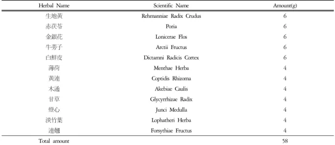
Herbal Name Scientific Name Amount(g)
生地黃 Rehmanniae Radix Crudus 6
赤茯苓 Poria 6
金銀花 Lonicerae Flos 6
牛蒡子 Arctii Fructus 6
白鮮皮 Dictamni Radicis Cortex 6
薄荷 Menthae Herba 4
黃連 Coptidis Rhizoma 4
木通 Akebiae Caulis 4
甘草 Glycyrrhizae Radix 4
燈心 Junci Medulla 4
淡竹葉 Lophatheri Herba 4
連翹 Forsythiae Fructus 4
Total amount 58
Table 1. Composition of SoPungDoJeokTangKami (SPDJTK) CCR3 mAb와 anti-mouse CD4 mAb는 Santa-Cruz 사 (California, USA) 제품을, LSAB kit는 DAKO 사 (Glostrup, Denmark) 제품을 사용하였으며, 기타 일반 시약은 특급 시약을 사용하였다.
(2) 기기
본 실험에 사용된 기기는 열탕추출기 (대웅, Korea), rotary vaccum evaporator (Büchi B-480, Switzerland), freeze dryer (EYELA FDU-540, Japan), CO2 incubator (Forma scientific Co., USA), clean bench (Vision scientific Co., Korea), autoclave (Sanyo, Japan), micro-pipet (Gilson, France), water bath (Vision scientific Co., Korea), vortex mixer (Vision scientific Co., Korea), spectrophotometer (Shimazue, Japan), centrifuge (Sigma, USA), deep-freezer (Sanyo, Japan), 자동혈구측정기 (MS9-5, France), Quantitative Real-Time RT-PCR (Applied Biosystems, USA), ice-maker (Vision scientific Co., Korea), homogenizer (OMNI, USA), plate shaker (Lab-Line, USA), VarioMACS (Bergisch Gladbach, Germany), FACScalibur (BD, USA) 및 ELISA leader (Molecular Devices, USA) 등을 사용하였다.
2) 동물
수컷 7 주령의 SPF(specific pathogen-free) NC/Nga mouse (15 ~ 20 g)는 Charles River Japan (Yokohama, Japan)사에서 공급받았다. 동물은 실험 당일까지 고형 사료 (항생제 무첨가, 삼양사료 Co.)와 물을 충분히 공 급하고 온도 22±2℃, 습도 55±15%, 12시간 (light-dark cycle)의 환경에서 1 주간 적응시킨 후 실험에 사용하 였다.
3) 약물
(1) 消風導赤湯加味
본 실험에 사용한 消風導赤湯加味의 구성은《新編 中醫皮膚病學》21)에 준하였으며, 사용한 약재들은 대 전대학교 둔산 한방병원에서 구입하여 정선하여 사용 하였고 그 내용과 분량은 다음과 같다(Table 1).
(2) 아토피 크림
본 실험의 아토피 크림에 사용된 베이스 크림과 아 로마 오일은 Aroma Korea. Co.; A712, 89-4, Gyeongun-dong, Jongno-gu, Seoul, Korea에서 구입하여 사용하였으며 그 배합은 다음과 같다(Table 2).
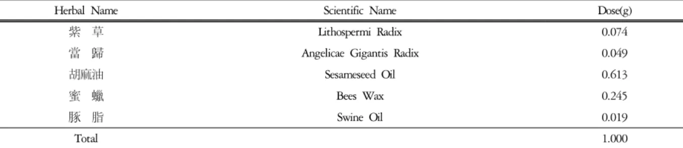
Herbal Name Scientific Name Dose(g)
紫 草 Lithospermi Radix 0.074
當 歸 Angelicae Gigantis Radix 0.049
胡麻油 Sesameseed Oil 0.613
蜜 蠟 Bees Wax 0.245
豚 脂 Swine Oil 0.019
Total 1.000
Table 3. Composition of Jawoongo
Scheme 1. Experimental Design for the Induction of Atopic Dermatitis like NC/Nga Mouse (3) 紫雲膏
본 실험에 사용한 紫雲膏의 구성은 《大田大學校 韓方病院 處方集》31)에 준하였으며, 사용한 약재들은 대전대학교 둔산 한방병원에서 구입하여 정선하여 사 용하였고 그 내용과 1 g 당 조성은 다음과 같다(Table 3).
(4) 消風導赤湯加味 추출물 분리
消風導赤湯加味 2 첩 분량에 증류수 2,000 ㎖를 가 하여 열탕 추출기에서 3 시간 추출하여 얻은 액을 흡입 여과하여 이를 감압 증류장치 (rotary vaccum evaporator) 로 농축하여, 이를 다시 동결 건조기(freeze dryer)를 이용하여 완전 건조한 消風導赤湯加味 추출물 23.4 g 을 냉동 보관(-84℃)하면서 적당한 농도로 희석하여 사용하였다.
2. 方法
1) 피부염 유도 및 시료처리
7 주령의 NC/Nga 생쥐를 1 주일 동안 적응시키고, 이미 피부염이 발생된 18 주령의 NC/Nga 생쥐와 2 주간 같은 공간에서 동시 사육하여 항원감작을 시킨 후 눈에서 capillary 관을 이용하여 100 ㎕의 혈액을 채혈하였다. 그 후 마취제인 chloral hydrate (10%)로 마취하고 등 쪽 목 부위를 깨끗하게 제모한 후 피부 의 미세 상처가 치유되도록 24 시간 방치하였다. 그 리고 중앙실험동물에서 제공하는 Biostir Mite antigen cream {이하 BMAC : Dermato -phagoides farinae crude extract (Biosir INC, Hyogo, Japan)}을 항원으로 제조된 것으로 진드기 항원 0.5% Tween 20이 포함된
ointment base로 제작되었다}을 1 주에 2 회 3 주간 (10 ~ 13 주령) 등과 목 부분에 고르게 도포하였고, 도포 2 시간 전에 4% SDS 용액을 분무하여 피부염이 잘 유발되도록 피부층을 파괴하여 이를 대조군으로 사용하였다 (Scheme 1). 도포 시작 2주 후 (12 주령) 등 부분에 피부염이 충분히 유발되면서 긁는 행동이 심 화되면 육안평가를 실시하였다. 정상군 (NC/Nga-Wild Type, 이하 NC/Nga-WT)은 7 주령 NC/Nga 생쥐를 15 주령까지 SPF 조건에서 사육하였다.
2) 약물처리 및 치료평가
실험은 7 주령 NC/Nga 생쥐를 15 주령까지 SPF 조 건에서 사육한 정상군(NC/Nga-WT)과 BMAC를 도포 한 대조군 (control, CT), BMAC를 도포하고 FK506 (tacrolimus)을 도포한 양성대조군 (positive control, PC), 그리고 BMAC를 도포하고 SPDJTK 또는 AJ+SPDJTK 를 투여한 실험군으로 나누어 실시하였으며, 각 군당 개체 수는 6 마리로 하였다.
BMAC는 총 3 주간 (10 주령 ~ 13 주령) 월요일과 목요일에 4% SDS 용액을 분무하고 2 시간 경과 후, 등에 200 ㎕씩 도포하였다. 그리고 BMAC 도포 시작 2 주 후 (12 주령) 피부 발진을 확인하고, 12 주령부터 15 주령까지 3 주간 매일 1 회 오후 3 ~ 4 시 사이에 각각 SPDJTK (348 ㎎/㎏)를 경구 투여 하였으며, AJ (아토피크림 도포 1 시간 후 紫雲膏를 도포함, 200 ㎕ /마리)와 SPDJTK (348 ㎎/㎏)를 병용투여 하였다. 또 한 양성 대조 약물로 FK506 0.3%도 12 주령부터 15 주령까지 3 주간 매일 1 회 등부분에 골고루 도포하
였다. 실험 종료 후 (15 주령)에 임상적 육안 평가를 실시한 다음 혈액을 채혈하고 등 부위의 피부를 절제 하여 10% 포르말린 용액에 담아 보관하였다.
3) Clinical skin score 측정
임상적 육안 평가는 아토피 피부염에서 일반적으 로 사용되는 Yamamoto M의 방법33)을 약간 변형하여 시행하였고, 7 주령 NC/Nga 생쥐를 15 주령까지 SPF 조건에서 사육한 NC/Nga-WT군과 BMAC를 도포한 대조군 (CT), BMAC를 도포하고 FK506를 도포한 양 성대조군 (PC), 그리고 SPDJTK와 AJ+SPDJTK를 각 각 투여한 실험군으로 나누어 실시하였다 (15 주령).
육안평가 항목은 erythema/hemorrhage, scarring/dryness, excoriation/erosion, edema, lichenification 5 가지 항목으로 하 고, 육안평가 결과는 각각 평가한 점수의 총 합으로 나타냈다. 각각의 항목은 없음 (0), 약함 (1), 중증도 (2), 심함 (3)으로 채점하였으며 최소 0 점에서 최고 15 점 사이의 점수를 측정하였다.
4) 백혈구 분석
최종 임상적 육안 평가를 실시한 후, 심장천자법으 로 채혈한 혈액을 바이오톡스텍(주)(청주, 충청북도) 에 의뢰하여 혈액의 백혈구 중 호중구, 호산구, 호염 기구 및 림프구, 단핵구의 세포수를 측정 하였다. 측 정은 자동혈구측정기(MS9-5, MELET SCHLOESING, France)로 Fonio법34)에 준하여 Minos -ST로 시행하였다.
5) 혈청 내 IgE와 IgG1 측정
NC/Nga 생쥐 8 주령, 12 주령, 15 주령의 눈에서 capillery 관을 이용하여 약 100 ㎕의 혈액을 채혈한 후 원심분리기 6,500 rpm에서 20 분간 원심분리한 후 30
㎕의 혈청을 분리하여 IgE 수준을 측정하였고, IgG1 수준은 15 주령의 NC/Nga 생쥐를 ethylether로 흡입 마취한 다음 심장천자법으로 혈액을 분리한 후, 각각 의 혈청을 취하여 -70℃에 냉동보관하였다. NC/Nga 생쥐의 혈청 내 IgE와 IgG1 농도는 enzyme-linked immuno-sorbent assay로 측정하였다. IgE는 NC/Nga 생 쥐 8 주령, 12 주령, 그리고 15 주령에서 채혈한 혈청 5 ㎕ (1/10 dilution)와 dilution buffer 45 ㎕를 혼합하여 96 well plate의 각 well에 분주하였고, IgG1은 15주령 에서 채혈한 혈청 50 ㎕ (1/10 dilution)과 dilution buffer 50 ㎕를 혼합하여 각 well에 분주하였다. 각각 2 시간 동안 25℃ 실온에서 방치한 후 2 회 washing 완
충용액으로 세척한 다음 각각 antibody biotin-IgE conjugated와 antibody biotin-IgG1 conjugated를 넣고 2 시간 방치하였다. 다시 2 회 수세 후 완충용액으로 세 척한 다음 antibody Avidin-HRP conjugeted 100 ㎕를 처리하고 1 시간 실온에서 방치한 후 다시 세척하였 다. TMB 기질을 100 ㎕씩 분주하고 암소에서 30 분 간 방치한 후 100 ㎕의 stop 용액을 처리한 후 ELISA leader 450 ㎚에서 각각 IgE와 IgG1에 대한 흡광도를 측정하였다35).
6) ALN 세포분리 및 cytokine 측정
약물투여 종료 후(15 주령) ALN을 적출하여 100 mesh로 ALN 세포를 분리하였다. 전날 BMAC 10 ㎍/
㎖을 96 well plate에 coating하여 4℃ 냉장고에서 overnight한 다음 D-PBS로 2 회 수세하였다. 분리한 ALN 세포는 ACK 용액(8.3 g NH4Cl, 1 g KHCO3, in 1 ℓ of demineralized water + 0.1 mM EDTA)으로 RBC를 제거한 후 BMAC가 coating된 각각의 well에 5×10⁵ 세포씩 5% FBS-DMEM 배양액에서 48 시간 동 안 배양한 후, 원심분리기 2,000 rpm에서 3 분간 원심 분리한 후 200 ㎕의 배양상층액을 얻었다. 배양상층 액내의 IL-4 (BioSource, USA), IFN-γ (BioSource, USA), IL-5 (BioSource, USA), IL-13 (R&D system, USA)의 수 준 측정은 enzyme-linked immuno-sorbent assay로 측정 하였다. 각 well에 배양상층액 50 ㎕를 분주하고, 2 시 간 동안 25℃ 실온에서 방치한 후 2 회 washing 완충 용액으로 세척한 다음 각각 antibody biotin-IL-4 conjugated, antibody biotin-IL-5 conjugated, 그리고 antibody biotin-IL-13 conjugated를 넣고 2 시간 방치하였 다. 다시 2 회 수세 후 완충용액으로 세척한 다음 antibody Avidin- HRP conjugeted 100 ㎕를 처리하고 1 시간 실온에서 방치한 후 다시 세척하였다. TMB 기 질을 100 ㎕씩 분주하고 암소에서 30 분간 방치한 후 100 ㎕의 stop 용액을 처리한 후 ELISA leader 450 ㎚ 에서 흡광도를 측정하였다.
7) ALN, DLN, PBMCs 및 등피부조직에서 형광 유세포 분석
약물투여 종료 후(15 주령) NC/Nga 생쥐에서 ALN 과 DLN을 각각 적출하여 100 mesh로 세포를 분리하 여 D-PBS로 5 분간 원심분리 (1,700 rpm)하고 2 회 세 척한 후 cell strainer (FALCON)에 통과시켜 세포 이외 의 분해되지 않은 조직이나 불순물을 제거하였다.
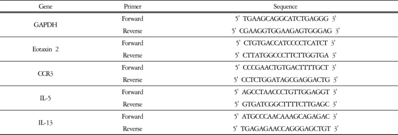
Gene Primer Sequence
GAPDH Forward 5' TGAAGCAGGCATCTGAGGG 3'
Reverse 5' CGAAGGTGGAAGAGTGGGAG 3'
Eotaxin 2 Forward 5' CTGTGACCATCCCCTCATCT 3'
Reverse 5' CTTATGGCCCTTCTTGGTGA 3'
CCR3 Forward 5' CCCGAACTGTGACTTTTGCT 3'
Reverse 5' CCTCTGGATAGCGAGGACTG 3'
IL-5 Forward 5' AGCCTAACCCTGTTGGAGGT 3'
Reverse 5' GTGATCGGCTTTTCTTGAGC 3'
IL-13 Forward 5' ATGCCCAACAAAGCAGAGAC 3'
Reverse 5' TGAGAGAACCAGGGAGCTGT 3'
Table 4. Primer Sequence
PBMCs는 실험을 종료한 후 NC/Nga 생쥐에서 heparin 을 처리한 3 ㎖ 주사기로 채혈한 후 미리 준비한 10
㎖의 ACK용액에 혼합하여 실온에서 5 분 동안 처리 하여 적혈구를 제거하였다. 2 회 1%의 FBS가 함유된 PBS (FACS buffer)로 세척한 후 cell strainer에 통과시켜 세포 이외의 불순물을 제거하였다. 등피부조직은 잘게 chopping한 후 collagenase 1 ㎎/㎖ (in 2% FBS + RPMI 1640)를 넣고 37℃ shaker (180 rpm, 20 min) 배양기에 서 배양한 후 상층액을 회수하는 방법으로 4 회 반복 하였다.
분리한 ALN, DLN, PBMCs, 그리고 등피부조직 침 윤세포의 총세포수를 측정한 다음 모든 조직의 세포 등을 5 × 10⁵ 세포로 조정한 후 4℃에서 면역 형광염 색 (immunofluorescence staining)을 실시하였다. 각각에 anti-CD3e -PE, anti-CD19-FITC, anti-CD4-FITC, anti-CD8-FITC, anti-CD23-FITC, anti-CD49b-FITC, anti-CCR3-PE, 그리고 anti-B220-PE를 넣고 30 분간 얼 음에서 반응시켰다. 반응 후 3 회 이상 PBS로 수세한 후 flow cytometry의 CellQuest 프로그램을 이용하여 CD3+, CD19+, CD4+, CD8+, CCR3+, 그리고 B220+CD23+, CXCR5+ 세포수를 백분율 (%)로 분 석한 후 총세포수를 적용하여 ALN, DLN, PBMCs, 그 리고 등피부조직에서의 절대 세포수(absolute number) 를 산출하였다.
8) Quantitative real-time PCR in dorsal skin &
ALN
Atopic dermatitis-like skin NC/Nga 생쥐의 등 피부조 직과 ALN를 적출하여 각각에 RNAzolB 500 ㎕를 넣 고 용해될 때까지 homogenizer로 분쇄하였다. 이 조직 분쇄 혼합 부유액에 chloroform (CHCl3) 50 ㎕를 첨가
한 후 15 초간 다시 혼합하였다. 이를 얼음에 15 분간 방치한 후 13,000 rpm에서 원심 분리한 후 약 200 ㎕ 의 상층액을 회수하여 2-propanol 200 ㎕와 동량 혼합 후 천천히 흔들고 얼음에서 15 분간 방치하였다. 이 를 다시 13,000 rpm에서 원심 분리한 후 80% EtOH로 수세하고 3 분간 vaccum pump에서 건조하여 RNA를 추출하였다. 추출한 RNA는 DEPC를 처리한 20 ㎕의 증류수에 녹여 heating block 75℃에서 불활성화 시킨 후 first strand cDNA합성에 사용하였다. Quantitative real-time PCR은 7500 Real-Time PCR system을 이용하 여 수행하였다36).
Mouse Olionucleotid의 염기배열은 다음과 같다 (Table 4).
Cytokine 유전자 발현은 SYBRⓇ Green PCR Master mix를 사용하였고, internal standard는 GAPDH로 Taqman probe를 사용하였으며, primer의 최종농도가 200 nM 이 되게 반응시켰다. Eotaxin2와 CCR3 mRNA 발현은 등피부조직에서 관찰하였고, Th2 mediate인 IL-5와 IL-13 mRNA 유전자 발현량 분석은 ALN에서 cDNA 를 합성하여 분석하였다.
Quantitative real-time PCR의 조건은 pre- denaturation 은 2 min at 50℃, 10 min 94℃, 그리고 40 cycles을 0.15 min at 95℃, 1 min at 60℃에서 수행하였다. 대 조군, FK506 도포군, SPDJTK 투여군과 AJ+SPDJTK 투여군은 internal standard로 GAPDH를 사용하였고 target group의 Quantitative PCR은 y = x(1+e)n
x = starting quantity y = yield
n = number of cycles
e = efficiency로 계산하여 RQ (relative quantitative) 값을 측정하였다.
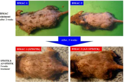
Fig. 1. Topical application of SPDJTK, AJ+SPDJTK treatment of atopic dermatitis skin lesions in NC/Nga mouse induced by BMAC for 3 weeks.
Shown are back of BMAC-ointment NC/Nga mouse, BMAC plus SPDJTK(348 mg/kg)-orally adminstration in NC/Nga mouse and BMAC plus AJ(200 ㎕/mouse)-ointment plus SPDJTK(348 mg/kg)-orally adminstration in NC/Nga mouse for 3 weeks.
9) 피부조직 채취 및 염색
약물 투여 종료 후(15 주령) ALN과 등 쪽 목 부분 의 피부를 떼어내어 10% paraform- aldehyde에서 24 시간 동안 포르말린에 고정하였다. 그 조직을 파라핀 으로 포매하였고, 5 ㎛ 두께로 block을 만들었다. 그 조직부분은 염증을 일으키는 epidermis, dermis, keratinocytes, neutrophils/eosinophil 그 외 다른 세포와 부종을 식별하는 hematoxyline/eosin(H&E) 염색과 비만 세포를 염색하는 toluidine blue 염색으로 비만세포의 침윤을 광학현미경 (Nikon, Japan, ×200)으로 관찰하였다37).
10) Immunohistochemical staining
약물 투여 종료 후(15 주령) 면역화학조직염색을 위하여 등쪽 피부를 적출하여 10% 포르말린 용액에 고정한 다음 파라핀 블록을 만든 후 rat anti-mouse CCR3 mAb와 rat anti-mouse CD4 mAb를 사용하였다.
조직절편을 4 ㎛ 두께로 세절하고 probe-on plus slide에 부착시켜 건조시켰다. 그리고 탈 파라핀 (deparaffinized) 후 함수시킨 다음 0.01 M citrate buffer (pH 6.0)를 이 용해 microwave oven에 15 분간 전 처리하였다. 조직 내 과산화효소의 작용을 억제하기 위하여 3% H2O2 에 10 분간 처리한 후, 조직 내의 항원과 비 특이적 단백결합을 억제하기 위해 정상 혈청으로 단백질을 차단시켰다. 그리고 일차 단일항체에 1 시간 동안 부 착시킨 다음 완충액으로 수세하였다. LSAB kit를 이 용하여 PE-conjugated goat anti-rat IgG에 30 분간 반응
시키고, 3 회 tris-buffered saline with 0.1% tween 20(TBST)용액으로 수세한 후 잘 건조하였다. 현미경 은 형광위상차현미경을 사용하여 ×400 배율로 관찰 하였다38).
3. 통계처리
다양한 실험으로부터 얻은 결과는 mean± standard error로 기록하였고, 유의성 검증은 Student's t-test 분 석방법을 이용하여 결정하였다39).
Ⅲ. 結 果
1. BMAC를 이용한 아토피 피부염 유발 및 SPDJTK와 AJ+SPDJTK 투여 3 주 후 등피부 변화
BMAC를 2 주간(총 4 회) 400 ㎎ 정도 도포한 결과, NC/Nga 생쥐 등피부부위에 사람 아토피 피부염과 같은 증상인 erythema/hemorrhage, scarring/dryness, excoriation/erosion, edema, lichenification이 나타났다(Fig.1 상단사진). BMAC 를 1 주 더 도포하여 3 주간(총 6 회) 600 ㎎ 정도 도 포하고 SPDJTK, 그리고 AJ+SPDJTK를 3 주간 처리 한 결과, SPDJTK만 투여한 실험군보다는 AJ+SPDJTK 를 병용투여한 실험군에서 NC/Nga 생쥐 등피부의 erythema/hemorrhage, scarring/ dryness, excoriation/erosion, edema, lichenification이 대조군에 비하여 현저하게 감 소하였다(Fig. 1 하단사진).
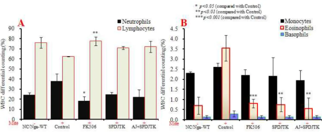
Fig. 3. WBC differential counting in atopic dermatitis skin lesions in NC/Nga mouse.
Atopic dermatitis NC/Nga mouse was induced by BMAC treatment in the dorsal skin, non treated BMAC(SPF-normal, NC/Nga-WT), BMAC treatment for 3 weeks(Control), BMAC treatment for 3 weeks with FK506(0.3%)-ointment(FK506), BMAC treatment for 3 weeks with SPDJTK(348 mg/kg) -orally administration(SPDJTK), and BMAC treatment for 3 weeks with AJ(200 ㎕/mouse)-ointment plus SPDJTK(348 mg/kg)-orally administration(AJ+SPDJTK) for 3 weeks. Blood was collected from the retro-orbital plexus under ether anesthesia and heparinized immediately thereafter. Cell contents were measured by hematology(BD, USA).
Fig. 2. Clinical skin features and severity of atopic dermatitis skin lesions in NC/Nga mouse induced by BMAC for 3 weeks.
Atopic dermatitis NC/Nga mouse was induced by BMAC treatment in the dorsal skin, non treated BMAC(SPF-normal, NC/Nga-WT), BMAC treatment for 3 weeks(Control), BMAC treatment for 3 weeks with FK506(0.3%)-ointment(FK506), BMAC treatment for 3 weeks with SPDJTK(348 mg/kg) -orally administration(SPDJTK), and BMAC treatment for 3 weeks with AJ(200
㎕/mouse)-ointment plus SPDJTK(348 mg/kg)-orally administration(AJ+SPDJTK) for 3 weeks. A total clinical severity score for AD-like lesions was defined as the sum of the individual scores graded as 0(none), 1 (mild), 2(moderate) and 3(severe) for each of five signs and symptoms(erythema/hemorrhage, scarring/dryness, edema, excoriation/erosion and lichenification) on the three parts of the body: ear, face and head and back. Each point represents the mean±SE of six mice.
2. Clinical skin score에 미치는 영향
대조군(Control)의 clinical skin score는 NC/Nga -WT군 (정상군)에 비하여 10 배 이상 증가하였고 FK506 도 포군(양성대조군)은 대조군에 비해 감소하였으며 (p<0.001), SPDJTK 단독처리군보다 AJ+SPDJTK 병 용투여군에서(p<0.05) 대조군에 비해 clinical skin score가 유의성 있게 감소하였다(Fig. 2).
3. 백혈구에 미치는 영향
실험 종료 후(15 주령) 백혈구(WBC)의 neutrophil과
eosinophils 비율은 NC/Nga-WT군에 비하여 대조군 (Control)이 현저하게 증가하였다. FK506 도포군은 대 조군에 비하여 NC/Nga-WT군에 가깝게 유의성 있게 감소하였고(p<0.05, p<0.001), SPDJTK 투여군, 그리고 AJ+SPDJTK 병용투여군의 neutrophil의 비율은 대조 군에 비하여 감소하였지만 유의성은 없었고, eosinophils 비율은 유의성 있게 감소하였다 (p<0.01)(Fig. 3A, Fig.
3B).
그리고 WBC 중 lymphocytes 비율은 대조군에서 NC/Nga-WT군에 비하여 약간 감소하였으나 FK506
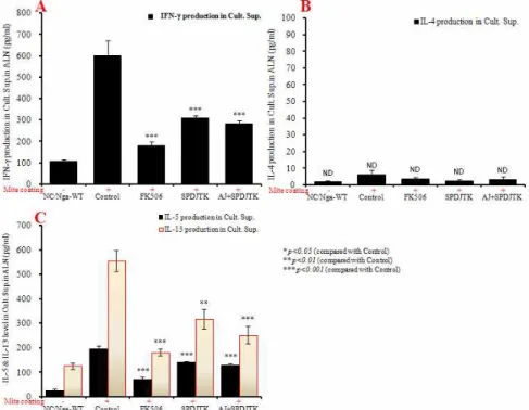
Fig. 5. Culture supernatant IL-4, IL-5, IL-13, and IFN-γ level in ALN in atopic dermatitis skin lesions in NC/Nga mouse.
Atopic dermatitis NC/Nga mouse was induced by BMAC treatment in the dorsal skin, non treated BMAC(SPF-normal, NC/Nga-WT), BMAC treatment for 3 weeks(Control), BMAC treatment for 3 weeks with FK506(0.3%)-ointment(FK506), BMAC treatment for 3 weeks with SPDJTK(348 mg/kg) -orally administration(SPDJTK), and BMAC treatment for 3 weeks with AJ(200
㎕/mouse)-ointment plus SPDJTK(348 mg/kg)-orally administration(AJ+SPDJTK) for 3 weeks. ALN from mouse at 15 weeks of age were re-stimulated with Mite extract(1 ㎍/㎖) for 48 hrs. IL-4, IL-5, IL-13, and IFN-γ levels were measured by a sandwich ELISA using an mouse ELISA kit I(Biosource, USA).
Fig. 4. Serum IgE & IgG1 elevation and development of atopic dermatitis skin lesions in NC/Nga mouse.
Atopic dermatitis NC/Nga mouse was induced by BMAC treatment in the dorsal skin, non treated BMAC(SPF-normal, NC/Nga-WT), BMAC treatment for 3 weeks(Control), BMAC treatment for 3 weeks with FK506(0.3%)-ointment(FK506), BMAC treatment for 3 weeks with SPDJTK(348 mg/kg) -orally administration(SPDJTK), and BMAC treatment for 3 weeks with AJ(200
㎕/mouse)-ointment plus SPDJTK(348 mg/kg)-orally administration(AJ+SPDJTK) for 3 weeks. Blood was collected from the retro-orbital plexus under ether anesthesia and heparinized immediately thereafter. Plasma samples were obtained by centrifugation and stored at -20°C until use. Total IgE and IgG1 levels were measured by a sandwich ELISA using an ELISA kit(Shibayagi, Japan). Each point represents the mean±SE of six mice.
도포군은 유의성 있게 증가하였고(p<0.01), SPDJTK 투여군, AJ+SPDJTK 병용투여군에서는 차이가 없었다.
monocytes와 basophils의 WBC 내 비율을 분석한 결과, 모든 실험군에서 차이가 나타나지 않았다(Fig. 3A, Fig. 3B).
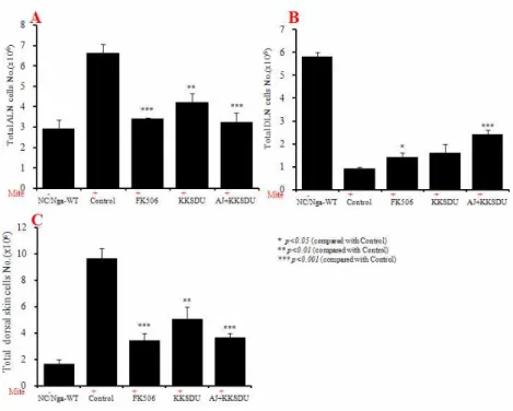
Fig. 6. Total cell number of ALN, DLN and dorsal skin in atopic dermatitis skin lesions in NC/ Nga mouse.
Atopic dermatitis NC/Nga mouse was induced by BMAC treatment in the dorsal skin, non treated BMAC(SPF-normal, NC/Nga-WT), BMAC treatment for 3 weeks(Control), BMAC treatment for 3 weeks with FK506(0.3%)-ointment(FK506), BMAC treatment for 3 weeks with SPDJTK(348 mg/kg) -orally administration(SPDJTK), and BMAC treatment for 3 weeks with AJ(200
㎕/mouse)-ointment plus SPDJTK(348 mg/kg)-orally administration(AJ+SPDJTK) for 3 weeks.
4. 혈청 IgE와 IgG1 수준에 미치는 영향
혈청 IgE 수준은 8 주령 NC/Nga 생쥐에서 자연적 으로 증가가 되고 대조군(Control)은 15 주령에서 NC/Nga-WT군과 차이가 없이 IgE 수준이 증가되었 다. 그리고 FK506 도포군, SPDJTK 투여군, 그리고 AJ+SPDJTK 병용투여군의 혈청내 IgE 수준은 12 주 령 이후 감소하여 15 주령에서는 대조군에 비해 유의 성 있게 감소되었다(p<0.001)(Fig. 4A).
혈청 IgG1의 수준은 FK506 도포군에서 대조군에 비해 유의성 있게 감소되었고(p<0.001), SPDJTK 투여 군, 그리고 AJ+SPDJTK 병용투여군 또한 대조군에 비해 유의성 있게 감소되었다(p<0.001)(Fig. 4B).
5. ALN에서 cytokine level에 미치는 영향
IFN-γ의 생산량은 NC/Nga-WT군에 비하여 BMAC 를 도포한 대조군(Control)이 증가한 결과를 얻었다.
그리고 FK506도포군은 대조군에 비하여 유의성있게 감소되었다(p<0.001). 또한 SPDJTK 투여군, 그리고 AJ+SPDJTK 병용투여군은 대조군에 비하여 유의성 있게 감소하였다( p<0.001)(Fig. 8A). 반면, ALN 세포 배양상층액 중 IL-4 생산량을 측정한 결과는 모든 실
험군에서 차이가 나타나지 않았다(Fig. 5B).
IL-5와 IL-13의 생산량을 측정한 결과, NC/Nga-WT 군에 비하여 대조군이 각각 증가되었다. 그리고 FK506 도포군은 대조군에 비하여 유의성있게 감소되 었다(p<0.001). SPDJTK 투여군, 그리고 AJ+SPDJTK 병용투여군 또한 대조군에 비하여 유의성 있게 감소 되었다(p<0.01, p<0.001)(Fig. 5C).
6. ALN, DLN과 등피부조직의 총 세포수에 미치는 영향 ALN과 등피부부위의 총 세포수를 측정한 결과, NC/
Nga-WT군에 비하여 대조군(Control)이 현저하게 증가 하였다. 그리고 FK506 도포군, SPDJTK 투여군, 그리고 AJ+SPDJTK 병용투여군에서는 대조군에 비하여 유 의성 있게 감소하였다(p<0.01, p<0.001) (Fig. 6A, Fig. 6C).
DLN의 총 세포수를 측정한 결과, ALN과는 반대로 NC/Nga-WT군에 비하여 대조군이 현저하게 감소하 였다. FK506 도포군, 그리고 AJ+SPDJTK 병용투여군 은 대조군에 비하여 유의성있게 증가하였다(p<0.05, p<0.001). 그리고 SPDJTK만을 투여한 실험군은 대조 군에 비하여 총 세포수가 증가하였으나 유의성은 나 타나지 않았다(Fig. 6B).
Fig. 7. Effects of SPDJTK & AJ+SPDJTK treatment on T and B cell changes of absolute numbers in ALN cells in NC/Nga mouse.
Atopic dermatitis NC/Nga mouse was induced by BMAC treatment in the dorsal skin, non treated BMAC(SPF-normal, NC/Nga-WT), BMAC treatment for 3 weeks(Control), BMAC treatment for 3 weeks with FK506(0.3%)-ointment(FK506), BMAC treatment for 3 weeks with SPDJTK(348 mg/kg) -orally administration(SPDJTK), and BMAC treatment for 3 weeks with AJ(200
㎕/mouse)-ointment plus SPDJTK(348 mg/kg)-orally administration(AJ+SPDJTK) for 3 weeks. NC/Nga mouse ALN cells (2×10⁵ cells/㎖) were isolated from ALN, and the ALN cells were washed twice and analyzed by flow cytometry. Absolute numbers of CD3+, CD19+(A) and CD4+, CD8+(B) in NC/Nga mouse.
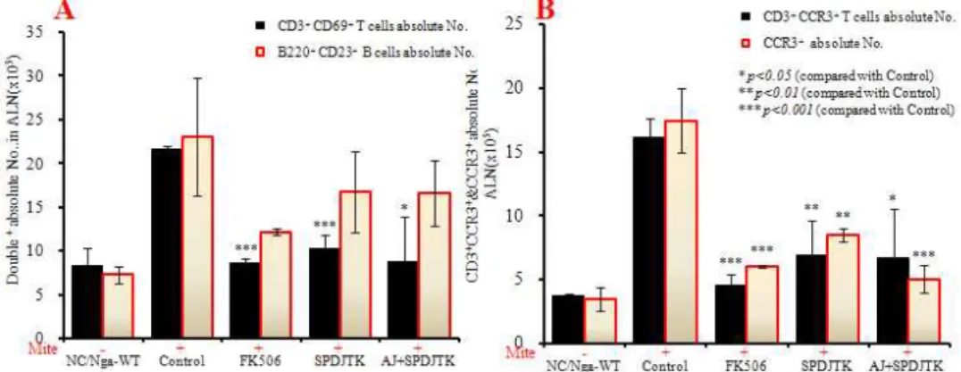
Fig. 8. Effects of SPDJTK & AJ+SPDJTK treatment on CD3+CD69+, B220+CD23+ and CD3+ CCR3+, CCR3+
changes of absolute numbers in ALN cells in NC/Nga mouse.
Atopic dermatitis NC/Nga mouse was induced by BMAC treatment in the dorsal skin, non treated BMAC(SPF-normal, NC/Nga-WT), BMAC treatment for 3 weeks(Control), BMAC treatment for 3 weeks with FK506(0.3%)-ointment(FK506), BMAC treatment for 3 weeks with SPDJTK(348 mg/kg) -orally administration(SPDJTK), and BMAC treatment for 3 weeks with AJ(200
㎕/mouse)-ointment plus SPDJTK(348 mg/kg)-orally administration(AJ+SPDJTK) for 3 weeks. NC/Nga mouse ALN cells (2×10⁵ cells/㎖) were isolated from ALN, and the ALN cells were washed twice and analyzed by flow cytometry. Absolute numbers of CD3+CD69+, B220+CD23+(A) and CD3+CCR3+, CCR3+(B) in NC/Nga mouse.
7. ALN에서 형광유세포분석을 통한 T & B 세포수에 미치는 영향
1) CD3+, CD19+ & CD4+, CD8+ absolute number ALN에서 활성 CD3+ T세포의 절대 세포수는 NC/Nga-WT군에 비하여 대조군(Control)이 현저하게 증가하였고, FK506 도포군은 대조군에 비하여 유의 성있게 감소하였다(p<0.001). 또한 SPDJTK 투여군에 서는 대조군에 비하여 유의성 있는 감소를 나타내었 지만(p<0.001) AJ+SPDJTK 병용투여군에서는 유의 성은 없었다(Fig.7A). ALN에서 활성 CD19+ B세포의 절대 세포수는 NC/Nga-WT군에 비하여 대조군이 현
저하게 증가하였고, FK506 도포군은 대조군에 비하여 유의성있게 감소하였다(p<0.01). 또한 SPDJTK 투여군, 그리고 AJ+SPDJTK 병용투여군도 ALN에서 CD19+
B 절대 세포수가 모두 대조군에 비하여 감소하였으 나, 유의성은 없었다 (Fig. 7A).
ALN에서 활성 CD4+ Th 세포와 활성 CD8+ c/sT 세포에 대한 절대 세포수는 NC/Nga-WT군에 비하여 대조군이 현저하게 증가하였고, FK506 도포군, SPDJTK 투여군, AJ+SPDJTK 병용투여군에서는 대조 군에 비하여 유의성있게 감소하였다(p<0.01, p<0.001) (Fig. 7B).
Fig. 9. Effects of SPDJTK & AJ+SPDJTK treatment on CD4+CXCR5+ gated cells and changes of absolute numbers in ALN cells in NC/Nga mouse.
Atopic dermatitis NC/Nga mouse was induced by BMAC treatment in the dorsal skin, non treated BMAC(SPF-normal, NC/Nga-WT), BMAC treatment for 3 weeks(Control), BMAC treatment for 3 weeks with FK506(0.3%)-ointment(FK506), BMAC treatment for 3 weeks with SPDJTK(348 mg/kg) -orally administration(SPDJTK), and BMAC treatment for 3 weeks with AJ(200
㎕/mouse)-ointment plus SPDJTK(348 mg/kg)-orally administration(AJ+SPDJTK) for 3 weeks.. NC/Nga mouse ALN cells (2×10⁵ cells/㎖) were isolated from ALN, and the ALN cells were washed twice and analyzed by flow cytometry. Absolute number of CD4+CXCR5+ in NC/Nga mouse.
Fig. 10. Effects of SPDJTK & AJ+SPDJTK treatment on the percentage of granulocytes gated cells in PBMCs in NC/Nga mouse.
Atopic dermatitis NC/Nga mouse was induced by BMAC treatment in the dorsal skin, non treated BMAC(SPF-normal, NC/Nga-WT), BMAC treatment for 3 weeks(Control), BMAC treatment for 3 weeks with FK(0.3%)-ointment(FK), BMAC treatment for 3 weeks with SPDJTK(348 mg/kg) -orally administration(SPDJTK), and BMAC treatment for 3 weeks with AJ(200
㎕/mouse)-ointment plus SPDJTK(348 mg/kg)-orally administration(AJ+SPDJTK) for 3 weeks. NC/Nga mouse PBMCs (2×10⁵ cells/㎖) were isolated from Blood and the PBMCs were washed twice and analyzed by flow cytometry. Total cell content(%) of granulocytes gated cells in NC/Nga mouse.
2) CD3+CD69+, B220+CD23+ & CD3+ CCR3+, CCR3+ absolute number
ALN에서 CD3+CD69+ T 세포와 B220+ CD23+
B 세포의 절대 세포수는 NC/Nga-WT군에 비하여 대 조군(Control)이 모두 증가하였고, FK506 도포군, SPDJTK 투여군, 그리고 AJ+SPDJTK 병용투여군의 CD3+
CD69+ 절대 세포수는 대조군에 비하여 유의성있게 감소하였다(p<0.01, p<0.001). 그리고 FK506 도포군,
SPDJTK 투여군, 그리고 AJ+SPDJTK 병용투여군의 B220+CD23+ B 세포 수는 모두 대조군에 비하여 감 소하였으나 유의성은 없었다(Fig. 8A).
ALN에서 활성 CD3+CCR3+세포와 CCR3+세포에 대한 절대 세포수는 NC/Nga-WT군에 비하여 대조군이 증가 하였고, FK506 도포군, SPDJTK 투여군, AJ+SPDJTK 병용투여군에서는 대조군에 비하여 현저하게 유의성 있는감소를 나타내었다(p<0.05, p<0.01, p<0.001)(Fig. 8B).
Fig. 11. Effects of SPDJTK & AJ+SPDJTK treatment on the percentage of T & B gated cells in PBMCs in NC/Nga mouse.
Atopic dermatitis NC/Nga mouse was induced by BMAC treatment in the dorsal skin, non treated BMAC(SPF-normal, NC/Nga-WT), BMAC treatment for 3 weeks(Control), BMAC treatment for 3 weeks with FK506(0.3%)-ointment(FK506), BMAC treatment for 3 weeks with SPDJTK(348 mg/kg) -orally administration(SPDJTK), and BMAC treatment for 3 weeks with AJ(200
㎕/mouse)-ointment plus SPDJTK(348 mg/kg)-orally administration(AJ+SPDJTK) for 3 weeks. NC/Nga mouse PBMCs (2×10⁵ cells/㎖) were isolated from Blood and the PBMCs were washed twice and analyzed by flow cytometry. Total cell content(%) of CD3+ & CD19+(A), CD4+ & CD8+(B), CD3+CD69+ and B220+CD23+(C), CD3+CCR3+ and CCR3+(D) in NC/Nga mouse.
3) CD4+CXCR5+ absolute number
ALN에서 활성화된 T세포를 관찰한 결과 CD4+
CXCR5+ 절대 세포수는 NC/Nga-WT군에 비하여 대 조군(Control)이 현저하게 증가하였고, FK506 도포군, SPDJTK 투여군, AJ+SPDJTK 병용 투여군에서는 모 두 대조군에 비하여 유의성 있게 감소하였다(p<0.05, p<0.001)(Fig. 9).
8. PBMCs에서 세포수에 미치는 영향 1) PBMCs에서 granulocyte의 변화
PBMCs에서 granulocytes의 빈도(%)는 NC/Nga-WT 군에 비하여 대조군(Control)이 증가하였고, FK506 도 포군은 대조군에 비하여 유의성 있게 감소하였다 (p<0.001). SPDJTK 투여군, 그리고 AJ+SPDJTK 병용 투여군의 granulocytes의 빈도수(%)는 대조군에 비하 여 유의성 있게 감소하였다.(p<0.01, p<0.001) (Fig. 10).
2) PBMCs에서 형광유세포분석을 통한 T & B 세 포수에 미치는 영향
PBMCs의 CD3+ T세포, CD4+ Th세포, CD8+ c/s T세포, CD3+/CD69+세포, B220+ /CD23+, CD3+
/CCR3+세포, 그리고 CCR3+세포는 NC/Nga-WT군에 비하여 대조군(Control)에서 총활성세포 빈도(%)가 모 두 현저하게 증가하였다. FK 도포군의 CD3+ T세포, CD8+ c/s T세포, CD3+/CD69+세포, B220+ /CD23+, 그리고 CCR3+ 세포의 총활성세포 빈도(%)는 대조군 에 비하여 유의하게 감소하였고(p<0.01, p<0.001), CD4+ Th 세포와 CD3+CCR3+ 세포는 감소하였으나 유의성은 없었다. 또한 SPDJTK 투여군의 CD3+ T세 포, CD4+ Th세포, CD8+ c/s T세포, CD3+/ CD69+, 그리고 CD3+/CCR3+ 세포의 총활성세포 빈도(%)는 대조군에 비하여 감소하였지만 CD3+/CD69+세포에 서만 유의성이 나타났고(p<0.05), AJ+SPDJTK 병용투 여군에서는 모두에서 유의성있게 감소하였다(p<0.01,
Fig. 12. Effects of SPDJTK & AJ+SPDJTK treatment on T cell and B cell changes of absolute numbers in DLN cells in NC/Nga mouse.
Atopic dermatitis NC/Nga mouse was induced by BMAC treatment in the dorsal skin, non treated BMAC(SPF-normal, NC/Nga-WT), BMAC treatment for 3 weeks(Control), BMAC treatment for 3 weeks with FK506(0.3%)-ointment(FK506), BMAC treatment for 3 weeks with SPDJTK(348 mg/kg) -orally administration(SPDJTK), and BMAC treatment for 3 weeks with AJ(200
㎕/mouse)-ointment plus SPDJTK(348 mg/kg)-orally administration(AJ+SPDJTK) for 3 weeks. NC/Nga mouse PBMCs (2×10⁵ cells/㎖) were isolated from Blood and the PBMCs were washed twice and analyzed by flow cytometry. Total cell content(%) of CD3+ & CD19+(A), CD4+ & CD8+(B), CD3+CD69+ and B220+CD23+(C), CD3+CCR3+ and CCR3+(D) in NC/Nga mouse.
p<0.001). 그리고 CCR3+ 세포는 SPDJTK 투여군과 AJ+SPDJTK 병용투여군 모두에서 감소하였고 유의 성이 나타났다(p<0.05, p<0.01)(Fig. 11).
PBMCs에서 CD19+ B세포의 총활성세포 빈도(%) 는 NC/Nga-WT군에 비하여 대조군이 현저하게 감소 하였고, FK506 도포군에서는 대조군에 비하여 유의 성있게 증가하였다(p<0.001). 그리고 SPDJTK 투여군 과 AJ+ SPDJTK 병용투여군에서도 대조군에 비하여 유의성 있는 증가를 나타내었다(p<0.01, p< 0.001) (Fig. 11A).
9. DLN에서 형광유세포분석을 통한 T & B 세포수에 미치는 영향
DLN내 총 CD3+, CD19+과 CD4+, CD8+의 절대 세포수는 NC/Nga-WT군에 비하여 대조군이 현저하 게 감소하였고, FK506 도포군의 CD3+(p<0.001)와 CD4+(p<0.05), CD8+(p<0.001) 의 절대 세포수는 대조 군에 비하여 유의성있게 증가하였다. SPDJTK 투여
군, AJ+SPDJTK 병용투여군의 CD3+, CD4+의 절대 세포수는 대조군에 비하여 유의성 있게 증가하였고 (p<0.001), CD19+의 절대 세포수는 각 투여군 모두 유의성 있게 증가하였으나, CD8+ 절대 세포수는 AJ+SPDJTK 병용투여군 대조군에 비하여 증가되었으 나 유의성은 없었다 (Fig. 12A, B).
DLN내 CD3+CD69+ T cell의 절대 세포수와 B220+CD23+ B cell의 절대 세포수는 NC/ Nga-WT 군에 비하여 대조군이 현저하게 감소하였고, 모든 실 험군은 대조군과 큰 차이가 없었으며 유의성도 없었 다(Fig. 12C).
CD3+CCR3+ 절대 세포수는 SPDJTK 투여군, AJ+SPDJTK 병용투여군 모두 대조군에 비하여 유의 성 있게 감소하였으며(p<0.001), CCR3+의 절대 세포 수는 FK506 도포군(p<0.05), SPDJTK 투여군(p<0.01), AJ+SPDJTK 투여군(p<0.001)은 대조군에 비하여 유 의성 있게 감소하였다 (Fig. 12D).
Fig. 13. Effects of SPDJTK & AJ+SPDJTK treatment on changes of absolute numbers in dorsal skin cells in NC/Nga mouse.
Atopic dermatitis NC/Nga mouse was induced by BMAC treatment in the dorsal skin, non treated BMAC(SPF-normal, NC/Nga-WT), BMAC treatment for 3 weeks(Control), BMAC treatment for 3 weeks with FK506(0.3%)-ointment(FK506), BMAC treatment for 3 weeks with SPDJTK(348 mg/kg) -orally administration(SPDJTK), and BMAC treatment for 3 weeks with AJ(200
㎕/mouse)-ointment plus SPDJTK(348 mg/kg)-orally administration(AJ+SPDJTK) for 3 weeks. NC/Nga mouse dorsal skin cells(2×10⁵ cells/㎖) were isolated from dorsal skin, and the dorsal skin cells were washed twice and analyzed by flow cytometry.
Absolute numbers of CD4+CD25+, CD3+CD69+(A), and CCR3+, CD11b+Gr-1+(B) in NC/Nga mouse.
Fig. 14. Effects of SPDJTK & AJ+SPDJTK treatment on Foxp3 mRNA expression in dorsal skin tissue in NC/Nga mouse.
Atopic dermatitis NC/Nga mouse was induced by BMAC treatment in the dorsal skin, non treated BMAC(SPF-normal, NC/Nga-WT), BMAC treatment for 3 weeks(Control), BMAC treatment for 3 weeks with FK506(0.3%)-ointment(FK506), BMAC treatment for 3 weeks with SPDJTK(348 mg/kg) -orally administration(SPDJTK), and BMAC treatment for 3 weeks with AJ(200
㎕/mouse)-ointment plus SPDJTK(348 mg/kg)-orally administration(AJ+SPDJTK) for 3 weeks. Foxp3 mRNA synthesized by real-time PCR was analyzed. The amount of Taqman probe was measured at the end of each cycle. The cycle number at which the emission intensity of the sample rises above the baseline is referred as to the RQ(relative quantitative) and is proportional to the target concentration. Real-time PCR was performed in duplicate and analyzed by a Applied Biosystems 7500 Real-Time PCR system. Each point represents the mean±SE of six mice.
10. 등피부조직에서 형광유세포분석을 통한 T & B 세 포수에 미치는 영향
등피부조직에서 CD3+ 세포의 절대 세포수를 측정 한 결과 NC/Nga-WT군에 비하여 대조군이 현저하게 증가하였고, FK506 도포군은 대조군에 비하여 6배 이 상 유의성있게 감소하였다(p<0.001). SPDJTK 투여군, 그리고 AJ+ SPDJTK 병용투여군 또한 대조군에 비하 여 유의성있는 감소를 나타내었다(p<0.001)(Fig. 13A).
CCR3+, CD3+/CD69+세포, CD11b+Gr-1+ MSC
세포의 절대 세포수를 측정한 결과는 NC/Nga-WT군 에 비하여 대조군이 현저하게 증가하였고, FK506 도 포군은 대조군에 비하여 각각 유의성있게 감소하였다 (p<0.001). SPDJTK 투여군, 그리고 AJ+SPDJTK 병용 투여군 또한 대조군에 비하여 유의성있는 감소를 나 타내었다(p<0.001)(Fig. 13A, Fig. 13B).
11. 등피부조직에서 foxp3 mRNA 유전자 발현 분석 등피부조직에서 대조군(Control)에 대한 foxp3 mRNA
Fig. 15. Effects of SPDJTK & AJ+SPDJTK treatment on Eotaxin2, CCR3, IL-5, and IL-13 mRNA expression in NC/Nga mouse.
Atopic dermatitis NC/Nga mouse was induced by BMAC treatment in the dorsal skin, non treated BMAC(SPF-normal, NC/Nga-WT), BMAC treatment for 3 weeks(Control), BMAC treatment for 3 weeks with FK506(0.3%)-ointment(FK506), BMAC treatment for 3 weeks with SPDJTK (348 mg/kg)-orally administration (SPDJTK), and BMAC treatment for 3 weeks with AJ (200 ㎕/mouse)-ointment plus SPDJTK (348 mg/kg)-orally administration(AJ+SPDJTK) for 3 weeks. Total RNAs were extracted in dorsal skin tissue or ALN, and eotaxin 2, CCR3, IL-5, and IL-13 mRNA synthesized by real-time PCR was analyzed. The amount of SYBR Green was measured at the end of each cycle. The cycle number at which the emission intensity of the sample rises above the baseline is referred as to the RQ(relative quantitative) and is proportional to the target concentration. Real-time PCR was performed in duplicate and analyzed by a Applied Biosystems 7500 Real-Time PCR system.
의 유전자발현 상대정량 값(RQ)은 NC/Nga-WT군에 비하여 대조군과 FK506 도포군에서는 차이가 나타나 지 않았으나 SPDJTK 단독투여군, 그리고 AJ+
SPDJTK 병용투여군에서는 대조군에 비하여 각각 유 의성있게 증가하였다(p<0.001)(Fig. 14).
12. 등피부조직과 ALN에서 염증유전자 발현 분석 등피부조직에서 대조군(Contro)에 대한 Eotaxin2 mRNA와 CCR3 mRNA 유전자발현의 상대정량 값 (RQ)은 NC/Nga-WT군에 비하여 대조군이 각각 현저 하게 증가하였고, FK506 도포군에서는 대조군에 비 하여 유의성있는 억제효과를 나타내었다(p<0.001). 그 리고 SPDJTK 투여군, 그리고 AJ+SPDJTK 병용투여 군 또한 대조군에 비하여 각각 유의성있는 억제효과 를 나타내었다(p<0.001)(Fig. 15A).
ALN에서 IL-5 mRNA와 IL-13 mRNA 유전자발현의 대조군에 대한 상대정량 값(RQ)을 분석한 결과, NC/Nga-WT군에 비하여 대조군이 각각 유전자 발현 이 증가한 결과를 얻었고, FK506 도포군의 RQ값은 대조군에 비하여 유의성있는 억제효과를 나타내었다 (p<0.001). 또한 SPDJTK투여군, 그리고 AJ+SPDJTK 병용투여군의 IL-5 mRNA와 IL-13 mRNA의 유전자 발현의 RQ값은 대조군에 비하여 유의성있는 억제효 과를 나타내었다(p<0.01, p<0.001)(Fig. 15B).
13. 등피부조직에 미치는 영향 1) 등피부조직
등피부조직에 H&E 염색과 toluidine blue 염색을 실 시한 결과, NC/Nga-WT군에서는 epidermis가 얇게 분 포하고 비만세포는 거의 관찰되지 않았다(Fig. 16A, Fig. 17A). 그러나 대조군에서는 epidermis가 두께가 hyperplasia로 현저하게 확장되고(long red arrow), 그 주 변에 hyperkeratosis, acanthosis, hypergranulosis, parakeratosis, 그리고 비만세포(Fig. 17B, red arrow)의 침윤 등이 NC/Nga-WT군에 비하여 현저하게 증가되 었다(Fig. 16B, Fig. 17B).
FK506 도포군(양성대조군)에서는 대조군에 비하여 일부는 NC/Nga-WT군에 가깝게 epidermis의 두께가 줄 어들었고, 나머지 일부는 epidermis가 두께(red arrow) 와 그 주변에 hyperkeratosis, acanthosis, hypergranulosis, parakeratosis 그리고 비만세포(Fig.17C, red arrow)의 침 윤 등이 현저하게 감소하였다(Fig. 16C, Fig. 17C).
SPDJTK 단독투여군(Fig.16D, Fig.17D)에서는 대조 군에 비하여 일부에서 epidermis의 두께가 줄어들었 고, 나머지 일부에서는 epidermis가 두께(red arrow)와 그 주변에 hyperkeratosis, acanthosis, hypergranulosis, parakeratosis 그리고 비만세포(Fig. 17D, red arrow)의 침윤 등이 감소하였다.
AJ+SPDJTK 병용투여군에서는 대조군에 비하여
Fig. 16. Histological features of dorsal skin group in atopic dermatitis-like NC/Nga mouse.
Atopic dermatitis NC/Nga mouse was induced by BMAC treatment in the dorsal skin, A; non treated BMAC(SPF-normal, NC/Nga-WT), B; BMAC treatment for 3 weeks(Control), C; BMAC treatment for 3 weeks with FK506(0.3%)-ointment(FK506), D; BMAC treatment for 3 weeks with SPDJTK(348 mg/kg)-orally administration(SPDJTK), and E; BMAC treatment for 3 weeks with AJ (200 ㎕/mouse)-ointment plus SPDJTK(348 mg/kg)-orally administration(AJ+SPDJTK) for 3 weeks. NC/Nga skin biopsy were stained with hematoxylin and eosin(H&E) and shows the thickening of the epidermis(red arrow) by bright microscope(×200).
Data represent individual values and the average value of four individual mice in each group.
Fig. 17. Histological status of the skin stained with toluidine blue of dorsal skin group in NC/Nga mouse.
Atopic dermatitis NC/Nga mouse was induced by BMAC treatment in the dorsal skin, A; non treated BMAC(SPF-normal, NC/Nga-WT), B; BMAC treatment for 3 weeks(Control), C; BMAC treatment for 3 weeks with FK506(0.3%)-ointment(FK506), D; BMAC treatment for 3 weeks with SPDJTK(348 mg/kg)-orally administration(SPDJTK), and E; BMAC treatment for 3 weeks with AJ (200 ㎕/mouse)-ointment plus SPDJTK(348 mg/kg)-orally administration(AJ+SPDJTK) for 3 weeks. NC/Nga skin biopsy were stained with toluidine blue staining and shows the degranulated mast cells in the dermis(red arrow) by bright microscope(×200). Data represent individual values and the average value of four individual mice in each group.
epidermis의 두께가 NC/Nga-WT군에 가깝게 현저하게 줄어, 그 주변에 hyperkeratosis, acanthosis, hypergranulosis, parakeratosis 그리고 비만세포(Fig.17E, red arrow)의 침 윤 등이 거의 없었다(Fig. 16E, Fig. 17E).
2) ALN
ALN에 H&E 염색을 실시하여 조직에 침윤된 inflammatory lymphocytes cells(ILC)와 plasma cells(PC)를 관찰한 결과, NC/Nga -WT군(Fig. 18A)에 비하여 대조 군(Fig. 18B)에서 ILC와 PC의 침윤이 현저하게 증가된
Fig. 18. Histological features of ALN group in atopic dermatitis skin lesions in NC/Nga mouse.
Atopic dermatitis NC/Nga mouse was induced by BMAC treatment in the dorsal skin, A; non treated BMAC(SPF-normal, NC/Nga-WT), B; BMAC treatment for 3 weeks(Control), C; BMAC treatment for 3 weeks with FK506(0.3%)-ointment(FK506), D; BMAC treatment for 3 weeks with SPDJTK(348 mg/kg)-orally administration(SPDJTK), and E; BMAC treatment for 3 weeks with AJ (200 ㎕/mouse)-ointment plus-SPDJTK(348 mg/kg)-orally administration(AJ+SPDJTK) for 3 weeks. NC/Nga ALN biopsy were stained with hematoxylin and eosin(H&E), and the NC/Nga control(B) shows ALN in the infiltration of the Inflammatory Lymphocytes Cells (ILC, blue arrow) and plasma Cells(PC)(arrows) by bright microscope(×200).
Fig. 19. Histological status of the tissue stained with toluidine blue of ALN group in atopic dermatitis skin lesions in NC/Nga mouse.
Atopic dermatitis NC/Nga mouse was induced by BMAC treatment in the dorsal skin, A; non treated BMAC(SPF-normal, NC/Nga-WT), B; BMAC treatment for 3 weeks(Control), C; BMAC treatment for 3 weeks with FK506(0.3%)-ointment(FK506), D; BMAC treatment for 3 weeks with SPDJTK(348 mg/kg)-orally administration(SPDJTK), and E; BMAC treatment for 3 weeks with AJ (200 ㎕/mouse)-ointment plus SPDJTK(348 mg/kg)-orally administration(AJ+SPDJTK) for 3 weeks. NC/Nga ALN biopsy were stained with toluidine blue staining and shows the degranulated mast cells in the dermis(red arrow) by bright microscope(×200). Data represent individual values and the average value of four individual mice in each group.
것이 관찰되었다. FK506 도포군(Fig. 18C)의 ILC와 PC는 대조군에 비하여 크기가 현저하게 줄어들었고 SPDJTK 투여군(Fig. 18D)에서는 대조군에 비하여 약간 줄어들 었다. 그리고 AJ+SPDJTK 병용투여군(Fig. 18E)의 ILC 와 PC는 대조군에 비하여 정상군에 가깝게 감소했다.
ALN 조직에 침윤된 비만세포(red arrow)를 관찰하기 위해 toluidine blue 염색을 실시한 결과, NC/Nga-WT
군(Fig. 19A)에 비하여 대조군(Fig. 19B)에서 비만세포 들이 증가하여 cluster를 형성된 상태로 관찰되었다.
FK506 도포군(Fig.19C)에서는 비만세포들이 대조군에 비하여 현저하게 감소되었으며, SPDJTK 투여군(Fig.
19D), 그리고 AJ+SPDJTK 병용투여군(Fig. 19E)의 비 만세포 또한 감소하였으나 두 실험군 사이의 큰 차이 는 없었다.
Fig. 20. Immunohistochemical staining of the skin stained with CD4+ Th cells of dorsal skin in atopic dermatitis skin lesions in NC/Nga mouse.
Atopic dermatitis NC/Nga mouse was induced by BMAC treatment in the dorsal skin, A; non treated BMAC(SPF-normal, NC/Nga-WT), B; BMAC treatment for 3 weeks(Control), C; BMAC treatment for 3 weeks with FK506(0.3%)-ointment(FK506), D; BMAC treatment for 3 weeks with SPDJTK(348 mg/kg)-orally administration(SPDJTK), and E; BMAC treatment for 3 weeks with AJ (200 ㎕/mouse)-ointment plus SPDJTK(348 mg/kg)-orally administration(AJ+SPDJTK) for 3 weeks. Following 3 weeks, mouse dorsal skin biopsy were stained with anti-mouse CD4mAb respectively. Dorsal skin biopsy were stained with anti-mouse CD4mAb, used LSAB2 HRP. Rabbit/mouse(DAB) kit and shows the CD4+ T cells in the dermis(red arrow) by bright microscope(×400).
Fig. 21. Immunohistochemical staining of the tissue stained with CCR3+ cells of ALN in atopic dermatitis skin lesions in NC/Nga mouse.
Atopic dermatitis NC/Nga mouse was induced by BMAC treatment in ALN, A; non treated BMAC(SPF-normal, NC/Nga-WT), B;
BMAC treatment for 3 weeks(Control), C; BMAC treatment for 3 weeks with FK506(0.3%)-ointment(FK506), D; BMAC treatment for 3 weeks with SPDJTK(348 mg/kg)-orally administration(SPDJTK), and E; BMAC treatment for 3 weeks with AJ (200 ㎕ /mouse)-ointment plus SPDJTK(348 mg/kg)-orally administration(AJ+SPDJTK) for 3 weeks. Following 3 weeks, mouse ALN biopsy were stained with anti-mouse CD4mAb respectively. ALN biopsy were stained with anti-mouse CCR3mAb, used LSAB2 HRP.
Rabbit/mouse(DAB) kit and shows the CCR3+ cells in the dermis(red arrow) by bright microscope(×400).
14. 등피부조직과 ALN에서 면역화학조직염색 후 세포 의 변화
1) 등피부조직에서 CD4+ Th 세포의 변화 면역화학조직 염색 후 등피부조직에 침윤된 CD4+
Th 세포(brown color, arrow)를 관찰한 결과, NC/Nga- WT군(Fig. 20A)에 비하여 대조군(Fig. 20B)에서 CD4+
Th세포들이 epidermis 아래 부분에 cluster를 형성된 상 태로 현저하게 증가되었다. FK506 도포군(Fig. 20C),