排毒丸의 아토피피부염 병태모델에서의 면역 억제 효능
이종협․김선빈․김동희*
대전대학교 한의과대학 병리학교실
Effects of Baedokhwan on
Immune Modulation in Atopic Dermatitis Model of NC/Nga Mice
Jong Hyub Lee, Seon Bin Gim, Dong Hee Kim*
Department of Pathology, College of Oriental Medicine, Daejeon University
Atopic dermatitis induced NC/Nga mice were used to investigate the efficacy of BDH(Baedokhwan) on the recovery of dermatitic symptoms through its influence on the immune related factors and histological changes. First of all, BDH treated group showed improvement of atopic dermatitis with naked eye observation, and significant decrease of clinical index(CI) was observed after 14 weeks. And Infiltration of leukocytes was suppressed in BDH treated group, and the thickness of hypertrophied epidermis and dermis were decreased. In dorsal skin, BDH treated group showed significant decrease of the ratio of CD3+, CD11b+/Gr-1+ immune cells by 52.8%, 25.2, respectively. And also significant decrease the level of IL-5 mRNA and IL-13 mRNA by 44.4%, 28.0, respectively. In PBMC and serum, BDH treated group showed an decrease of CD4+/CD45+, B220+/CD23+, CD4+, CD8+, CD4+/CD25+ immune cells by 35.0%, 12.6%, 42.7%, 31.6% and 55.6%, respectively, and the level of histamine was decreased by 39.0%. The results above indicated that BDH clinically used for atopic dermatitis treatment has objective validity, and therefore can be provided as the basic data for anti-allergic and anti-inflammatory studies.
Key words : Baedokhwan, NC/Nga, atopy dermatitis, TNCB, cinical index, cytokine, histamine
* 교신저자 : 김동희, 대전시 동구 용운동 96-3, 대전대학교 한의과대학
․E-mail : [email protected], ․Tel : 042-280-2623
․접수 : 2010/09/20 ․수정 : 2010/09/30 ․채택 : 2010/10/14
서 론
아토피피부염(Atopic dermatitis)은 紅斑, 浮腫, 瘙痒症 및 鱗 屑 등의 임상 증상을 특징으로 하는 만성 피부질환 1-3) 으로, 유전 적․환경적․면역학적 요인 등 다양한 원인에 의해 발생한다 1-5) . 보건복지부 자료 6) 에 따르면 2001년에 비해 2005년에는 661% 유병율 증가를 나타내었고, 이 등 7) 의 연구 결과에 따르면 K시 유치원생 1,076명중 40.15%, 초등학생 7,271명중 33.56%가 아토피피부염 병력이 나타남으로써 이에 대한 조기 진단과 치료 의 필요성이 절실히 요구되고 있다.
아토피피부염은 유·소아기 때부터 나타나기 시작하여 장기 적으로 지속되며, 나이가 들면서 90% 자연치유 된다는 보고도 있지만, 10%는 성인이 되어도 지속되며 7,8) , 최근 환경 및 식품 유 해인자 등의 원인으로 인하여 성인 아토피 환자 발생율은 꾸준
한 증가 추세에 있다. 아울러 아토피피부염은 천식이나 알레르기 비염과 연관된 알레르기 질환으로 진행되는 특징으로 인하여 9) , 환자 삶의 질 저하와 함께 심리적 문제에 따르는 여러 가지 악영 향을 유발시킴으로써 10) , 이에 대한 조기 진단과 치료의 필요성이 절실히 요구되고 있다.
현재 이를 치료하기 위한 다양한 의학적 접근이 시도되고 있는 가운데, 양방에서는 항히스타민제, 면역억제제, 항생제 및 스테로이드제 등이 주된 약물 치료법으로 사용되고 있으나, 장기 사용에 따른 내성 증가 및 부작용으로 인하여 최근에는 천연물 유래 치료제 개발에 대한 높은 관심이 대두되고 있다 1,11-13) .
한의학에서 아토피피부염은 대표적 증상인 '紅斑', '浮腫', '
瘙痒症' 및 '鱗屑' 등의 증상에 준하여 濕疹, 奶癬, 浸淫瘡, 胎熱,
血風瘡, 四彎風 등을 가장 유사한 범주로 인식 14-16) 하고 있으며,
임상에서도 이들의 치료 처방 및 치료법에 준하여 치료가 이루
어지고 있다. 실험 연구 역시 관련 처방 17-21) 및 구성 약물 22-30) 을
중심으로 이루어지고 있으며, 임상에서 사용되고 있는 다양한 가
감방에 대한 실험적 연구 역시 활발하게 이루어지고 있다.
본 실험 역시 한의학적 치료의 객관성 확보를 위하여 현재 임상에서 아토피 피부염에 활용되고 있는 排毒丸을 시료로 NC/Nga 생쥐 동물 병태 모델에서 임상지수, 면역세포 변화, 사 이토카인 mRNA 발현, 혈청 histamine 및 IgE 생성량 등을 측 정하여 객관적인 효능을 규명하고자 하였다.
재료 및 방법
1. 재료 1) 동물
실험동물인 수컷 6주령의 NC/Nga Mice는 중앙실험동물에 서 공급 받아 실험 당일까지 고형사료 (항생제 무첨가, 삼양사료 Co., korea)와 물을 충분히 공급하고 온도 22 ± 2℃, 습도 55 ± 15%, 12시간-12시간 (light-dark cycle)의 환경에서 1주간 적응시 킨 후 실험에 사용하였다. 동물 실험은 실험 전 대전대학교 동물 실험윤리위원회 (IACUC: Institutional Animal Care and USE Committee)의 승인(승인번호: DJUARB 2010-002)을 받았다.
2) 약물
본 실험에 사용한 排毒丸 (Baedokhwan, 이하 BDH)의 구성 약재들은 대전대학교 부속한방병원에서 구입하여 정선하여 사용 하였고, 그 내용과 분량 (1첩)은 다음과 같다(Table 1).
Table 1. The Prescription of BDH (1첩)
韓 藥 名 生 藥 名 用量 (g)
烏 藥 Linderae Radix 8
葛 根 Pueraiae Radix 8
麻 黃 Ephedrae Herba 6
桂 枝 Cinnamomi Ramulus 6
當 歸 Angelicae Gigantis Radix 4
白 芷 Angelicae Dahuricae Radix 4
蘇 葉 Perilla Herba 4
乾 薑 Zingiberis Rhizoma 4
桃 仁 Persicae Semen 4
紅 花 Carthami Flos 4
甘 草 Glycyrrhizae Radix 2
大 棗 Jujubae Fructus 5
Total mount 59
3) 시약
본 실험에 사용된 시약 중 collagenase, Diethyl pyrocarbonate (DEPC), trypsin-EDTA, acetic acid, tris-base, tris-HCl, paraformaldehyde, dulbecco's phosphate buffered saline (D-PBS), Dexamethasone, DPPH (1,1-diphenyl-2-picrylhydrazyl) 등은 (Sigma, USA), 우태아혈청 (fetal bovine serum, FBS) DMEM, antibiotics (Gibco, USA), TNCB(2,4,6-trinitrochlorobenzene)는 (동아제약, Korea), anti-CD4-FITC, anti-CD69-FITC, anti-CD23-FITC, anti-CD11b-FITC, anti-CD8-PE, anti-CD25-PE, anti-B220-PE, anti-Gr-1-PE, anti-CD3-PE, anti-CD45-PE, Histamine ELISA kit (Biosource, USA) 등의 제품을 구입하여 사용하였으며, 기타 일 반 시약은 특급 시약을 사용하였다.
4) 기기
본 실험에 사용된 기기는 열탕추출기 (대웅, Korea), Rotary vacuum evaporator (Büchi B-480, Switzerland), microwave oven (LG, Korea), freeze dryer (EYELA FDU-540 Co., Japan), CO 2 incubator (Forma scientific Co., USA), autoclave, deep -freezer (Sanyo Co., Japan), clean bench, water bath, vortex mixer, heating block, ice-maker (Vision scientific Co., Korea), plate shaker (Lab-line Co., USA), spectrophotometer (Shimadzu Co., Japan), centrifuge (Hanil Co., Korea), homogenizer (OMNI Co., USA), flow cytometer (Becton Dickinson, USA), Applied Biosystems 7500 Fast Real-Time PCR system (Applied Biosystems, USA), 48-well microchamber (Neuroprobe, Gaithersburg, MD, USA), ELISA reader (Molecular Devices, Co., USA), fluorescence contrast microscope (Nikon, Japan) 등 을 사용하였다.
2. 방법 1) 시료 추출
BDH 3첩 분량에 증류수 1,500 ㎖을 가하여 열탕 추출기에서 3시간 추출한 후 얻은 추출액을 다시 흡입 여과하여, 이를 감압 증류 장치로 농축하였다. 농축액을 다시 동결 건조기를 이용하여 완전 건조한 다음 얻어진 분말 22.5 g을 냉동 보관 (-80℃) 하면 서 적당한 농도로 희석하여 사용하였다.
2) 피부염 유도 및 시료 처리
7 주령이 된 NC/Nga 생쥐의 등 부위를 깨끗하게 제모한 후 제모가 끝나면 피부의 미세 상처가 치유되도록 24시간 방치하였 다. 1% TNCB 용액 (아세톤 : 올리브오일 = 3 : 1) 200 ㎕를 등 부위에 도포하고, 4일 후 0.2% TNCB 용액 150 ㎕를 다시 도포하 였으며, 2일 후부터는 1주일에 2번씩 0.2% TNCB 용액 150 ㎕를 4주간 등 부위에 도포하였다. 실험은 4개의 군으로 나누었으며 정상군, 대조군에는 증류수를, 양성대조군인 Dexamethasone (이 하, Dexa) 투여군은 0.5 ㎎/㎏, 실험군인 BDH 투여군은 6.3 ㎎ /25 g/day 농도로 6주간 경구 투여하였다.
3) 관능 평가
약물 처리 후 2주, 4주, 6주 때에 아토피성 피부염에서 일반 적으로 사용되는 임상적 육안 평가법으로 관능평가를 실시하였 다. 평가 항목은 홍반 (Erythema), 가려움과 건조 피부 (Pruritus
& Dry skin), 부종과 혈종 (Edema & Hematoma), 짓무름 (Excoriation), 태선화 (Lichenification)로 5가지이다. 각각의 항목 은 없음 (0), 약함 (1), 중증도 (2), 심함 (3)으로 채점하였다.
4) 면역세포 분리
실험 종료 후 TNCB로 유발된 NC/Nga 생쥐에서 배부 일정
량의 피부 조직을 잘게 chopping 한 후 collagenase 1 ㎎/㎖ (in
2% FBS + RPMI 1640)을 넣고 37℃ shaker (140 rpm, 20min.) 배
양기에서 배양하여 상층액을 회수하는 방법으로 4회 반복하였
다. 이들 세포들에 ACK 용액 (8.3 g NH 4 Cl, 1 g KHCO 3 in 1L
of demineralized water + 0.1 mM EDTA)을 실온에서 5분 동안
처리하여 적혈구를 용해시키고, 다시 D-PBS로 2회 세척한 후
cell strainer를 통과시킨 세포를 유세포 분석에 사용하였다.
PBMC는 심장 채혈 후 ACK 용액을 넣어 적혈구를 용해시키고 2번 세척한 후 cell strainer를 통과 시켜 얻은 세포를 유세포 분 석에 사용하였다.
5) 형광 유세포 분석
분리한 세포를 5 × 10 5 cells/㎖의 농도로 조정한 후 4℃에서 면역형광염색을 실시하였다. 각각에 anti-CD4-FITC, anti-CD69-FITC, anti-CD23-FITC, anti-CD11b-FITC, anti-CD8-PE, anti-CD25-PE, anti-B220-PE, anti-Gr-1-PE, anti-CD3-PE, anti-CD45-PE 를 넣고 30분간 4℃에서 반응시켰다.
반응 후 3회 이상 인산완충 생리식염수로 수세하고 flow cytometry를 이용하여 측정하고 분석하였다.
6) 배부 피부 조직에서Quantitative real-time-PCR 측정 (1) NC/Nga 생쥐의 배부 피부조직에서 RNA 분리
NC/Nga 생쥐의 배부 피부조직을 적출하여 각각에 RNAzolB 500 ㎕를 넣고 용해될 때까지 homogenizer로 분쇄하 였다. 이 조직분쇄 혼합 부유액에 chloroform (CHCl 3 ) 50 ㎕를 첨가한 후 15 초간 다시 혼합하였다. 이를 얼음에 15 분간 방치 한 후 13,000 rpm에서 원심 분리한 후 약 200 ㎕의 상층액을 회 수하여 2-propanol 200 ㎕와 동량 혼합 후 천천히 흔들고 얼음에 서 15분간 방치하였다. 이를 다시 13,000 rpm에서 원심 분리한 후 80% EtOH로 수세하고 3 분간 vacuum pump에서 건조하여 RNA를 추출하였다. 추출한 RNA는 DEPC를 처리한 20 ㎕의 증 류수에 녹여 heating block 75℃에서 불활성화 시킨 후 first strand cDNA합성에 사용하였다.
(2) Quantitative real time PCR은 7500 Real-Time PCR system 을 이용하여 수행하였다. Mouse Olionucleotid의 염기배열은 다 음과 같다(Table 2).
Cytokine 유전자 발현은 SYBRⓇ Green PCR Master mix를 사용하였고, internal standard는 GAPDH로 Taqman probe를 사 용하였으며, primer의 최종 농도는 200 nM이 되게 반응시켰다.
IL-5와 IL-13 mRNA 유전자 발현량 분석은 배부 피부조직에서 cDNA를 합성하여 분석하였다. Quantitative real time PCR의 조 건은 pre-denaturation은 2 min at 50℃, 10 min 94℃, 40 cycles 을 0.15 min at 95℃, 1 min at 60℃에서 수행하였다. 실험약물 투여군의 대조군은 internal standard로 GAPDH를 사용하여 IL-5와 IL-13의 상대정량값(RQ)을 측정하였다. RQ (relative quantitative)값은 아래와 같이 측정하였다.
y = x(1 + e)n
x = starting quantity, y = yield, n = number of cycles, e = efficiency
Table 2. Primer Sequence
Gene Primer Sequence
GAPDH Probe-VIC 5‘-6-FAM-AACAGCCCAGAATAATGAATAAAGTTCCTCAATTGTTAC-TAMRA-3′
IL-5 Forward 5' AGCCTAACCCTGTTGGAGGT 3' Reverse 5' GTGATCGGCTTTTCTTGAGC 3' IL-13 Forward 5' ATGCCCAACAAAGCAGAGAC 3' Reverse 5' TGAGAGAACCAGGGAGCTGT 3'
7) 조직 검사
실험 종료 후에, 배부의 피부를 떼어내어 10% formaldehyde
에서 24시간 고정하였다. 고정이 끝난 조직을 파라핀으로 포매하 였고, 5 ㎛ 부분의 두께로 block을 만들었다. 이를 염증을 일으키 는 epidermis, dermis, keratinocytes, neutrophils/eosinophil 그 외 다른 세포와 부종을 식별하는 hematoxyline/eosin (H&E) 염 색과 비만세포 (mast cells)를 염색하는 toluidine blue 염색을 실 시하여 조직 내 비만세포의 침윤을 관찰하였다.
8) 혈청 내 histamine 농도 측정
Histamine 농도는 ELISA kit를 이용하여 다음과 같이 측정 하였다. 각 well에 혈청을 50 ㎕씩 분주하고 biotin-conjugated antibody를 50 ㎕씩 가하여 혼합한 후 2시간 동안 37℃에서 반응 시키고 washing buffer를 이용하여 4회 세척하였다. 이를 다시 100 ㎕의 streptavidin-HRP Working solution을 가하여 30분 동 안 실온에서 반응시킨 후 washing buffer를 이용하여 4회 세척하 였다. 100 ㎕의 stabilized chromogen을 가하여 암소에서 30분간 방치한 후 100 ㎕의 stop 용액을 처리하고 ELISA reader로 450
㎚에서 흡광도를 측정하였다.
9) 통계처리
다양한 실험으로부터 얻은 결과는 mean ± standard error (S.E.)로 기록하였고, 유의성 검증은 Student's t-test 분석법을 이 용하여 결정하였다.
결 과
1. NC/Nga 생쥐의 피부염 지수에 미치는 영향
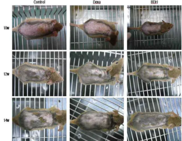
사진에서 보는 바와 같이 각각의 실험군은 최종 6주 후에 상 대적으로 외형 조직상 차이를 발견할 수 있었다. Fig. 1 은 10주, 12주, 14주의 피부염 유발 정도와 개선 정도를 나타내주는 사진 으로, BDH 투여군이 대조군에 비해서 피부염 유발 정도가 현저 하게 떨어졌음을 확인할 수 있다. 피부염 지수는 피부염의 심화 정도를 관능적 방법에 의하여 측정한 결과로, 대조군은 7.67 ± 1.21, 양성대조군인 Dexa 투여군은 3.33 ± 1.03, BDH 투여군은 2.67 ± 1.03 로 나타나 양성대조군과 BDH 투여군 모두에서 대조 군에 비해 유의성있는(**p<0.01) 감소를 나타내었다(Fig. 2).
Fig. 1. Comparison of skin manifestation in NC/Nga mice between
control and experimental group.
Atopic dermatitis was induced by TNCB treatment in the dorsal skin.Fig. 2. Effects of BDH on clinical skin features and severity in TNCB-induced NC/Nga mice.
Clinical skin index of dermatitis was defined as the sum of the individual scores graded as 0 (none), 1 (mild), 2 (moderate) and 3 (severe) for each of five signs and symptoms (itch, erythema/hemorrhage, edema, excoriation /erosion and scaling/dryness) ; Symptoms were evaluated by skin dryness, eruption and wound on the three parts of the body; ear, face and back. The results represent the mean ± S.E. Statistically significant value was calculated by compared with control group by student's t-test (**p<0.01).2. 배부 피부내 면역세포 변화에 미치는 영향 1) CD3+ 세포 비율에 미치는 영향
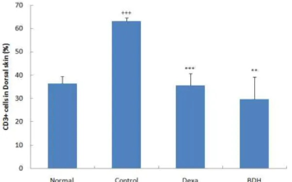
정상군은 36.45 ± 2.95 (%), 대조군은 63.10 ± 1.50 (%)로 나 타나 대조군에서 정상군에 비하여 유의성 있는 (+++p<0.001) 증 가를 나타내었다. Dexa 투여군은 35.55 ± 5.15 (%), BDH 투여군 은 29.80 ± 9.40 (%)로 나타나, 대조군에 비하여 유의성 있게 (***p<0.001, **p<0.01) 감소하였다(Fig. 3).
Fig. 3. Effect of BDH on CD3+ cell rate of Dorsal skin in TNCB-induced atopic dermatitis model of NC/Nga mice.
NC/Nga mice model followed by the treatment of BDH for 6 weeks. At the end of the experiment, the cells from Dorsal skin stained with anti-CD3 and positively stained cells were analyzed by flow cytometry. The results represent the mean ± S.E. Statistically significant value was calculated by compared with normal group by student's t-test (+++p<0.001). Statistically significant value was calculated by compared with control group by student's t-test (***p<0.001, **p<0.01).2) CD11b+/Gr-1+ 세포 비율에 미치는 영향
정상군은 11.45 ± 0.95 (%), 대조군은 33.35 ± 0.85 (%)로 나 타나 대조군에서 정상군에 비하여 유의성 있게 (+++p<0.001) 증 가를 나타내었고, Dexa 투여군은 23.75 ± 1.65 (%), BDH 투여군 은 24.95 ± 2.45 (%)로 나타나 대조군에 비하여 유의성 있게 (***p<0.001, **p<0.01) 감소하였다(Fig. 4).
3. 배부 피부에서의 사이토카인 mRNA 발현에 미치는 영향 1) IL-5 mRNA 발현에 미치는 영향
IL-5 mRNA 유전자 발현은 대조군의 RQ 값이 1.103 ± 0.103 일 때, 정상군은 0.673 ± 0.076으로 나타나, 정상군에 비하여 유의 성 있게 (++p<0.01) 증가하였다. Dexa 투여군은 0.797 ± 0.049, BDH 투여군은 0.613 ± 0.014로 나타나, 대조군에 비하여 유의성 있게 (**p<0.01, ***p<0.001) 감소하였다(Fig. 5).
Fig. 4. Effect of BDH on CD11b+/Gr-1+ cell rate of Dorsal skin in TNCB-induced atopic dermatitis model of NC/Nga mice.
NC/Nga mice model followed by the treatment of BDH for 6 weeks. At the end of the experiment, the cells from Dorsal skin stained with anti-CD11b and anti-Gr-1 and positively stained cells were analyzed by flow cytometry. The results represent the mean ± S.E. Statistically significant value was calculated by compared with normal group by student's t-test (+++p<0.001). Statistically significant value was calculated by compared with control group by student's t-test (***p<0.001, **p<0.01).Fig. 5. Effect of BDH Extract on the level of IL-5 mRNA in the dorsal skin of TNCB-induced atopic dermatitis model of NC/Nga mice.
Skin was obtained by biopsy and used for preparing total RNA. Isolated RNAs were analyzed by Real Time-PCR using gene specific oligomers. The results represent the mean ± S.E. Statistically significant value was calculated by compared with normal group by student's t-test (++p<0.01). Statistically significant value was calculated by compared with control group by student's t-test (**p<0.01, ***p<0.001).2) IL-13 mRNA 발현에 미치는 영향
IL-13 mRNA 유전자 발현은 대조군의 RQ 값이 1.060 ± 0.060일 때, 정상군은 0.809 ± 0.040으로 나타나, 정상군에 비하여 유의성 있게 (++p<0.01) 증가하였다. Dexa 투여군은 0.721 ± 0.027, BDH 투여군은 0.764 ± 0.116으로 나타나, 대조군에 비하 여 유의성 있게 (***p<0.001, *p<0.05) 감소하였다(Fig. 6).
4. 조직 변화에 미치는 영향
1) Hematoxylin and eosin 염색 변화
BDH 투여가 피부 조직에 미치는 영향을 알아보기 위하여
실험 종료 후 배부의 피부 일부를 채취하여 H&E 염색을 실시하
였다. H&E 염색을 한 대조군의 배부 피부 조직은 epidermis와
dermis가 부종으로 현저하게 확장되었고, 백혈구의 침윤도 관찰 된 반면 (Fig. 7B), BDH 투여군은 epidermis와 dermis의 두께 및 백혈구의 침윤이 상대적으로 감소하였다(Fig. 7D).
Fig. 6. Effect of BDH Extract on the level of IL-13 mRNA in the dorsal skin of TNCB-induced atopic dermatitis model of NC/Nga mice.
Skin was obtained by biopsy and used for preparing total RNA. Isolated RNAs were analyzed by Real Time-PCR using gene specific oligomers. The results represent the mean ± S.E. Statistically significant value was calculated by compared with normal group by student's t-test (++p<0.01). Statistically significant value was calculated by compared with control group by student's t-test (***p<0.001, *p<0.05).Fig. 7. Histologic examination of dorsal skin lesion in TNCB-induced NC/Nga mice.
NC/Nga mice model followed by the treatment of BDH for 6 weeks. Dorsal skin biopsies was stained with hematoxylin and eosin (H&E) (A;Normal, B; Control, C; Dexa, D; BDH) for examining inflammatory cells. The stained tissues were observed by bright microscopy (Nikon, Japan, orignal magnification, x100).
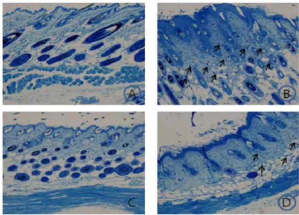
2) Toluidine blue 염색 변화
BDH 투여가 피부 조직에 미치는 영향을 알아보기 위하여 실험 종료 후 배부의 피부 일부를 채취하여 toluidine blue 염색 을 실시하였다. Toluidine blue 염색을 한 대조군의 dermis 주변 에 비만세포가 많이 침윤된 반면(Fig. 8B), BDH 투여군은 대조군 에 비하여 비만세포의 침윤이 감소되었다(Fig. 8D).
5. PBMC 내 면역세포 변화에 미치는 영향 1) CD4+/CD45+ 세포 비율에 미치는 영향
정상군은 4.73 ± 0.31 (%), 대조군은 14.20 ± 0.42 (%)로 나타 나 대조군에서 정상군에 비하여 유의성 있는 (+++p<0.01) 증가 를 나타내었고, Dexa 투여군은 9.14 ± 0.74 (%), BDH 투여군은 9.22 ± 0.12 (%)로 나타나 대조군에 비하여 유의성 있게 (***p<0.001) 감소하였다(Fig. 9).
Fig. 8. Histologic examination of dorsal skin lesion in TNCB-induced NC/Nga mice.
NC/Nga mice model followed by the treatment of BDH for 6 weeks. Dorsal skin biopsies was stained with toluidine blue (A; Normal, B; Control, C; Dexa, D; BDH) for examining mast cells. The stained tissues were observed by bright microscopy (Nikon, Japan, orignal magnification, x100).Fig. 9. Effect of BDH on CD4+/CD45+ cell rate of PBMC in TNCB-induced atopic dermatitis model of NC/Nga mice.
NC/Nga mice model followed by the treatment of BDH for 6 weeks. At the end of the experiment, the cells from PBMC stained with anti-CD4 and anti-CD45 and positively stained cells were analyzed by flow cytometry. The results represent the mean ± S.E.Statistically significant value was calculated by compared with normal group by student's t-test (+++p<0.001). Statistically significant value was calculated by compared with control group by student's t-test (***p<0.001).
Fig. 10. Effect of BDH on B220+/CD23+ cell rate of PBMC in TNCB-induced atopic dermatitis model of NC/Nga mice.
NC/Nga mice model followed by the treatment of BDH for 6 weeks. At the end of the experiment, the cells from PBMC stained with anti-B220 and anti-CD23 and positively stained cells were analyzed by flow cytometry. The results represent the mean ± S.E.Statistically significant value was calculated by compared with normal group by student's t-test (+++p<0.001). Statistically significant value was calculated by compared with control group by student's t-test (**p<0.01, *p<0.05).
2) B220+/CD23+ 세포 비율에 미치는 영향
정상군은 14.30 ± 1.60 (%), 대조군은 31.80 ± 1.90 (%)로 나
타나 대조군에서 정상군에 비하여 유의성 있는 (+++p<0.001) 증 가를 나타내었고, Dexa 투여군은 25.20 ± 1.40 (%), BDH 투여군 은 27.80 ± 0.50 (%)로 나타나 대조군에 비하여 유의성 있는 (**p<0.01, *p<0.05) 감소를 나타내었다(Fig. 10).
3) CD4+ 세포 비율에 미치는 영향
정상군은 9.28 ± 1.63 (%), 대조군은 15.61 ± 0.60 (%)로 나타 나 대조군에서 정상군에 비하여 유의성 있는 (++p<0.01) 증가를 나타내었고, Dexa 투여군은 8.60 ± 1.00 (%), BDH 투여군은 8.95
± 1.65 (%)로 대조군에 비하여 유의성 있게 (***p<0.001,
**p<0.01) 감소하였다(Fig. 11).
Fig. 11. Effect of BDH on CD4+ cell rate of PBMC in TNCB-induced atopic dermatitis model of NC/Nga mice.
NC/Nga mice model followed by the treatment of BDH for 6 weeks. At the end of the experiment, the cells from PBMC stained with anti-CD4 and positively stained cells were analyzed by flow cytometry. The results represent the mean ± S.E. Statistically significant value was calculated by compared with normal group by student's t-test (++p<0.01).Statistically significant value was calculated by compared with control group by student's t-test (***p<0.001, **p<0.01).
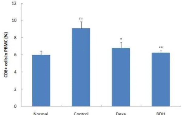
4) CD8+ 세포 비율에 미치는 영향
정상군은 6.00 ± 0.44 (%), 대조군은 9.14 ± 0.76 (%)로 나타 나 대조군에서 정상군에 비하여 유의성 있는 (++p<0.01) 증가를 나타내었고, Dexa 투여군은 6.80 ± 0.70 (%), BDH 투여군은 6.25
± 0.25 (%)로 대조군에 비하여 유의성 있게 (*p<0.05, **p<0.01) 감소하였다(Fig. 12).
Fig. 12. Effect of BDH on CD8+ cell rate of PBMC in TNCB-induced atopic dermatitis model of NC/Nga mice.
NC/Nga mice model followed by the treatment of BDH for 6 weeks. At the end of the experiment, the cells from PBMC stained with anti-CD8 and positively stained cells were analyzed by flow cytometry. The results represent the mean ± S.E. Statistically significant value was calculated by compared with normal group by student's t-test (++p<0.01).Statistically significant value was calculated by compared with control group by student's t-test (*p<0.05, **p<0.01).
5) CD3+/CD69+ 세포 비율에 미치는 영향
정상군은 3.80 ± 0.67 (%), 대조군은 7.20 ± 0.70 (%)로 나타 나 대조군에서 정상군에 비하여 유의성 있는 (++p<0.01)증가를 나타내었고, Dexa 투여군은 4.80 ± 1.10 (%), BDH 투여군은 6.06
± 0.26 (%)로 대조군에 비하여 감소하였다(Fig. 13).
Fig. 13. Effect of BDH on CD3+/CD69+ cell rate of PBMC in TNCB-induced atopic dermatitis model of NC/Nga mice.
NC/Nga mice model followed by the treatment of BDH for 6 weeks. At the end of the experiment, the cells from PBMC stained with anti-CD3 and anti-CD69 and positively stained cells were analyzed by flow cytometry. The results represent the mean ± S.E.Statistically significant value was calculated by compared with normal group by student's t-test (++p<0.01).
6) CD4+/CD25+ 세포 비율에 미치는 영향
정상군은 0.72 ± 0.02 (%), 대조군은 2.32 ± 0.16 (%)로 나타 나 대조군에서 정상군에 비하여 유의성 있는 (+++p<0.001) 증가 를 나타내었고, Dexa 투여군은 1.35 ± 0.08 (%), BDH 투여군은 1.03 ± 0.23 (%)로 나타나 모두 대조군에 비하여 유의성 있게 (***p<0.001) 감소하였다(Fig. 14).
Fig. 14. Effect of BDH on CD4+/CD25+ cell rate of PBMC in TNCB-induced atopic dermatitis model of NC/Nga mice.
NC/Nga mice model followed by the treatment of BDH for 6 weeks. At the end of the experiment, the cells from PBMC stained with anti-CD4 and anti-CD25 and positively stained cells were analyzed by flow cytometry. The results represent the mean ± S.E.Statistically significant value was calculated by compared with normal group by student's t-test (+++p<0.001). Statistically significant value was calculated by compared with control group by student's t-test (***p<0.001).
7) CD11b+/Gr-1+ 세포 비율에 미치는 영향
정상군은 23.35 ± 2.15 (%), 대조군은 41.70 ± 4.30 (%)로 나
타나 대조군에서 정상군에 비하여 유의성 있는 (++p<0.01) 증가
를 나타내었고, Dexa 투여군은 28.05 ± 1.45 (%), BDH 투여군은
36.70 ± 4.20 (%)로 대조군에 비하여 Dexa 투여군에서 유의성 있
게 (**p<0.01) 감소하였다(Fig. 15).
Fig. 15. Effect of BDH on CD11b+/Gr-1+ cell rate of PBMC in TNCB-induced atopic dermatitis model of NC/Nga mice.
NC/Nga mice model followed by the treatment of BDH for 6 weeks. At the end of the experiment, the cells from PBMC stained with anti-CD11b and anti-Gr-1 and positively stained cells were analyzed by flow cytometry. The results represent the mean ± S.E.Statistically significant value was calculated by compared with normal group by student's t-test (++p<0.01). Statistically significant value was calculated by compared with control group by student's t-test (**p<0.01).
6. 혈청내 histamine 생성량에 미치는 영향
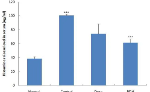
정상군은 38.5 ± 2.9 (ng/㎖), 대조군은 100.5 ± 1.9 (ng/㎖) 으로 나타나 정상군에 비하여 유의성 있는 (+++p<0.001) 증가를 나타내었다. Dexa 투여군은 74.1 ± 14.6 (ng/㎖), BDH 투여군은 61.3 ± 5.6 (ng/㎖)로 나타나, 대조군에 비하여 BDH 투여군에서 유의성 있게 (***p<0.001) 감소하였다(Fig. 16).
Fig. 16. Effect of BDH on the level of histamine in the serum of TNCB-induced atopic dermatitis model of NC/Nga mice.
The levels of histamine were determined using a commercially available ELISA kit. The results represent the mean ± S.E. Statistically significant value was calculated by compared with normal group by student's t-test (+++p<0.001). Statistically significant value was calculated by compared with control group by student's t-test (***p<0.001).고 찰
아토피피부염은 유전적․환경적․면역학적 요인 등과 관련 있는 만성 피부질환으로 홍반, 부종, 소양증, 인설 등이 특징으로 나타난다 1-5) . 유전적으로는 아토피피부염 환자의 50~70%가 가족 력을 가지고 있음이 보고된 바가 있으며 31,32) , 1916년에 Cooke와 Van der Veer는 양부모가 알레르기 질환을 가질 경우 자식의 발 병률은 75%, 한 부모가 알레르기 질환을 가질 경우 자식의 발병
률은 50%라고 보고하였으며, 특히 어머니의 아토피피부염 병력 이 가장 큰 위험요인이 될 수 있다고 하였다 33,34) . 유전인자 중에 서는 특히 5q31-33, 3q21 등 염색체와의 연관성이 보고되었으며, 독일과 스칸디나비아인을 대상으로 한 연구에서는 혈청의 IgE 농도와 3q21과의 연관성도 보고되었다 34-36) .
환경적 요인으로 인간의 주거환경과 관련하여 발생할 수 있 는 다양한 항원들 외에 식품 알레르기, 대기환경 등이 관여하는 것으로 보고되고 있다 37-41) .
현재 이러한 유전적, 환경적 요인과 더불어 면역학적 이상이 주요 병인으로 인식되면서 치료제 개발을 위한 실험에서 면역조 절능에 관한 다양한 실험이 이루어지고 있다.
아토피피부염은 내인성 아토피피부염(intrinsic or non-allergic atopic dermatitis, IAD)과 외인성 아토피피부염 (extrinsic of allergic atopic dermatitis, EAD)으로 구분할 수 있 는데, 현재 외인성 아토피피부염이 70~80%를 차지하며 이는 IgE 항체와 연관성을 가진다 42) .
IgE 항체는 아토피피부염 환자에서 외부의 단백 항원에 반 응하는 항체로, 아토피피부염 환자의 약 75~80%에서 혈중 IgE 및 호산구 증가가 나타나기에 35,43) , 일반적으로 IgE 발현량은 아 토피피부염을 진단할 수 있는 중요한 척도로 사용되어 진다. IgE 는 B세포가 발현하는 면역글로불린으로 Th2 사이토카인인 IL-4 에 의해 생성되는데 1) , IL-4가 B세포에서 IgE의 생성을 증가시키 고 IgE가 비만세포를 자극하여 히스타민을 포함한 염증물질의 방출을 유도해 피부에 가려움증 등의 증상이 유발된다 44,45) .
아토피피부염이 진행되면서 T세포의 활성화가 일어나 helper T cell로 변하게 되는데, 이 때 발현되는 사이토카인에 따 라 helper T cell은 Th1과 Th2 세포로 분화하게 된다 46) . 즉 아토 피피부염은 Th2 반응에 치우친 면역반응으로 Th2에서 발현되는 사이토카인인 IL-4, IL-5, IL-10, IL-13 등이 많아지고, Th1에서 발 현되는 사이토카인인 IFN-γ와 IL-2가 상대적으로 적어지면서 인 체 내 면역학적 불균형으로 발생한다고 보고되고 있다 11,35,47) . 이 와 같은 반응은 급성 아토피피부염일 경우 나타나는 것으로 앞 서 IL-4와 IL-13은 B세포의 동형전환을 유발하여 IgE 생성을 증 가시키며, IL-5는 호산구를 동원하고, IL-10은 Th1이나 대식세포 의 IFN-γ의 생산을 억제시키는 것으로, 정상일 경우에는 IL-4가 Th1 반응을 억제하고 IFN-γ는 Th2 반응을 억제하여 상호 균형 을 유지하나, 아토피피부염이 진행되면서 위와 같은 면역반응이 진행되는 것이다 46,48,49) . 그러나 아토피피부염이 만성화 될 경우에 는 급성일 경우 증가되었던 IL-4, IL-5의 수는 적으면서 IFN-γ, IL-12, GM-CSF, Chemokine 등은 급성기에 비해 증가함으로써 Th2반응이 Th1반응으로 전환된다고도 보고도 있다 35,50,51) .
본 연구에서는 임상 활용방인 배독환의 객관적 효능 평가를 위하여 실험 연구에서 가장 기본적인 면역학적 지표를 측정 검 사하였다. 배독환은 烏藥, 葛根, 麻黃, 桂枝, 當歸, 白芷, 蘇葉, 乾 薑, 桃仁, 紅花, 甘草, 大棗로 구성되었는데, 본 실험과 관련된 이 들 구성 약물의 항염, 면역증강 및 항산화 효과 등은 이미 많은 실험적 보고가 되어 있다.
이 등 22) 은 “烏藥이 토끼의 수축혈관에 미치는 영향”을 통해
烏藥의 順氣시키는 약리작용에 의한 혈관이완효능을 보고하였 고, 김 등 23) 은 “갈근 추출물에 의한 염증성 Cytokine 생성 억제 및 Prostaglandin E2 활성 저해에 관한 연구”를 통해 갈근의 항 염증 및 소염효과를 보고하였으며, 김 등 24) 은 “마황의 면역작용 에 미치는 효과”에서 마황이 세포성 및 체액성 면역 반응에 대해 억제적으로 작용함을 보고 하였다. 박 등 25) 은 계지의 항염 효과 를, 박 등 26) 은 당귀의 면역증강제 개발 가능성을, 이 등 27) 은 백지 의 항알러지, 항산화 효과, 강 등 28) 은 蘇葉의 알레르기성 염증 반 응에 유의한 효과를 각각 보고한 바가 있다. 이 밖에 황 등 29) 은
“桃仁 및 紅花를 포함하는 方藥合編處方에 대한 考察”에서 어혈 을 제거하고 혈행을 촉진시키는 도인을 홍화와 함께 사용했을 때 치료 제고 효과를, 金 등 30) 은 “桃仁 및 紅花 藥鍼의 鎭痛․抗 血栓 效能에 關한 硏究”에서 도인과 홍화의 항혈전 효과를 보고 하였다.
현재 아토피피부염의 양방학적 치료제로는 스테로이드제, 항히스타민제, 항생제, 면역억제제 등이 있는데 52,53) , 본 실험에서 는 알레르기 및 염증 치료제로 사용하면서 다른 실험군과 동일 한 방법의 처치가 가능한 용해성 있는 dexamethasone을 양성대 조군으로 사용하였다. Dexamethasone은 cytokine 생산을 억제하 는 스테로이드제로 이러한 부신피질호르몬은 면역세포들의 파괴 나 기능저지, 재분배의 작용이 있는 것으로 생쥐, 토끼, 햄스터 등의 종에서 흉선, 비장, 임파선에 민감하다고 알려져 있다 54,55) . 동물 병태 모델에 사용한 NC/Nga 생쥐는 SPF(specific pathogen free) 조건하에서는 정상적인 상태를 유지하나, 일반적 환경에서는 가려움으로 인한 긁기와 습진성 피부염의 증상이 나 타나는 아토피 피부염 질환 동물 모델이다 56,57) . NC/Nga 생쥐의 아토피피부염 발병은 면역학적 이상이 사람에게 발생되는 병변 과 유사하지만 자연 유발률이 50% 이하이면서 발병정도와 발병 시기가 다양해 아토피치료제 개발과 관련하여 실험 효능을 평가 하기에는 재현성이 낮다는 단점이 있다 58-60) . 따라서 본 실험에서
는 NC/Nga 등 부위를 제모하고, TNCB
(2,4,6-trinitrochlorobenzene)를 일정기간 동안 반복 도포하여 아 토피피부염을 유발시켰는데, TNCB의 적용은 피부가 두꺼워지고 부종이 생기면서 손상된 피부에 염증세포를 침윤시키는 등의 증 상이 나타나는 것으로 아토피피부염의 병변과 유사하게 나타난
다 61,62) . 본 실험 대조군에서는 기존의 보고대로 피부 병태 현상이
뚜렷하게 나타나 TNCB로 유발된 NC/Nga 생쥐의 약물 투여 기 간에 따른 피부 상태 개선 정도와 피부염 지수의 관능 평가를 정 확하게 측정할 수 있었다(Fig. 1). 외형상 피부염 발생 정도는 사 진에서 보는 바와 같이 상대적인 차이를 나타냈으며, 피부염 임 상 지수에서도 12주가 지나고 부터는 대조군도 자체 피부개선효 과가 나타났으나, BDH 투여군에서 12주와 14주 때 대조군에 비 해 각각 41.2%, 65.1%로 유의성 있게 감소하였다(Fig. 2).
H&E 염색과 toluidine 염색을 통한 조직변화에서도 임상지 수나 외형상 나타난 피부 증상 결과와 부합되는 결과가 도출되 었는데, 대조군의 배부 조직이 부종으로 확장되거나 비만세포의 침윤이 확인된 반면 (Fig. 7B, 8B), BDH 투여군에서는 대조군에 비해 표피, 진피의 두께와 비만세포의 침윤이 상대적으로 감소되
었음을 확인할 수 있었다(Fig. 7D, 8D).
CD(cluster of defferentiation) 들은 T세포의 결정표지인자 로 63) , 세포-세포 상호작용 및 부착을 증진하는 것과 림프구를 활 성화시켜 신호를 전달하는 기능을 한다 64) . 그 중 CD45+는 T 및 B 세포 항원 수용체 매개 신호 전달을 조절하는데 중요한 역할 을 수행하는 것으로 알려져 있으며 64) , PBMC 내의 CD4+/CD45+
는 BDH 투여군이 대조군에 비해 35.0% 유의성 있게 감소한 것 으로 나타났다(Fig. 9).
면역반응의 조절에 관여하는 T 세포를 보조 T 림프구 (helper T cell)라고 부르며 직접 다른 세포를 죽이는 기능을 가 진 T 세포를 세포독성 T 림프구(cytotoxic T cell)라고 부른다.
helper T cell의 대부분은 세포 표면에 TCR(T cell receptor) 외에 CD4 분자를 공통으로 가지고 있으며, cytotoxic T cell은 CD8 분 자를 가지고 있다 63) .
아토피 피부 병변에 분포하는 T 세포의 주된 구성은 CD4+
세포이며 CD4+ 세포에서 생성되는 사이토카인에 따라 Th1과 Th2 세포로 구분되는데, 아토피 피부염은 Th2 세포 반응이 Th1 보다 우세할 경우 및 보조 T 세포(CD4+ T cell)에 대한 억제 T 세포(CD8+ T cell)비의 감소 등이 병의 악화에 영향을 준다는 가 설이 제시 되었다 35) .
CD4+T 세포는 여러 종류의 사이토카인을 생산하여 B cell이 효율적으로 항체를 생산하도록 도와주고, 면역 반응의 행동 세포 를 활성화시키는 역할을 하며 65,66) , CD8+ T 세포는 사이토카인의 생산보다는 세포 살해 활성(cytotoxic activity)을 가지고 있는 altered self-cell을 제거하는 역할을 한다 67) .
PBMC에서 CD4+ 세포 비율은 정상군에 비해 대조군에서 68.2% 유의성 있게 증가하였으나, Dexa 투여군과 BDH 투여군에 서는 각각 44.9%, 42.7%로 유의성 있게 감소하였다(Fig. 11). 또 CD8+ 세포 비율은 정상군에 비해 대조군에서 52.3% 유의성 있 게 증가한 반면, Dexa 투여군과 BDH 투여군에서는 각각 25.6%, 31.6% 유의성 있게 감소하여(Fig. 12) BDH 투여로 인해 이상적 인 면역 반응 활성이 억제되었음을 시사하였다.
CD23은 B세포 활성 분자로 IgE 생성 및 염증 반응에 관계 되고 68) , B220+/CD23+는 성숙한 B 세포를 나타내는 것으로 69,70) , PBMC에서 B220+ /CD23+ 세포 비율은 정상군에 비해 대조군에 서 122.4% 유의성 있게 증가함으로써 IgE 생성 증가 및 염증 반 응이 활발히 진행되었음을 확인할 수 있었으며, Dexa 투여군과 BDH 투여군에서는 각각 20.8%, 12.6% 유의성 있게 감소함으로 써 (Fig. 10) 본 시료의 아토피 피부염의 개선 효과를 확인할 수 있었다.
활성화된 T 세포를 의미하는 CD3+/CD69+세포 71) 는 PBMC
내에서 정상군에 비해 대조군이 89.5% 유의성 있게 증가하였고,
BDH 투여군에서는 15.8% 감소하였다(Fig. 13). CD3 분자는 T 림
프구 항원 수용체에 항원이 결합된 신호를 세포안으로 전달하는
기능을 하는 것으로 72) , 배부 피부에서도 CD3+세포 비율은 정상
군에 비해 대조군이 73.1% 유의성 있는 증가를 나타내었으나
BDH 투여군에서 52.8% 유의성 있는 감소를 나타내어 본 시료의
면역조절 작용은 PBMC와 배부 피부 모두에서 확인할 수 있었다
(Fig. 3).
T 세포 중에는 면역반응을 억제하는 세포들이 존재한다는 것이 확인되어 이를 조절 T 세포 (regulatory T cell) 라고 하며 작동 T 세포와는 다르게 이를 억제하는데, 구성적으로 CD25를 발현하고 TCR 이외에 대부분 CD4분자와 CD25 분자를 가지고 있어서 CD4+/CD25+ regulatory T 세포라고도 부른다 64,73) .
PBMC 내의 CD4+/CD25+는 정상군에 비해 대조군에서 231.9% 유의성 있는 증가를 나타내었고, BDH 투여군에서는 55.6%로 유의성 있게 감소하였다(Fig. 14). CD4+/CD25+ 관련된 동물 실험에서는 기존의 실험 결과에서도 대조군에 비하여 감소 하는 것으로 나타났으나, 임상 보고에서 아토피피부염 환자에서 CD4+/CD25+가 감소되었다는 연구 결과 74) 와는 서로 상반된 결 과로, 향 후 이와 관련된 다양한 연구를 통하여 해석이 필요할 것으로 사료된다.
그 밖에 과립세포를 나타내는 CD11b+/Gr-1+ 세포는 PBMC 와 배부 피부에서 대조군은 정상군에 비해 각각 78.6%, 191.3%로 유의성 있게 증가하였으며, BDH 투여군에서는 12.0%, 25.2%로 감소하여 BDH 투여가 피부 조직으로의 면역세포 침윤을 억제한 것으로 나타났다(Fig. 4, 15).
앞서 기술한 바와 같이 아토피피부염은 Th2 반응에 치우친 면역반응으로 Th2에서 발현되는 사이토카인인 IL-4, IL-5, IL-10, IL-13 등이 많아지고, Th1에서 발현되는 사이토카인인 IFN-γ와 IL-2가 상대적으로 적어지면서 나타나는 인체 내 면역학적 불균 형으로 발생한다. 본 실험에서는 배부 피부내의 염증 관련 사이 토카인인 IL-5, 13의 mRNA 발현량을 검색하였는데, 모두 대조 군에 비해 각각 44.4%, 28.0% 유의성 있는 감소를 나타내었다 (Fig. 5, 6).
Histamine은 IgE가 비만세포를 자극하여 염증 물질과 함께 방출되는 것으로 44,45) , 대조군에서 정상군에 비해 161.0% 유의성 있게 증가하였으며, BDH 투여군에서 39.0% 유의성 있게 감소하 여 염증 반응이 감소되었음을 확인할 수 있었다(Fig. 16).
이상의 결과로 BDH 투여는 임상에서 뿐만 아니라 NC/Nga 생쥐를 이용한 실험적 연구에서도 아토피 피부염 유관인자에 대 한 유의성 있는 결과가 도출됨으로써 면역 억제 효능이 인정되 었다. 향 후 개별 약물 효능과 약물 상호 작용에 대한 보완적 연 구가 요구되며, 이를 통하여 보다 효과적인 치료 처방에 대한 근 거가 제공될 수 있을 것으로 사료된다.
결 론
한의학적 치료의 객관성 확보를 위하여 현재 임상에서 아토 피 피부염에 활용되고 있는 排毒丸을 시료로 NC/Nga 동물 병 태 모델에서 임상지수, 면역세포 변화, 사이토카인 mRNA 발현, 조직학적 변화 및 혈청 histamine 생성량 등을 측정하여 다음과 같은 결론을 얻었다. 먼저 최종 결론이라고 할 수 있는 피부염 임상 지수에서는 대조군에 비하여 65.1%로 유의성 있게 감소시 켰으며, 육안적으로도 뚜렷한 피부 병변 차이를 나타내었다. 조 직 검사에서는 대조군에 비하여 백혈구와 비만세포가 피부로 침
윤되는 것을 억제하고 비후해진 epidermis와 dermis의 두께를 감소시켰다.
다음으로 면역반응에 대한 효과를 확인하기 위하여 배부 피 부내 CD3+, CD11b+/Gr-1+ 면역세포율을 검색한 결과 각각 52.8%, 25.2% 유의성 있게 감소되었으며, IL-5 mRNA, IL-13 mRNA 발현 역시 각각 44.4%, 28.0%로 유의성 있게 감소되었다.
PBMC 내 CD4+/CD45+, B220+/CD23+, CD4+, CD8+, CD4+/CD25+ 면역 세포율 역시 각각 35.0%, 12.6%, 42.7%, 31.6%, 55.6% 유의성 있게 감소시켰으며, 혈청 내 histamine 생 성량은 39.0%의 유의성 있는 감소가 나타났다.
이상의 결과로 임상활용 처방인 배독환의 항아토피피부염 효능이 객관적으로 규명됨으로써 임상에서의 폭넓은 활용이 기 대되며, 향 후 개별 약물 효능과 약물 상호 작용에 대한 보완적 연구를 통하여 보다 효과적인 치료 처방에 대한 근거가 제공될 수 있을 것으로 사료된다.
감사의 글
본 연구는 지식경제부 지정 대전대학교 난치성면역질환의 동서생명의학연구 지역혁신센터의 지원에 의한 것임.
참고문헌